Nanotechnology-Based Antimicrobial and Antiviral Surface Coating Strategies


1
Faculty of Engineering and Natural Sciences, Bahcesehir University, Istanbul 34353, Turkey
2
Institute of Pharmaceutical Biology, Goethe University Frankfurt, 60438 Frankfurt, Germany
3
Department of Biomedical Technologies, Graduate School of Natural and Applied Sciences, Ege University, İzmir 35100, Turkey
*
Author to whom correspondence should be addressed.
Prosthesis 2021, 3(1), 25-52; https://0-doi-org.brum.beds.ac.uk/10.3390/prosthesis3010005
Submission received: 6 January 2021
/
Revised: 22 January 2021
/
Accepted: 27 January 2021
/
Published: 1 February 2021
(This article belongs to the Special Issue Antiviral and Antimicrobial Surface Design Strategies)
Abstract
:Biocontamination of medical devices and implants is a growing issue that causes medical complications and increased expenses. In the fight against biocontamination, developing synthetic surfaces, which reduce the adhesion of microbes and provide biocidal activity or combinatory effects, has emerged as a major global strategy. Advances in nanotechnology and biological sciences have made it possible to design smart surfaces for decreasing infections. Nevertheless, the clinical performance of these surfaces is highly depending on the choice of material. This review focuses on the antimicrobial surfaces with functional material coatings, such as cationic polymers, metal coatings and antifouling micro-/nanostructures. One of the highlights of the review is providing insights into the virus-inactivating surface development, which might particularly be useful for controlling the currently confronted pandemic coronavirus disease 2019 (COVID-19). The nanotechnology-based strategies presented here might be beneficial to produce materials that reduce or prevent the transmission of airborne viral droplets, once applied to biomedical devices and protective equipment of medical workers. Overall, this review compiles existing studies in this broad field by focusing on the recent related developments, draws attention to the possible activity mechanisms, discusses the key challenges and provides future recommendations for developing new, efficient antimicrobial and antiviral surface coatings.
1. Introduction
Treatment of infectious diseases is presently tackling a crisis. Therapeutic options against bacterial pathogens have been limited by common antibiotic resistance. On the other side, the recurrent emergence of viral pathogens also poses a significant threat [1].
Adhesion and colonization of microorganisms on implanted medical instruments including catheters, knee and hip implants and pacemaker leads are the main health care problems that affect patient life-quality [2]. They exhibit high risks of local and systemic infections after implantation. Microorganism binding limits the lifetime and functionality of medical devices, as well [3].
The increased utilization of patient-specific devices due to availability and incorporation of new technologies may help to solve recently faced world-wide medical challenges [4]. Recently, synthetic biomaterials have demonstrated some exciting possibilities in the field of medicine. Their field of application ranges from medical devices, pharmaceuticals and tissue replacement therapies to engineered nanorobots developed for cellular intervention [5,6]. Bulk properties of a material are important to initially establish material suitability for an application. For example, porosity of a scaffold is important for proper migration of cells and blood perfusion to deeper sites of the implant. In addition to bulk properties, surface properties including both physical features and chemistry are essential for the functionality of many biomedical devices [4]. Using topographical modifications and chemical surface modifications, such as covalently bound coatings, bacterial colonization and biofilm formation on surfaces have been decreased, to minimize the probability of infection spreading from medical devices [7].
Additionally, in recent years, research has shown the antiviral activity of nanomaterials on several viruses. The SARS-Cov-2 (severe acute respiratory syndrome-related coronavirus-2), also known as COVID-19, pandemic spread globally over the duration of 2020 and has not yet seen an end. In addition to SARS-CoV-2, several viral pandemics, including influenza A virus subtypes H2N2 and H3N3, human immunodeficiency virus (HIV) and SARS, have been documented in recent history, while Middle East respiratory syndrome-related coronavirus (MERS-CoV) and Ebola viruses are still regarded in the pre-pandemic stage [8]. Existing anti-microbial technologies and virus inactivation systems can be improved to develop antiviral products, which may find a solution for this fatal infection [6,9]. Recently, we have witnessed a rousing development: Sahin and his colleagues invented a lipid nanoparticle-formulated mRNA vaccine suggesting a high potential to protect against COVID-19 [10]. In this manner, to control the spreading of the current pandemic situation, nanosized materials are also useful for exhibiting antiviral performance on surfaces through various ways of action, such as physical contact, generation of reactive oxygen species (ROS), catalytic oxidation, photothermal effects and metal ion release. The antiviral nanomaterials can be applied as spray agents to manufacture antiviral surfaces that help to manage the spreading of viruses. Consequently, they can be used in a broad range of applications, such as air purifier filters, ventilation systems, medical textiles [11].
This review gives an overview of anti-microbial and anti-viral coating materials by mainly focusing on studies published in the last five years and giving some key examples from the earlier literature. We aim to propose possible mechanisms for the development of new anti-bacterial/-viral approaches using the nanoscience platform.
2. Coating Strategies
The initial role of surface coatings in industrial applications was to provide protection from corrosion and mechanical resistance [12]. Recently, with the advancement in nanoscience, polymer-/nanocomposite-based coatings have been developed and utilized for several purposes including biomedical applications, such as antibacterial surfaces [13,14,15,16].
The polymer coating methods can be summarized as the following categories: simple solution and dip coatings, cast-coating, Doctor-blading, spraying method and spin coating technologies. In the simple drop-casting coating method, a polymer solution is dropped and coated on a substrate and allowed to evaporate (Figure 1a). The dip coating technique includes immersion of the substrate in polymer melt or solution, then withdrawing and solvent evaporation, followed by drying (Figure 1b) [13]. Free polymeric films can also be obtained by the cast-coating technique, where a polymeric solution is cast onto a nonstick mold with a desired shape, and subsequently the solvent is evaporated (Figure 1c) [17].
Doctor blade, also known as tape casting, is one of the commonly employed methods for generating thin films on large surface areas. With a constant relative movement of the blade on the substrate, the polymer solution spreads on the substrate and forms a thin film, consequently a gel-layer after drying (Figure 1d) [14]. The spraying method is also a fast method utilized for polymer coating, particularly advantageous for coating three-dimensional solid objects. Melted or dissolved polymer is sprayed onto a surface in this technique. In the nozzle of the spray head, the polymer solution is atomized and dispersed to the surface with a continuous droplet flow (Figure 1e). Finally, in the spin coating technique, a small drop of coating material is dropped onto the center of the substrate, which is spun at the chosen speed to spread the coating material by centrifugal force and to achieve high quality and fine film (Figure 1f) [13].
In addition to polymer coatings, incorporation of inorganic/organic nanoparticles into a coating material has been developed as an alternative way to further develop the features of the existing surface coating to meet the promptly changing demands of medical applications. Previously, numerous surface treatment techniques, involving electroplating, electroless plating, and chemical conversion coating, was studied to improve surface functionality. For instance, Jiang et al. worked a novel silane-TiO2 dual-functional coating material that is prepared by controlled addition of nanoparticles on stainless steel (Figure 2). In this study, dispersed nano-SiO2 showed a high contact angle value, which increased the hydrophobicity of surface, and the TiO2 nanoparticles provided additional protection due to their photocatalytic activity [12].
Moreover, many surface-coating types, such as non-ionic or charged coatings, chemically functionalized coatings, and hydrophilic/hydrophobic coatings, have been revealed to influence properties of nanoparticles. These coatings, especially, have been preferred to improve the therapeutic function, colloidal stability to prevent agglomeration, and biocompatibility of nanomaterials. Once NPs encounter with biological fluids, proteins adsorb on them through electrostatic, dispersive, and covalent interactions, leading to the formation of a so named “protein corona” mainly determining the biological activity of the particle [15]. For instance, polyethylene glycol (PEG) coating has decreased protein adsorption via steric repulsion forces and led to longer circulation times and improved biodistribution [19].
In this part of the review, we focused on the variety of materials, which could be employed as coating materials, to improve antibacterial and antiviral properties for potential medical applications.
3. Coated Surfaces
3.1. Metal-Based Nanomaterial Coatings
In combating drug-resistant pathogens, biomedical devices modified with antimicrobial metal nanoparticles offer a strong microbicidal approach and have gained significant consideration in both the pharmaceutical and academic industries. Antimicrobial nanomaterials can be categorized into three major group; intrinsically antimicrobial ones, anti-microbial agent carriers, and those that occupy either of these functional features [16].
Many studies have shown that metal ions and metal-based materials, including the nanoparticles of gold (Au-NPs) [20], silver (Ag-NPs) [21,22], magnesium oxide (MgO-NPs) [23], copper oxide (CuO-NPs) [24], titanium oxide (TiO2-NPs) and zinc oxide (ZnO-NPs) [25] could be used to generate antimicrobial coatings. However, the knowledge about their long-term effects on human health and the environment is limited. The possible accumulation in organs and uncontrolled release of metal ions should be carefully investigated, and protective coatings might be useful in this context. Among the metal-oxide particles, MgO and ZnO have been recently reported as biocompatible nanoparticles with biocompatible degradation by-products, owing to their usage as trace elements in the human body [25,26]. The possible antibacterial mechanisms metal-oxide nanoparticles are not completely revealed yet. Findings have shown that ion concentrations, oxidative stress and membrane damage are the possible mechanisms of action against bacteria [23].
In a present work [27], the monolithic ZnO and composite ZnO with carbon (ZnO-C) and ZnO with copper (ZnO-Cu) were sputter-deposited using a vacuum coating technique, magnetron sputtering. All sputter surfaces were ethanol-sterilized and used for the antimicrobial test. In this study, Pseudomonas aeruginosa (P. aeruginosa) and Staphylococcus aureus (S. aureus) were selected as a resistant and a sensitive strain to Zn2+ ions, respectively. The coated surfaces were either submersed into bacterial solutions or were placed in direct contact with bacteria in solid medium, as well as the experiment were conducted under three light conditions: visible light, no light and UV light (365 nm). Visible light exposure particularly increased antimicrobial effect of the nanocomposite surfaces, and under UV pre-treatment, the antimicrobial activity of all surfaces increased because of the ROS generation. The ZnO-C nanocomposite coatings reported as the most efficient surfaces against the resistant P. aeruginosa inhibition.
Though the last decade has seen great progress in metal nanoparticles and their antibacterial efficiencies, it is also worth and timely to emphasize on the antiviral properties of metal nanoparticles. Inactivation of viruses before their binding to the host cells is the most direct way to control the spreading of viral infections. For example, heparan sulfate (HS) proteoglycans, which are expressed on the surface of almost all eukaryotic cell types, are the most conserved targets for viruses like Herpes simplex virus (HSV), HIV-1, human papilloma virus (HPV). Recently, Au-NPs were modified with mercaptoethanesulfonate based on its mimicry of HS were demonstrated to impede viral attachment, cellular entrance, and spreading [8,28]. Metal NPs including Fe or Cu in the ionic form can be a catalyzator in the generation of free radicals (ROS) that oxidize the capsid proteins and thus preventing the viral infection at early stage. Polyethylenimine (PEI) modified AgNPs can attach and deliver siRNA, which exhibited improved capabilities for cellular uptake and stopping Enterovirus 71 (EV71) virus infection [29]. In another key study, the addition of AgNPs to neutralizing antibodies has considerably improved the potential of neutralizing for antibodies in prevention of cell-associated HIV-1 transmission and infection [30].
3.2. Polymer-Based Surfaces
Polymers, with various chain length scales, have been the subject of a broad range of biosystems. Controllable surface chemistries and mechanical properties have made polymers favored materials for incorporating them into diverse molecular and supramolecular organizations. The bio-passive polymer layers, which are formed on the treated surfaces, facilitate minimum protein adsorption to occur, and consequently inhibit the bacterial adhesion [2]. Polymers, such as polyurethane (PU) and poly- (ethylene glycol) (PEG), have been considered to diminish in vitro adhesion of bacteria. However, the in vivo efficacy fluctuates usually with polymer composition, the length of the chains [31,32], surface chemistry [33], and among bacterial species [34,35].
The dentistry is one of the fields that utilizes polymers as bio-adhesives and antimicrobial agents, and for the controlled release of intracanal drugs. Bio-adhesive nanomaterials have been demonstrated to be beneficial for reaching inaccessible sites of teeth and roots. Nguyen et al. reported that pectin-coated liposomes could be created naturally on tooth enamel by forming charge complexes with adsorbed the hydroxyapatite (HA) in vitro, and they can remain on the surface as protective biofilms. They also showed that the negatively charged liposomes have the most durability in saliva [36,37]. Moreover, metal-based nanoparticles such as silver nanoparticles (Ag-NPs), zirconium oxide nanoparticles (ZrO2-NPs) or platinum nanoparticles (Pt-NPs) was incorporated into polymethyl methacrylate (PMMA) to decrease bacterial or fungal colonization on denture bases or tooth prosthesis, thanks to their anti-adhesive properties. A possible mechanism for the prevention of biofilm formation by a polymeric film coating is represented in Figure 3. NP coated surfaces resist on colonization owing to its metal ion content, while uncoated surfaces cannot resist on the biofilm formation [37].
In addition to implants, surgical sutures are also optimal surfaces for the bacterial adhesion and subsequent surgical site infections, which can conclude in severe complications after surgical interventions. Due to the recent increase in antibiotic-resistant bacterial strains, exploring inherently anti-bacterial polymeric sutures have gained importance. These polymeric materials are advantageous for providing a long-term antibacterial activity and reduced cytotoxic effects on the applied tissue, as well as good tissue adhesiveness for healthy wound closure following surgery. In addition, if they are biodegradable, it is possible to skip the removal step of sutures once healing is completed [38,39]. Antimicrobial effects of biopolymers can be improved by altering functional groups to control charge density, hydrophilicity or by incorporating other anti-microbial molecules [38]. For instance, Reinbold et al. developed a coating for surgical sutures composed of an antibacterial substance totarol, a natural diterpenoid isolated from Podocarpus totara tree together with as a biodegradable polymeric drug delivery system, poly (lactide-co-glycolide acid) (PLGA). The results of agar diffusion test confirmed that the PLGA/totarol-coated sutures were effective against S. aureus infection over a period of 15 days, and biocompatibility of coated-sutures were confirmed on murine fibroblasts by a 3-(4,5-dimethylthiazol-2-yl)-2,5-diphenyltetrazolium bromide (MTT) assay [40].
As mentioned in the selected examples, antibacterial polymers have been used in numerous areas for decades. The systematic optimization of the polymer composition, length of chain, charge, hydrophobicity, cost-effectiveness, scalability, and biocompatibility are crucial for the effectiveness of these systems. In this section, we focus on the main design principles polymeric antibacterial and antiviral surfaces as well.
3.2.1. Antimicrobial Agent Coupled Polymers
As an alternative to polymers that reduce the microbial adhesion, polymers coupled with antimicrobial agents including antibiotics, quaternary ammonium compounds, guanides, phosphonium salts are widely preferred to kill microbes upon contact. There are various methods to combine antimicrobial agents with polymeric materials. Among them, covalent binding of antimicrobial agents onto polymer backbone presents better uniformity and mechanical stability compared to surface physisorption approaches [2].
Antimicrobial peptides are evolutionarily ancient weapons produced by many species including microorganisms, plants, invertebrates and animals [41]. They are predominately polypeptides that contain less than 50 amino acids with an overall cationic charge [2]. Antimicrobial peptides are known to particularly target bacterial membranes, which are organized to have negatively charged phospholipid heads at the outside of the lipid bilayer. In contrast to conventional antibiotics such as penicillin, which are readily bypassed by microbes, gaining resistance by a microbial strain against antimicrobial peptides is unlikely to happen [41]. Antimicrobial peptides exhibit selective attraction to the more negatively charged bacteria over human cells which is facilitated by the electrostatic binding of the cationic groups of peptides. Then, the peptides’ amphiphilic structure leads the incorporation of their hydrophobic sidechains into the lipid membrane that interrupts the membrane integrity, initiating the leakage of cellular components, disruption of membrane potential, and consequently cell death [42].
3.2.2. Cationic Polymers
Cationic polymers, bearing electropositive groups, have been used to generate bioactive coatings that kill microbes via contact-dependent manner without releasing any chemicals. They act like the cationic peptides, and they are comparatively less expensive and easier to synthesize. In biomedical applications, ammonium, phosphonium, sulfonium, pyridinium salts, and guanidines are the most used ones. Especially, quaternary ammonium salts have a broad antibacterial activity. These cations used to functionalize long hydrophobic alkyl chains of various polymers, which are later immobilized on the surface to ensure bactericidal action by contact [43]. Briefly, polycationic agents act by their adsorption, via the positively charged groups, onto negatively charged bacterial surfaces, which then causes an increase in cell permeability and disruption of the cell membrane [44].
Cationic antimicrobial polymers can be categorized regarding their origins, as natural or synthetic. Natural cationic polymers and their derivatives include chitosan, gelatin, dextran, cellulose and cyclodextrin. Moreover, poly(ethyleneimine) (PEI), poly-L-(lysine) (PLL), and poly [2- (N, N-dimethylamino) ethyl methacrylate] (PDMAEMA) are the most known synthetically produced cationic polymers. Interesting examples of natural and synthetic cationic polymers that possess antimicrobial and antiviral effects were chosen from the literature and listed in Table 1.
Natural cationic polymers are known as non-toxic, biocompatible and biodegradable [69]. Gelatin is one of the mostly used natural polymers, which is obtained by hydrolysis of animal originated collagen, and it can be applied to many medical and pharmaceutical applications [70]. Two kinds of gelatin are obtained: specifically, type A (by acid hydrolysis) and type B (by alkaline hydrolysis). At physiological pH, gelatin type A is positively charged, whereas gelatin type B possesses negative charges. It has the ability to form poly-ion complexes with positively or negatively charged therapeutics, depending on the type [51].
Dextrans, which are water-soluble polysaccharides composed of glucose units, are widely available and easy to process [71]. Cationic-derivatives of dextrans are mostly used in cosmetic applications [72]. Similarly, cyclodextrins (CDs) are produced from bacteria as sugar derivatives with hydrophilic and lipophilic parts [73]. Cationic derivatives of CDs have many advantages, owing to the monodisperse structure. It can be chemically modified easily, and its toxicity is considerably low [74]. It shows high affinity to viral vectors and nucleotides [75].
Another natural polymer, cellulose is a fibrous and water-insoluble plant- or bacteria-based polysaccharide, which is declared as the most common organic compound all over the world. Its cationic derivatives have many advantages, such as hydrophilicity and antibacterial properties [76]. Owing to its antibacterial properties, it is used in several applications of textile, food packaging and medical industry [77].
In addition to natural cationic polymers, synthetically produced homopolymers of positively charged amino acids, such as poly-l-lysine, have been used widely for nucleic acid delivery in viral diseases [78]. It is mainly classified as α-poly-lysine and ε-poly-lysine. However, ε-poly-lysine form is more preferred for being less toxic and hydrophilic. For this reason, it is widely used in several areas such as drug delivery, antimicrobial medical applications, the food industry and so on [79,80].
As described above, there are many natural and synthetic cationic polymers used as antimicrobial agents. Among them, chitosan and PEI have been the mostly studied cationic polymers in the literature for their antimicrobial properties [74]. For this reason, in this section, we focused on chitosan and PEI, as representative examples of antimicrobial cationic natural and synthetic polymers. These polymers were reviewed in detail and their most remarkable antimicrobial applications were explained.
Chitosan, also known as deacetylated chitin, is a cationic natural polymer which is composed of randomly β-(1–4)-linked d-glucosamine and N-acetyl-d-glucosamine molecules. It is the structural skeleton element of insects and the cell walls of fungus [81]. Chitosan is generally utilized in drug and/or gene delivery, water treatment, heavy metal remediation and functional foods due to its bioactivities with the aid of positively charged amino groups of chitosan chain. It has a pKa value of 6.5 and results in solubility in acidic media but insolubility in media with pH values of higher than 6.5. Moreover, the solubility of chitosan is correlated with the degree of deacetylation (DDA), molecular solubility in solutions with pH values up weight (MW) and the ionic strength of the solution. For example, when the DDA value of chitosan is 40%, it can go to 9.0, whereas it can be soluble only up to a pH 6.5 once the DDA of chitosan is 80% [82]. The DDA is a measure of free or increasing amino groups in a chitosan molecule and it is defined most accurately with infrared spectroscopy (IR) but also with pH metric and elemental analysis [83]. Molecular weight (MW) is defined as the mass value of one mole of a substance and it affects structure, solubility, viscosity and cytotoxicity, as well as strength, stability and drug release rate. The molecular weight of chitosan could be determined using the Mark–Houwink–Sakurada (MHS) equation theoretically but also with atomic force microscopy (AFM), Gel permeation chromatography and Langmuir–Blodget techniques [84,85].
Chitosan has been used in various fields, such as food processing, agriculture, textile, medical and cosmetic applications in its nanoparticle’s forms [86]. In Figure 4, chitosan’s chemical structure and applications of its nanoparticle forms have been summarized.
The most striking property of chitosan and its derivatives is the antimicrobial activity. The antimicrobial activity of chitosan changes with its molecular weight and concentration. Chitosan with low molecular weight has strong antibacterial and antitoxic properties [87]. The Mw of chitosan had a great effect on the encapsulation efficiency, size distribution, controlled release behavior and mucoadhesive properties. Low-molecular-weight (LMW; 40,000 Da), medium-molecular-weight (MMW; 480,000 Da) and high-molecular-weight (HMW; 850,000 Da) chitosan with the same degree of deacetylation (96%) were compared for the release of methotrexate in a study. Low molecular weighted chitosan has the best flowability and highest bulk density, but it has not enough with respect to adhesion and controlled release performance. Medium molecular weighted chitosan showed the strongest adhesion. High molecular weighted chitosan performed with lower adhesion and lower release [88]. The DDA parameter of chitosan is also related with material characterization such as crystallinity, elastic modulus tensile strength, and swelling properties. Higher DDA chitosan films exhibited a greater crystallinity, a higher elastic modulus and tensile strength and a lower swelling index than those with lower DDA [89]. Furthermore, chitosan-based materials exhibit other bioactivities such as analgesic and hemostatic effects.
In addition to the intrinsic antibacterial properties of chitosan, this natural polymer is functionalized with other antibacterial molecules to have superior properties to defeat resistant bacteria. For example, N-acetylcysteine (NAC), which is a drug that acts against both Gram-positive and Gram-negative bacteria by destroying intermolecular/intramolecular disulfide bonds of bacterial proteins, and avoids methicillin-resistant Staphylococcus aureus biofilm formation when immobilized on chitosan coatings [90].
Amankwaah et al. developed an edible chitosan film to control the infectivity of pathogenic viruses and bacteria by combining it with antimicrobial green tea extract (GTE). The activity of produced film was investigated against murine norovirus (MNV-1), Listeria innocua and E. coli K12. This work revealed that chitosan films with GTE content have the potential to decrease levels of both bacteria and viruses, promising to prevent spreading of bacteria/virus-caused foodborne diseases that have emerged as a worldwide public health problem [45]. This study provides only in vitro susceptibility test results; however, long-term effects usually differ in vivo, when granulocytes are present [91]. It is worth noting that, for the actualization of antimicrobial polymers, in vitro susceptibility tests should be correlated well with in vivo activity in animal models [92].
Chitosan-based coatings are also preferred to provide antifouling properties for implants. Buzzacchera et al. developed implantable sensor devices from chitosan, which were functionalized with methacrylate-based polymer brushes. The functionalization of the surface decreased the protein fouling, inhibited leukocyte adhesion and platelet activation. This technique could be an alternative way to functionalize the implantable devices and/or sensors with antifouling properties that improve hemocompatibility and device integration in tissue [48].
In another article, Kumar et al. reported their polypyrrole/chitosan-based bioactive composites. Chitosan addition to the composite resulted in increased surface hydrophilicity. Furthermore, the effects of the composite coatings on MG-63 human osteoblast cell growth were explored, and Monte Carlo simulations were carried out to determine interactions between metal surface and composite coatings. The composite exhibited in vitro biocompatibility and has the potential to be applied on 316L stainless steel implants [49].
Another application area of chitosan-based materials might be the virus purification or removal processes, which could be beneficial for viral vaccine manufacturing. Recently, Ciejka et al. developed a novel biopolymeric material in the form of nano/microspheres, which aimed to adsorb coronaviruses. The biopolymer was designed using chitosan (CHIT) with genipin, and chitosan nano/microspheres obtained (CHIT-NS/MS) with glycidyltrimethyl-ammonium chloride (GTMAC). The N-(2-hydroxypropyl)-3-trimethyl chitosan (HTCC-NS/MS) resulted as a product of the synthesis. Human coronavirus NL63 (HCoV-NL63), human coronavirus OC43 (HCoV-OC43) and mouse hepatitis virus (MHV) particles in aqueous virus suspensions were adsorbed on HTCC-NS/MS. Consequently, it has been seen that the developed surface can absorb HCoV-NL63 and MHV and but cannot absorb HCoV-OC43. It is very important that HCoV-NL63 virus is selectively adsorbed by HTCC-NS/MS in cell lysates. The results suggest the potential of the chitosan-based materials for the removal and purification of coronaviruses [46].
In a current study, Raghuwanshi et al. used chitosan nanoparticles for severe acute respiratory syndrome coronavirus (SARS-CoV) immunization at low nanoparticles doses. In this work, plasmid DNA-loaded biotinylated chitosan nanoparticles were used as an antigen of SARS-CoV. This study provided a new strategy for gene delivery to nasal resident dendritic cells. The nanoparticles were targeted by functionalizing with bifunctional fusion protein (bfFp) vectors. They showed intranasal administration of bfFp targeted formulations, which increased IgA and IgG levels. This study has importance for presenting unique results to design low dose vaccines against SARS or similar infections [47].
Hydrophobic polycations, such as poly (vinyl pyridines) or alkylated polyethylenimines (PEIs), have been covalently bound to numerous solid surfaces to efficiently inactivate bacteria and viruses without developing resistance. In one example study, Liu et al. covalently immobilized N, N-hexyl, methyl-PEI (HMPEI) using an atmospheric-pressure plasma liquid deposition method. They showed that HMPEI-coated glass slides generated by plasma exposure reduced the viral titer of human influenza A (H1N1) virus compared to control, as well as the bacterial titer of waterborne E. coli [93].
In addition, polyethyleneimine (PEI) is a well-known synthetic polymer, with a cationic charge due to the presence of positively charged amino groups [44]. Linear and branched PEIs have been usually preferred as non-viral vector systems for drug and gene delivery across cell membranes. Additionally, numerous studies have focused on their antibacterial activity [42]. For example, Khalil et al. indicated that the synergistic combination of PEI and antimicrobial drugs could be effective on the treatment of resistant Pseudomonas strains. From 10 antibiotic classes, 16 antibiotics were selected for bactericidal activity experiments. PEI was able to decrease the minimum inhibitory concentrations (MICs) of hydrophilic and hydrophobic compounds with some exceptions. This mechanism is explained by the characteristics of polycationic polyamine as a permeabilizer that increase the bacterial uptake. However, contrary to other permeabilizers such as poly-lysins and protamine, PEI does not stimulate LPS release from the bacterial outer membrane. It has probably a role in the redistribution of phospholipids from the inner to the outer layer of the outer membrane. This would allow the entry of hydrophobic antibiotics due to the increased bacterial membrane permeability [68].
In a study of Xu et al. [94], Polyethyleneimine (PEI)-capped silver nanoclusters (PEI-AgNCs) showed strong antibacterial activity against E. coli (Figure 5). If the PEI’s molecular weight decreases, PEI-AgNCs showed higher antibacterial properties.
Moreover, Azevedo et al. tested the antimicrobial activity of PEI and PEI-based nanoparticles against Gram-positive bacteria (S. epidermidis, S. aureus), Gram-negative bacteria (A. baumannii) and Candida albicans, and evaluated their activity on biofilm formation on polyurethane-based medical catheters. They showed that PEI inhibited growth of all microbial species; however, the efficacy biofilm formation inhibition induced by PEI was dependent on the sensitivity of strains and varied in between species. For example, PEI was more active against Gram-positive than Gram-negative bacterial biofilms owing to the distinct membrane properties, and a higher concentration of PEI was needed to inhibit bacterial growth compared to yeast growth. However, a higher concentration of PEI nanoparticles was required to reduce growth of all species, which is probably needed to permeabilize the cell membrane due to the difference in structures [44].
In another study, the structure–bioactivity relationship of unmodified PEI molecules using linear (L) or branched (B) PEIs with various molecular weights (500–12,000) and the amine contents was studied. Both PEIs showed selectivity against S. aureus over E. coli since disturbing bilayer integrity is easier, owing to the single-membrane structure of Gram-positive bacteria. However, L-PEIs caused the depolarization of S. aureus membrane. The toxicity of polymer to human cells was also explored on human red blood cells (RBCs). The PEIs were also selective to bacteria over RBCs [42]. This result can be explained by the relatively lower negative charge of RBC membranes compared to the bacterial cell surface. If cationic amphiphilic polymers are too hydrophobic, they have ability to non-selectively bind to the RBCs and cause hemolysis [95]. Another result reported by Gibney et al. was that the B-PEIs with low MW are less cytotoxic to human epithelial carcinoma (HEp-2) cells compared to L-PEIs [42]. Overall, the balance between cationic functionality and hydrophobicity of cationic polymers is important for their applicability as antimicrobial molecules.
3.2.3. Polyzwitterions
Polyzwitterions, also called “polybetaines”, are a special type of polyampholytes, which include zwitterionic parts as monomers. Polyampholytes have charged groups on different monomer units, while polyzwitterions have anionic and cationic groups on the same monomer unit. Polysulfobetaines, polyphosphobetaines and polycarbobetaines can be also listed under the polyampholyte family. In Figure 6a,b, their chemical structures and schematic representations are demonstrated [96].
The presence of charged groups in their structures gives different properties to polyzwitterions, such as the anti-polyelectrolyte effect (Figure 6c). This effect ensures the polymer coil collapse in the absence of additional counterions in aqueous solutions, which makes the polymer insoluble. Consequently, their water-swelling ability also changes. To solubilize the polymer, it is needed to add salt molecules that break ion-pairs. However, this effect is not detected for all polyzwitterions [98].
Polyzwitterionic surfaces are frequently known as protein- and cell-repellent materials, which repress accumulation of biological materials at the water interface. Therefore, polyzwitterion-modified surfaces have received growing interest as potent candidates for biomedical applications [98]. One of these application areas is the usage of them as antimicrobial agents. For example, Liu et al. designed a pH-sensitive polymer, poly (N′-citraconyl-2-(3-aminopropyl-N,N-dimethylammonium) ethyl methacrylate), or P(CitAPDMAEMA). P(CitAPDMAEMA), which has zwitterionic properties at physiological pH, and shows low hemotoxicity, as well as good biocompatibility. Conversion of the polymer from neutral to cationic form with increasing pH values resulted in the binding of bacteria with cationic charge, and significantly decreased the growth of S. aureus and E. coli. These obtained results point out the potential of the developed polymer as an antimicrobial agent [99].
In another study, poly (sulfobetaine acrylamide) (pSBAA)-based zwitterionic nanocomposite hydrogels were integrated with germicidal silver nanoparticles (AgNPs) for the aim of using at the infected chronic wounds’ treatment. The AgNP including nanocomposite hydrogels showed germicidal effects against Gram-negative P. aeruginosa and Gram-positive S. epidermidis. Secondly, bacteria infected diabetic rat models were utilized for the in vivo experiments of these polymers. This study suggests that these hydrogels may possess high potential for curing infected chronic wounds, as an alternative for commercial wound dressings [100].
In addition, multifunctional surface coatings were performed to improve the comfort and enhance antimicrobial properties of contact lenses. Liu et al. developed zwitterionic and antimicrobial metal-phenolic networks (MPNs) to significantly enhance the wettability of contact lenses and decrease their protein adsorptions. This coating showed a broad-spectrum and strong antimicrobial activity against infectious keratitis related pathogenic microbes. The coating on the contact lens, effectively decreases formation of biofilm even after 14 days. It is necessary to note that this coating was reported as biocompatible to human corneal epithelial cells for 48 h of treatment, and also the optical clarity was preserved [20].
An antimicrobial and cell-compatible surface-attached polymer network was developed by Kurowska et al., which was generated by coating with poly(oxonorbornene)-based zwitterions (PZI). The mentioned process was applicable to surfaces such as silicon, glass and polyurethane foam wound dressings. The time-dependent antimicrobial activity assay showed the high antimicrobial activity of the PZI, and surface plasmon resonance spectroscopy (SPR) assay was used to show that it was also highly protein-repellent. Biofilm formation studies confirmed that the material also decreased S. aureus and E. coli biofilm formation. PZI may be a great coating material in biomedical applications, especially against bacterial biofilms on medical devices or other surfaces [101].
Furthermore, the zwitterion-based nanomaterials can be used as bioactive platforms of biosensors to diagnose viral diseases. The strategy of design can be differed due to the requirements [6]. Horiguchi et al. developed gold nanoparticles (GNPs) with ligand/zwitterion hybrid layer to detect influenza A virus subtype H1N1, via resistive pulse sensing. The role of these surface on the GNPs is to retain the stability of dispersion and to determine the specific interactions. Detection of viruses by individual particle counting could be a new method for diagnosis [102].
Many antiviral surfaces, that benefit from existing naturally antimicrobial structures, are commercially available. However, currently, there is an instant need for developing materials to kill SARS-CoV-2 or other deadly viruses [103]. All these strategies listed in this review for developing effective antimicrobial surfaces can be re-considered to be utilized as antiviral surfaces. The COVID-19 outbreak was a reminder that the possibility of evolutionary development of viruses might cause deadly diseases in the future, as today; thus, it is a great requirement to develop effective, broad-range antivirals.
3.3. Surfactants
Surfactants are defined as surface-active synthetic chemicals that can reduce the surface tension and provide favorable conditions for mixing or dispersing. Another classification of surfactants has relied on their charge categorized as: anionic, cationic, non-ionic or amphoteric [104].
Surfactants have been combined with polymers that forms complex structures. In nanotechnology applications, surfactants have been commonly used to provide stability to nanoparticle dispersions to avoid the aggregation process [105]. In an example study, Fages et al. examined polymer-surfactant complexes to ensure dispersion of nanoparticles. The results showed that a good dispersion material was obtained, and a significant antimicrobial activity was observed against S. aureus with the usage of oleic acid (OA) [106].
By using cationic surfactants, Gifu and colleagues [107] developed polyacrylic-based antimicrobial film complexes with the aim of developing a coating material. These films were tested against numerous microorganisms and the results showed that the surfactant complex exhibits the best efficiency against S. aureus among the other tested bacteria species, and less sensitivity against C. albicans.
In another study, a group of researchers developed an antimicrobial formulation using a cationic surfactant from lysine amino acid, and hyaluronic acid as a biopolymer, for the purpose of producing viscose fabric surface coatings. This coating demonstrated good antimicrobial activities against both Gram-negative and Gram-positive bacteria, as well as pathogenic fungi. This study suggests that the developed coating materials could be promising for wound healing and medical textile applications [108].
Moreover, as reported by El-Nahhal et al., surfactants can be used as adhesive agents to bind metal nanoparticles to cotton fiber surfaces. They used sodium dodecyl sulfate (SDS) and alkyl hydroxy-ethyl dimethyl ammonium chloride, C16H36NOCl. C18H40NOCl (HY), as surfactants to enhance the coating stability and inhibit bacterial growth [109].
Despite the wide range of applications of surfactants, they have also some drawbacks, such as dose-dependent toxicity, non-biodegradability and consequently environmental accumulation problem. Hence, utilization of biological-based surfactants has gained interest in recent years, as biosurfactants. Biosurfactant is a type of surfactant that contains an amphiphilic part obtained from microbial products [110]. Biosurfactants usually consist of lipids, peptides and polysaccharide complexes. They have been widely used in the medical field with their antibacterial, antifungal and antiviral activities. These activities are critical for combating against many diseases. Furthermore, they have roles as therapeutics and anti-adhesive agents. When biosurfactants are coated on medical implants and surfaces, they provide sterilization by repelling bacteria, due to their anti-adhesive properties [111]. Biosurfactants have many advantages as biodegradability, low production costs, diversity and in situ applications; however, their disadvantages can be listed as high waste volume and high recovery costs [112].
In a study of Janek et al., Pseudofactin II was used as a biosurfactant and an anti-adhesive compound. This biosurfactant showed anti-adhesive properties against many microorganisms which can generate biofilms on implants, catheters and internal prostheses. They revealed that pseudofactin II has the potential to be utilized as a disinfectant or surface coating agent for different surfaces, such as glass, polystyrene and silicone surfaces [113]. Additionally, the antibacterial properties of several biosurfactants were investigated in a research by Diaz De Rienzo et al. As a result of the study, sophorolipids have been found as the candidate inhibitors of biofilms formed by Gram-negative and Gram-positive microorganisms [114].
Biosurfactants have not been only approved as the ideal antibacterial agent candidates, but they exhibit also potential against many virus types. For instance, sophorolipids are regarded as antiviral materials and cytokine stimulants [115]. Considering the all applications of biosurfactants as effective and safe cleaning solutions, they exhibit great potential against SARS-CoV-2. Owing to the ability of biosurfactants to disintegrate virus’ lipid membranes, encountered viruses can be fragmented, and consequently washed away from surfaces (Figure 7) [116].
Apparently, biosurfactants can be applied directly or non-directly on viral pathogens by benefiting from the nanotechnological tools. They are promising candidates to control spreading of viral pathogens by environmental, pharmaceutical, sterilization processes. For example, biosurfactants could be used as spray formulations for the sterilization of surfaces, or biosurfactant-based nanoparticle assisted systems can be used for laboratory diagnostics [117]. Biosurfactants can also be used to inhibit or reduce bacterial attachment and prevent biofilm formation. Properties of surfaces might be altered to avoid bacterial attachment through the direct application of biosurfactants. For example, rhamnolipids, a class of glycolipids, have been used as metal surface coatings and they have been found to inhibit the growth of Pseudomonas sp. in a concentration dependent manner [118]. Another glycolipid-based biosurfactant, which was produced using the Pseudomonas mosselii F01 bacterial strain, was used against corrosive bacterial strains to control biocorrosion of carbon steel (API 5LX). Minimal bactericidal concentration (MBC) of the glycolipid biosurfactant is the lowest for Bacillus subtilis (1280 μg/mL) compared to other used species, Sphaerodactylus parvus, Pseudomonas stutzeri and Acinetobacter baumannii (2560 μg/mL). This biosurfactant has been reported as a potent microbial inhibitor to minimize the corrosion problem in hypersaline environment [119]. As exemplified above, there is a need for developing new biosurfactants which are environmentally friendly and can be produced by benefitting from biological resources [120]. Beside the environmentally friendly applications, there is still a lot to discover in the field of surfactant-based coatings for generating functional and cost-effective products, most importantly for the specific-targeting of pathogenic species to ensure more accurate, effective, and competitive techniques for the future applications.
4. Modification of Surface Topography
The adhesion of microbes on material surfaces is of critical importance in different areas such as marine fouling on ship hulls, the food and beverage industry and the biocontamination of medical devices [121,122,123]. Bacteria bond on a solid surface and they generate colonies and then biofilms, which promote the development of pathogenic infections [122]. When a biofilm has occurred, their removal by antibiotics becomes noticeably more difficult; since the activity of antibiotics is generally limited to the top layer of the biofilm, whereas bottom layers are shielded and in the end develop antibiotic resistance [124]. Therefore, designing strategies that can block bacterial adhesion and at the same time kill bonded bacteria while diminishing bacterial colonization is essential.
4.1. Anti-Fouling Surface Structures
The antifouling coatings deposited on surfaces prevent first the absorption of proteins and then adhesion of cells on the surface. The most common non-adhesive coatings consist of self-assembled monolayers (SAMs) or polymer brushes mostly based on PEG. Despite the reported antifouling properties, PEG-based layers do not completely block the bacteria adhesion, and SAMs usually fail to succeed in long-term stability [122].
In recent years, biomimetic approaches that are inspired by naturally antibacterial surfaces such as lotus leaf, dragonfly wings, gecko and shark skin have attracted considerable attention to engineer nano/micro-scale structures [124,125,126,127], via techniques including pulsed laser irradiation, chemical etching, grit blasting, laser ablation in liquids, plasma-spray and photochemical reduction of surface processing [124,128,129].
Nano-pillar shaped structures (with diameter 50–250 nm, height 80–250 nm and pitch 100–250 nm), which can empierce and disintegrate bacterial membranes, have been defined as bactericidal surfaces. Whereas structures in the sub-micron range (0.5–5 µm, diameter and spacing) are optimized to minimize bacteria attachment by repulsive forces that reduces the possibility of forming bacterial biofilms (Figure 8) [124,125].
In current studies, structurally modified superhydrophobic surfaces have become especially attractive for being stable antibacterial surfaces. Additionally, these superhydrophobic surfaces exhibit intrinsic self-cleaning and water-repelling features that inhibit bacteria growth and prevent bacterial resistance commonly observed with antimicrobial chemical agents [130]. Due to the high surface roughness and low surface energy of superhydrophobic surfaces, water molecules form pearl-shaped drops on these surfaces and easily roll off once the surface is moved. During the rolling of droplets, the surface is cleaned of dust and dirt particles. This phenomenon is named as the self-cleaning effect or “lotus effect”. The self-cleaning effect is reported as the reason for diminished bacterial attachment to superhydrophobic surfaces [22].
Freschauf et al. developed a method to create superhydrophobic surfaces on consumer hard plastic materials benefitting from the buckling of metal coated shrink films for antibacterial applications. The antibacterial tests applied on polystyrene (PS), polyethylene (PE) and polycarbonate (PC) hard plastics prepared by this method, demonstrated promising results against E. coli bacteria [130].
In a study published in 2020, the effects of topography on bacterial growth were explored using polyetheretherketone (PEEK), a polymer with good biocompatibility and mechanical features but limited bacteria-killing capacity. Using colloidal lithography (self-assembled polystyrene (PS) spheres) and plasma etching, cone- or pillar-like micro/nano-arrays were fabricated on PEEK. The nanoarrays exhibited a bacterial killing mechanism by successfully damaging the cell membrane. Nano-cones with shaper tips demonstrated a better antibacterial effect than nano-pillars. When the size increased, regarding microarrays, the bacteria behave differently on the hybrid micro-structures. The lateral surface between the cones/pillars causes the formation of a tangential force on the attached bacteria, which prevents the adaptation of bacteria to the environment, opposite to nano-cones. Most of the residing E. coli cells stay on top of the rough surface of micro-arrays, which can be easily distorted. In brief, the nano- and micro-arrays kill bacteria with a different mode of action [131]. In another work by Pegalajar-Juradoa et al., colloidal arrays and plasma polymerization technique were combined as a fabrication method to generate antibacterial surfaces without altering surface chemistry. This study suggests that bacteria prefer to adhere on the nanostructured hydrophilic regions [132].
Despite the common success of superhydrophobic surfaces in reducing bacterial invasion, the minimization of adhesion may not always achieve the entire elimination of bacteria. The antibacterial effect of these surfaces is shown to be dependent on bacteria threshold value during initial stage of infection. Consequently, it has become important to produce dual-functional surfaces with both bacteria repellency and bactericidal activity as well. Many studies have reported various antibacterial agents, such as inorganic antibacterial metal-oxides nanoparticles (e.g., CuO-, ZnO-, TiO2- NPs), organic antibacterial agents (e.g., quaternary ammonium salts) and naturally antibacterial materials (e.g., chitosan) [24].
4.2. Fluorine-Containing Polymers
Fluorination is a surface treatment approach, particularly useful in medicinal chemistry. This technique also creates superhydrophobic surfaces with antimicrobial properties.
Heinonen et al. manufactured stainless steel by a combination of ceramic nanotopography, silver nanoparticles and hydrophobic fluorosilane to obtain a antibacterial surface efficient against both Gram-negative and Gram-positive bacteria [22].
Moreover, fluorination technology is preferred to produce antibacterial textiles due to its broad applications in hygiene, medicine, the hospital and so on. Incorporation of fluorine into the polymer networks has been shown to significantly improve antibacterial activity, particularly bacterial anti-adhesion. Due to the low surface energy-induced hydrophobicity of fluorine, bacteria suspensions in the aqueous environment are usually unsuccessful in wetting the surface and penetrating the fiber interior [133].
Privett et al. described a mild synthesis route for a superhydrophobic fluoroalkoxysilane coating that can be used to modify any surfaces regardless of shape or size. They demonstrated fluorinated silane xerogel surfaces compared to control, exhibited 2-fold more anti-adhesive effect against pathogenic S. aureus and P. aeruginosa bacterial strains [34].
5. Current Status of Virus Inactivating Surfaces
In recent years, several viruses have become apparent with their pandemic potential. The emergence of SARS-CoV in 2002, the pandemic of H1N1 influenza in 2009, followed by H5N1 and H5N7 influenza A virus subtypes, and subsequently the emergence of MERS-CoV in 2012 demonstrate the present hazard of these viruses [134,135,136]. At the end of 2019, a human coronavirus, which is now known as severe acute respiratory syndrome coronavirus 2 (SARS-CoV-2) (previously named as HCoV-19), appeared in Wuhan, China, and is now causing a pandemic [137].
These viral hazards share a few common features, despite the structural and epidemiological differences. One of their common properties is the transmission routes. They frequently contact with a host body via transmission of droplets containing viable viruses. The droplets larger than 5 µm diameter can travel less than 1 m and can be transferred to the host organism by direct hand contact of the infected individual or indirect contact through surfaces, whereas smaller droplets can travel longer distances and make contact with the nose, mouth or upper respiratory tract, and the airborne viral particles are inhaled by the host (Figure 9) [136]. Afterwards, the viral infection of cells starts with the adhesion of the virus to the host cell surface, mediated by the binding of a viral adhesion protein to the related cell surface receptor. Then, the virus can penetrate into the cell either by receptor-mediated endocytosis or direct fusion with the plasma membrane [138].
Previous studies revealed that viruses with pandemic potential such as influenza, MERS-CoV and SARS-CoV have the ability to survive for a long time on dry surfaces. For example, in their dried forms, SARS-CoV and human coronavirus HCoV-229E survived on Petri dishes for approximately six and three days, respectively. Furthermore, influenza and coronaviruses both have the capacity to survive on a variety of porous and non-porous material surfaces, containing plastics, metals, glass, paper, wood, medical equipment, and protective equipment such as respirators, gloves, and laboratory coats [136,139,140]. In 2005, Lai et al. investigated the survival period of SARS-CoV on different materials. They used paper, an ordinary laboratory coat made of cotton and a water-resistant disposable laboratory coat made of polypropylene material (35 g/m2) coated with a polyethylene film (15 g/m2) as surfaces for the experiments. It was reported that a fast loss of infectivity was demonstrated for paper and cotton material, while inactivation on the water-resistant surface took much longer [139].
MacIntyre et al. performed a trial to compare effects of face-mask material on infection rates of 1607 hospital healthcare workers. The participants were wearing either cloth (2-layered, cotton) or medical masks (3-layered, non-woven material), while performing their daily works. Employees with cloth masks suffered higher rates of influenza infection [140,141]. The examples show that addressing the choice of material used in medical equipment and cloths is crucial to prevent or diminish infection rates. The material should prevent the transmission of the virus and bacteria but at the same time must be comfortable for the wearer. Shape conformability and moisture repellency are the other set of important factors [141].
The virus contaminated materials need superior cleaning and disinfection processes to ensure effective prevention and control of infection. Especially in hospitals, a broad range of disinfectants are presently used, such as alcohol, quaternary ammonium compounds, hypo-chlorites (bleach) and hydrogen peroxide, though inactivation process is time and concentration dependent and can be affected by other factors such as the type of contaminated surface material, as exemplified above [136]. These materials might not always be resistant to harsh treatment with sanitizers, or these chemicals might not be present in every clinical location. On the other side, ineffectual cleaning processes may leave viral particles that can trigger infection. Therefore, the usage of surfaces with improved material properties such as biocidal or antifouling surfaces can decrease the frequency of infections spread by touching contaminated surfaces [1].
Numerous technologies readily exist to develop antimicrobial surfaces have the potentials to be extended to explore antiviral activities. For example, cationic pyridinum-type quaternary salts with adsorptive activities are known to exhibit antibacterial activities and are also effective in the removal of many pathogenic human viruses [142]. In 2015, Xue et al. developed water-soluble pyridinium-type polycations that show both antiviral and antibacterial activities against enveloped influenza virus and E. coli [143]. Additionally, metal-based antibacterial surface materials, including copper and copper-nickel alloys, have been shown to inactivate murine norovirus and human norovirus, and human coronavirus 229E (HuCoV-229E). A real-time quantitative PCR (RT-q-PCR) analysis ensured that the coronavirus genome on these surfaces is fragmented, confirming that inactivation is permanent [1].
Recently, the possible mechanism of action of typical polymer coatings, metal ions/oxides and functional nanomaterials was illustrated by Pemmada et al. (Figure 10). A broad range of polymers has been utilized as antiviral surfaces. The antiviral agents can be encapsulated into the polymer network to release the antivirals upon specific requirements (Figure 10a). Similarly, both the metal ions and metal oxides demonstrate similar antiviral mechanisms in controlling the spreading of different viral strains. For example, metal ions may adhere to the viral envelope and the membrane of cells, subsequently entering the interior, damaging viral DNA or RNA (Figure 10b). Considering the nanoscale size of viruses, it is also possible to develop varying hybrid nanomaterials functionalized with multiple cues, to achieve viricidal effects (Figure 10c) [9].
In 2020, it was reported that copper and cardboard materials are better to prevent SARS-CoV-2 spread compared to stainless steel and plastic surfaces, where viable virus particles were detected for up to three days [137].
There are also antimicrobial agents that have not yet been tested in corona viruses but have been determined to be effective in other virus types. For instance, a photo-activated copper and silver loaded titanium dioxide nanowire membrane was used for water disinfection against E. coli and bacteriophage MS2 [144], and zinc ions have been proven to inhibit the infectivity of picoma, rhino, herpes, toga and vaccinia viruses [145].
Moreover, these metal particles have been also combined with antimicrobial polymers such as cationic PEIs. Haldar et al. showed that influenza virus inactivated on a PEI painted glass slide within minutes [146]. In another study published in 2019, with the aim of producing safe drinking water, micro-filtration membranes were modified with PEI, silver and copper nanoparticles to impart antiviral properties. The membranes, which were tested against MS2 bacteriophage, offer a combination of virus elimination and inactivation [147].
These results highlight the potential of cationic salts/polymers, and metal oxides and their NPs, as antiviral agents to stop deadly viral infections.
6. Challenges and Future Perspectives
Antimicrobial coatings of polymers, polymeric composite and nanocomposite employed for various purposes in biomedical applications. For example, the nanomaterial coatings may well modify the surfaces of numerous metallic implants for their implementation in orthopedic applications. These coatings are promising to improve the host response in the long-term by supporting cell migration, proliferation and gene level regulation at the vicinity, through the adjustment of hydrophobicity and/or stiffness of the surfaces, as well as protecting the implant from microbial attack and biofilm formation [148].
Understanding the nature of coating materials and optimization of production parameters, such as coating thickness, surface geometry, functionality, and high performance is essential for commercialization. It is also essential to reveal the long-term stability mechanism of these coatings in vitro and in vivo conditions. In addition, if the material is loaded with antimicrobial agents, exploring the release kinetics from polymer coating is critical. Besides, the parameters, including the random aggregation of nanoparticles in coating material, and uniformity of coatings on large scale, still remain as immense challenges [13]. All in all, it is required to further design and fabricate novel polymer/nanocomposite coatings to develop successful long-term stable tools for biomedical industry.
In recent years, pandemic diseases have been a global public health issue. Hence, there is a necessity for new technologies to improve new antimicrobial and antiviral molecules, and other therapeutic approaches to limit their spreading. The polymer/nanocomposite-based coatings technologies presented in this report could be utilized as surface coatings to diminish the transmission of infectious diseases, as well as COVID-19, through surfaces. For instance, the use of ROS generating nanomaterials can find applications in surface coating and textile. The general broad virucidal efficacy of copper-iodide (CuI) nanoparticles confirmed for the H1N1 pandemic influenza [149] can be further examined for SARS-Cov-2, which could be used for enhancing the protection efficiency of face masks. However, the performance of ROS generating photocatalytic materials is considerably influenced by the light source, which may increase their application expense. Therefore, milder alternatives that work in room-temperature without additional energy exposaure deserve further attention.
Furthermore, COVID-19 patients have been shown to exhibit pneumonia-like symptoms, such as difficulty in breathing. Therefore, it is critical to support breathing with appropriate medical devices. Additive manufacturing or 3D printing using antimicrobial polymer blends can be used to produce critical medical devices or device pieces including connectors for ventilators [150]. This technology might provide alternative options to access critical medical devices and speed up their production process.
In summary, there are many antimicrobial compounds, polymers/composites and NPs with confirmed anti-bacterial, anti-fungal or anti-viral activity that can be directly applied onto surfaces or incorporated into coatings to prevent the risk of spreading. Besides, combining basic and real-time sensing skills to the antimicrobial surfaces could aid in identifying the pathogens present in the environment and ultimately helping public health experts in controlling infectious disease pandemics. Overall, the application of nanotechnology is important to prevent the spreading of pandemic diseases and would be essential for the future and long-term success of biomedical devices.
Author Contributions
P.E. and F.U.-K. wrote the paper and revised the entire manuscript. P.E. designed the manuscript layout. All authors have read and agreed to the published version of the manuscript.
Funding
This research received no external funding.
Institutional Review Board Statement
Not applicable.
Informed Consent Statement
Not applicable.
Conflicts of Interest
The authors declare no conflict of interest.
References
- Warnes, S.L.; Little, Z.R.; Keevil, C.W. Human Coronavirus 229E Remains Infectious on Common Touch Surface Materials. mBio 2015, 6. [Google Scholar] [CrossRef] [Green Version]
- Charnley, M.; Textor, M.; Acikgoz, C. Designed polymer structures with antifouling–antimicrobial properties. React. Funct. Polym. 2011, 71, 329–334. [Google Scholar] [CrossRef]
- Swartjes, J.J.T.M.; Sharma, P.K.; Kooten, T.G.v.; Mei, H.C.v.d.; Mahmoudi, M.; Busscher, H.J.; Rochford, E.T.J. Current Developments in Antimicrobial Surface Coatings for Biomedical Applications. Curr. Med. Chem. 2015, 22, 2116–2129. [Google Scholar] [CrossRef] [PubMed] [Green Version]
- Bose, S.; Robertson, S.F.; Bandyopadhyay, A. Surface modification of biomaterials and biomedical devices using additive manufacturing. Acta Biomater. 2018, 66, 6–22. [Google Scholar] [CrossRef] [PubMed]
- Erkoc, P.; Yasa, I.C.; Ceylan, H.; Yasa, O.; Alapan, Y.; Sitti, M. Mobile Microrobots for Active Therapeutic Delivery. Adv. Ther. 2019, 2, 1800064. [Google Scholar] [CrossRef] [Green Version]
- Muhammad, W.; Zhai, Z.; Gao, C. Antiviral Activity of Nanomaterials against Coronaviruses. Macromol. Biosci. 2020, 20, 2000196. [Google Scholar] [CrossRef]
- Li, W.; Zhang, H.; Li, X.; Yu, H.; Che, C.; Luan, S.; Ren, Y.; Li, S.; Liu, P.; Yu, X.; et al. Multifunctional Antibacterial Materials Comprising Water Dispersible Random Copolymers Containing a Fluorinated Block and Their Application in Catheters. Acs Appl. Mater. Interfaces 2020, 12, 7617–7630. [Google Scholar] [CrossRef]
- Reina, G.; Peng, S.; Jacquemin, L.; Andrade, A.F.; Bianco, A. Hard Nanomaterials in Time of Viral Pandemics. Acs Nano 2020, 14, 9364–9388. [Google Scholar] [CrossRef]
- Pemmada, R.; Zhu, X.; Dash, M.; Zhou, Y.; Ramakrishna, S.; Peng, X.; Thomas, V.; Jain, S.; Nanda, H.S. Science-Based Strategies of Antiviral Coatings with Viricidal Properties for the COVID-19 Like Pandemics. Materials 2020, 13, 4041. [Google Scholar] [CrossRef]
- Sahin, U.; Muik, A.; Derhovanessian, E.; Vogler, I.; Kranz, L.M.; Vormehr, M.; Baum, A.; Pascal, K.; Quandt, J.; Maurus, D.; et al. COVID-19 vaccine BNT162b1 elicits human antibody and TH1 T cell responses. Nature 2020, 586, 594–599. [Google Scholar] [CrossRef]
- Li, R.; Cui, L.; Chen, M.; Huang, Y. Nanomaterials for Airborne Virus Inactivation: A Short Review. Aerosol Sci. Eng. 2020. [Google Scholar] [CrossRef]
- Jiang, C.-c.; Cao, Y.-k.; Xiao, G.-y.; Zhu, R.-f.; Lu, Y.-p. A review on the application of inorganic nanoparticles in chemical surface coatings on metallic substrates. RSC Adv. 2017, 7, 7531–7539. [Google Scholar] [CrossRef] [Green Version]
- Kausar, A. Polymer coating technology for high performance applications: Fundamentals and advances. J. Macromol. Sci. Part A 2018, 55, 440–448. [Google Scholar] [CrossRef]
- Habibi, M.H.; Parhizkar, J. Cobalt ferrite nano-composite coated on glass by Doctor Blade method for photo-catalytic degradation of an azo textile dye Reactive Red 4: XRD, FESEM and DRS investigations. Spectrochim. Acta Part A Mol. Biomol. Spectrosc. 2015, 150, 879–885. [Google Scholar] [CrossRef] [PubMed]
- Jurasin, D.D.; Curlin, M.; Capjak, I.; Crnkovic, T.; Lovric, M.; Babic, M.; Horak, D.; Vinkovic Vrcek, I.; Gajovic, S. Surface coating affects behavior of metallic nanoparticles in a biological environment. Beilstein J. Nanotechnol. 2016, 7, 246–262. [Google Scholar] [CrossRef] [PubMed] [Green Version]
- Makvandi, P.; Wang, C.-y.; Zare, E.N.; Borzacchiello, A.; Niu, L.-n.; Tay, F.R. Metal-Based Nanomaterials in Biomedical Applications: Antimicrobial Activity and Cytotoxicity Aspects. Adv. Funct. Mater. 2020, 30, 1910021. [Google Scholar] [CrossRef]
- Felton, L.A. Characterization of coating systems. AAPS PharmSciTech 2007, 8, 258–266. [Google Scholar] [CrossRef]
- Wen, Y.; Xu, J. Scientific Importance of Water-Processable PEDOT–PSS and Preparation, Challenge and New Application in Sensors of Its Film Electrode: A Review. J. Polym. Sci. Part A Polym. Chem. 2017, 55, 1121–1150. [Google Scholar] [CrossRef] [Green Version]
- Owens, D.E., 3rd; Peppas, N.A. Opsonization, biodistribution, and pharmacokinetics of polymeric nanoparticles. Int. J. Pharm. 2006, 307, 93–102. [Google Scholar] [CrossRef]
- Zhang, Y.; Shareena Dasari, T.P.; Deng, H.; Yu, H. Antimicrobial Activity of Gold Nanoparticles and Ionic Gold. J. Environ. Sci. Health Part C 2015, 33, 286–327. [Google Scholar] [CrossRef]
- Erkoc, P. Sodium Borohydride and Essential Oils as Reducing Agents for the Chemically and Green Synthesis of Silver Nanoparticles: A Comparative Analysis. J. Turk. Chem. Soc. Sect. A Chem. 2021, 8, 1–8. [Google Scholar] [CrossRef]
- Heinonen, S.; Nikkanen, J.P.; Laakso, J.; Raulio, M.; Priha, O.; Levänen, E. Bacterial growth on a superhydrophobic surface containing silver nanoparticles. IOP Conf. Ser. Mater. Sci. Eng. 2013, 47, 012064. [Google Scholar] [CrossRef] [Green Version]
- Nguyen, N.T.; Grelling, N.; Wetteland, C.L.; Rosario, R.; Liu, H. Antimicrobial Activities and Mechanisms of Magnesium Oxide Nanoparticles (nMgO) against Pathogenic Bacteria, Yeasts, and Biofilms. Sci. Rep. 2018, 8, 16260. [Google Scholar] [CrossRef] [PubMed] [Green Version]
- Ren, T.; Yang, M.; Wang, K.; Zhang, Y.; He, J. CuO Nanoparticles-Containing Highly Transparent and Superhydrophobic Coatings with Extremely Low Bacterial Adhesion and Excellent Bactericidal Property. Acs Appl. Mater. Interfaces 2018, 10, 25717–25725. [Google Scholar] [CrossRef] [PubMed]
- Zhang, R.; Liu, X.; Xiong, Z.; Huang, Q.; Yang, X.; Yan, H.; Ma, J.; Feng, Q.; Shen, Z. Novel micro/nanostructured TiO2/ZnO coating with antibacterial capacity and cytocompatibility. Ceram. Int. 2018, 44, 9711–9719. [Google Scholar] [CrossRef]
- Noori, A.J.; Kareem, F.A. The effect of magnesium oxide nanoparticles on the antibacterial and antibiofilm properties of glass-ionomer cement. Heliyon 2019, 5, e02568. [Google Scholar] [CrossRef] [Green Version]
- Piedade, A.P.; Pinho, A.C.; Branco, R.; Morais, P.V. Evaluation of antimicrobial activity of ZnO based nanocomposites for the coating of non-critical equipment in medical-care facilities. Appl. Surf. Sci. 2020, 513, 145818. [Google Scholar] [CrossRef]
- Baram-Pinto, D.; Shukla, S.; Gedanken, A.; Sarid, R. Inhibition of HSV-1 attachment, entry, and cell-to-cell spread by functionalized multivalent gold nanoparticles. Small 2010, 6, 1044–1050. [Google Scholar] [CrossRef]
- Li, Y.; Lin, Z.; Xu, T.; Wang, C.; Zhao, M.; Xiao, M.; Wang, H.; Deng, N.; Zhu, B. Delivery of VP1 siRNA to inhibit the EV71 virus using functionalized silver nanoparticles through ROS-mediated signaling pathways. RSC Adv. 2017, 7, 1453–1463. [Google Scholar] [CrossRef] [Green Version]
- Lara, H.H.; Ixtepan-Turrent, L.; Garza Treviño, E.N.; Singh, D.K. Use of silver nanoparticles increased inhibition of cell-associated HIV-1 infection by neutralizing antibodies developed against HIV-1 envelope proteins. J. Nanobiotechnol. 2011, 9, 38. [Google Scholar] [CrossRef] [Green Version]
- Yang, C.; Gao, S.; Dagnæs-Hansen, F.; Jakobsen, M.; Kjems, J. Impact of PEG Chain Length on the Physical Properties and Bioactivity of PEGylated Chitosan/siRNA Nanoparticles in Vitro and in Vivo. Acs Appl. Mater. Interfaces 2017, 9, 12203–12216. [Google Scholar] [CrossRef] [PubMed]
- Mao, S.; Neu, M.; Germershaus, O.; Merkel, O.; Sitterberg, J.; Bakowsky, U.; Kissel, T. Influence of Polyethylene Glycol Chain Length on the Physicochemical and Biological Properties of Poly(ethylene imine)-graft-Poly(ethylene glycol) Block Copolymer/SiRNA Polyplexes. Bioconjug. Chem. 2006, 17, 1209–1218. [Google Scholar] [CrossRef] [PubMed]
- Noorisafa, F.; Razmjou, A.; Emami, N.; Low, Z.-X.; Korayem, A.H.; Kajani, A.A. Surface modification of polyurethane via creating a biocompatible superhydrophilic nanostructured layer: Role of surface chemistry and structure. J. Exp. Nanosci. 2016, 11, 1087–1109. [Google Scholar] [CrossRef] [Green Version]
- Privett, B.J.; Youn, J.; Hong, S.A.; Lee, J.; Han, J.; Shin, J.H.; Schoenfisch, M.H. Antibacterial fluorinated silica colloid superhydrophobic surfaces. Langmuir 2011, 27, 9597–9601. [Google Scholar] [CrossRef] [Green Version]
- Levison, M.E.; Levison, J.H. Pharmacokinetics and pharmacodynamics of antibacterial agents. Infect. Dis. Clin. N. Am. 2009, 23. [Google Scholar] [CrossRef] [Green Version]
- Nguyen, S.; Hiorth, M.; Rykke, M.; Smistad, G. Polymer coated liposomes for dental drug delivery—Interactions with parotid saliva and dental enamel. Eur. J. Pharm. Sci. 2013, 50, 78–85. [Google Scholar] [CrossRef]
- Rokaya, D.; Srimaneepong, V.; Sapkota, J.; Qin, J.; Siraleartmukul, K.; Siriwongrungson, V. Polymeric materials and films in dentistry: An overview. J. Adv. Res. 2018, 14, 25–34. [Google Scholar] [CrossRef]
- Zhong, Y.; Xiao, H.; Seidi, F.; Jin, Y. Natural Polymer-Based Antimicrobial Hydrogels without Synthetic Antibiotics as Wound Dressings. Biomacromolecules 2020, 21, 2983–3006. [Google Scholar] [CrossRef]
- James, B.; Ramakrishnan, R.; Aprem, A.S. Development of Environmentally Safe Biodegradable, Antibacterial Surgical Sutures Using Nanosilver Particles. J. Polym. Environ. 2021. [Google Scholar] [CrossRef]
- Reinbold, J.; Uhde, A.-K.; Müller, I.; Weindl, T.; Geis-Gerstorfer, J.; Schlensak, C.; Wendel, H.-P.; Krajewski, S. Preventing Surgical Site Infections Using a Natural, Biodegradable, Antibacterial Coating on Surgical Sutures. Moleculars 2017, 22, 1570. [Google Scholar] [CrossRef] [Green Version]
- Zasloff, M. Antimicrobial peptides of multicellular organisms. Nature 2002, 415, 389–395. [Google Scholar] [CrossRef] [PubMed]
- Gibney, K.A.; Sovadinova, I.; Lopez, A.I.; Urban, M.; Ridgway, Z.; Caputo, G.A.; Kuroda, K. Poly(ethylene imine)s as antimicrobial agents with selective activity. Macromol. Biosci. 2012, 12, 1279–1289. [Google Scholar] [CrossRef] [PubMed] [Green Version]
- Delaviz, Y.; Santerre, J.P.; Cvitkovitch, D.G. 11—Infection resistant biomaterials. In Biomaterials and Medical Device—Associated Infections; Barnes, L., Cooper, I.R., Eds.; Woodhead Publishing: Oxford, UK, 2015; pp. 223–254. [Google Scholar] [CrossRef]
- Azevedo, M.M.; Ramalho, P.; Silva, A.P.; Teixeira-Santos, R.; Pina-Vaz, C.; Rodrigues, A.G. Polyethyleneimine and polyethyleneimine-based nanoparticles: Novel bacterial and yeast biofilm inhibitors. J. Med Microbiol. 2014, 63, 1167–1173. [Google Scholar] [CrossRef]
- Amankwaah, C.; Li, J.; Lee, J.; Pascall, M.A. Antimicrobial Activity of Chitosan-Based Films Enriched with Green Tea Extracts on Murine Norovirus, Escherichia coli, and Listeria innocua. Int. J. Food Sci. 2020. [Google Scholar] [CrossRef] [PubMed]
- Ciejka, J.; Wolski, K.; Nowakowska, M.; Pyrc, K.; Szczubiałka, K. Biopolymeric nano/microspheres for selective and reversible adsorption of coronaviruses. Mater. Sci. Eng. C 2017, 76, 735–742. [Google Scholar] [CrossRef]
- Raghuwanshi, D.; Mishra, V.; Das, D.; Kaur, K.; Suresh, M.R. Dendritic Cell Targeted Chitosan Nanoparticles for Nasal DNA Immunization against SARS CoV Nucleocapsid Protein. Mol. Pharm. 2012, 9, 946–956. [Google Scholar] [CrossRef] [Green Version]
- Buzzacchera, I.; Vorobii, M.; Kostina, N.Y.; De Los Santos Pereira, A.; Riedel, T.; Bruns, M.; Ogieglo, W.; Möller, M.; Wilson, C.J.; Rodriguez-Emmenegger, C. Polymer Brush-Functionalized Chitosan Hydrogels as Antifouling Implant Coatings. Biomacromolecules 2017, 18, 1983–1992. [Google Scholar] [CrossRef] [Green Version]
- Kumar, A.M.; Suresh, B.; Das, S.; Obot, I.B.; Adesina, A.Y.; Ramakrishna, S. Promising bio-composites of polypyrrole and chitosan: Surface protective and in vitro biocompatibility performance on 316L SS implants. Carbohydr. Polym. 2017, 173, 121–130. [Google Scholar] [CrossRef]
- Yi, X.; He, J.; Wang, X.; Zhang, Y.; Tan, G.; Zhou, Z.; Chen, J.; Chen, D.; Wang, R.; Tian, W.; et al. Tunable Mechanical, Antibacterial, and Cytocompatible Hydrogels Based on a Functionalized Dual Network of Metal Coordination Bonds and Covalent Crosslinking. Acs Appl. Mater. Interfaces 2018, 10, 6190–6198. [Google Scholar] [CrossRef]
- Zheng, Y.; Liang, Y.; Zhang, D.; Sun, X.; Liang, L.; Li, J.; Liu, Y.-N. Gelatin-Based Hydrogels Blended with Gellan as an Injectable Wound Dressing. Acs Omega 2018, 3, 4766–4775. [Google Scholar] [CrossRef] [Green Version]
- Chen, M.; Tian, J.; Liu, Y.; Cao, H.; Li, R.; Wang, J.; Wu, J.; Zhang, Q. Dynamic covalent constructed self-healing hydrogel for sequential delivery of antibacterial agent and growth factor in wound healing. Chem. Eng. J. 2019, 373, 413–424. [Google Scholar] [CrossRef]
- Wu, Z.; Hong, Y. Combination of the Silver–Ethylene Interaction and 3D Printing To Develop Antibacterial Superporous Hydrogels for Wound Management. Acs Appl. Mater. Interfaces 2019, 11, 33734–33747. [Google Scholar] [CrossRef] [PubMed]
- Demircan, D.; Zhang, B. Facile synthesis of novel soluble cellulose-grafted hyperbranched polymers as potential natural antimicrobial materials. Carbohydr. Polym. 2017, 157, 1913–1921. [Google Scholar] [CrossRef] [PubMed]
- Dai, T.; Wang, C.; Wang, Y.; Xu, W.; Hu, J.; Cheng, Y. A Nanocomposite Hydrogel with Potent and Broad-Spectrum Antibacterial Activity. Acs Appl. Mater. Interfaces 2018, 10, 15163–15173. [Google Scholar] [CrossRef] [PubMed]
- Hoque, J.; Haldar, J. Direct Synthesis of Dextran-Based Antibacterial Hydrogels for Extended Release of Biocides and Eradication of Topical Biofilms. Acs Appl. Mater. Interfaces 2017, 9, 15975–15985. [Google Scholar] [CrossRef] [PubMed]
- Belbekhouche, S.; Bousserrhine, N.; Alphonse, V.; Carbonnier, B. From beta-cyclodextrin polyelectrolyte to layer-by-layer self-assembly microcapsules: From inhibition of bacterial growth to bactericidal effect. Food Hydrocoll. 2019, 95, 219–227. [Google Scholar] [CrossRef]
- Pan, Y.; Xue, Y.; Snow, J.; Xiao, H. Tailor-Made Antimicrobial/Antiviral Star Polymer via ATRP of Cyclodextrin and Guanidine-Based Macromonomer. Macromol. Chem. Phys. 2015, 216, 511–518. [Google Scholar] [CrossRef]
- Chiloeches, A.; Echeverría, C.; Cuervo-Rodríguez, R.; Plachà, D.; López-Fabal, F.; Fernández-García, M.; Muñoz-Bonilla, A. Adhesive antibacterial coatings based on copolymers bearing thiazolium cationic groups and catechol moieties as robust anchors. Prog. Org. Coat. 2019, 136, 105272. [Google Scholar] [CrossRef]
- Tu, Q.; Tian, C.; Ma, T.; Pang, L.; Wang, J. Click synthesis of quaternized poly(dimethylaminoethyl methacrylate) functionalized graphene oxide with improved antibacterial and antifouling ability. Colloids Surf. B Biointerfaces 2016, 141, 196–205. [Google Scholar] [CrossRef]
- Plachá, D.; Muñoz-Bonilla, A.; Škrlová, K.; Echeverria, C.; Chiloeches, A.; Petr, M.; Lafdi, K.; Fernández-García, M. Antibacterial Character of Cationic Polymers Attached to Carbon-Based Nanomaterials. Nanomaterials 2020, 10, 1218. [Google Scholar] [CrossRef]
- Fang, B.; Jiang, Y.; Nüsslein, K.; Rotello, V.M.; Santore, M.M. Antimicrobial surfaces containing cationic nanoparticles: How immobilized, clustered, and protruding cationic charge presentation affects killing activity and kinetics. Colloids Surf. B Biointerfaces 2015, 125, 255–263. [Google Scholar] [CrossRef]
- Alkekhia, D.; Shukla, A. Influence of poly-l-lysine molecular weight on antibacterial efficacy in polymer multilayer films. J. Biomed Mater. Res. A 2019, 107, 1324–1339. [Google Scholar] [CrossRef] [PubMed]
- Wang, F.; Wang, B.; Li, X.; Wu, Z.; He, Y.; Song, P.; Wang, R. Antimicrobial cationic acrylate-based hybrid coatings against microorganism contamination. Prog. Org. Coat. 2020, 142, 105576. [Google Scholar] [CrossRef]
- Bastarrachea, L.J.; Goddard, J.M. Self-healing antimicrobial polymer coating with efficacy in the presence of organic matter. Appl. Surf. Sci. 2016, 378, 479–488. [Google Scholar] [CrossRef] [Green Version]
- Gokkaya, D.; Topuzogullari, M.; Arasoglu, T.; Trabzonlu, K.; Ozmen, M.M.; Abdurrahmanoğlu, S. Antibacterial properties of cationic copolymers as a function of pendant alkyl chain length and degree of quaternization. Polym. Int. 2020. [Google Scholar] [CrossRef]
- Özçelik, H.; Vrana, N.E.; Gudima, A.; Riabov, V.; Gratchev, A.; Haikel, Y.; Metz-Boutigue, M.H.; Carradò, A.; Faerber, J.; Roland, T.; et al. Harnessing the multifunctionality in nature: A bioactive agent release system with self-antimicrobial and immunomodulatory properties. Adv. Healthc. Mater. 2015, 4, 2026–2036. [Google Scholar] [CrossRef]
- Khalil, H.; Chen, T.; Riffon, R.; Wang, R.; Wang, Z. Synergy between polyethylenimine and different families of antibiotics against a resistant clinical isolate of Pseudomonas aeruginosa. Antimicrob. Agents Chemother. 2008, 52, 1635–1641. [Google Scholar] [CrossRef] [Green Version]
- Farshbaf, M.; Davaran, S.; Zarebkohan, A.; Annabi, N.; Akbarzadeh, A.; Salehi, R. Significant role of cationic polymers in drug delivery systems. Artif. Cells Nanomed. Biotechnol. 2018, 46, 1872–1891. [Google Scholar] [CrossRef]
- Babu, R.J.; Annaji, M.; Alsaqr, A.; Arnold, R.D. Chapter 15—Animal-Based Materials in the Formulation of Nanocarriers for Anticancer Therapeutics. In Polymeric Nanoparticles as a Promising Tool for Anti-Cancer Therapeutics; Kesharwani, P., Paknikar, K.M., Gajbhiye, V., Eds.; Academic Press: Cambridge, MA, USA, 2019; pp. 319–341. [Google Scholar] [CrossRef]
- Mehvar, R. Dextrans for targeted and sustained delivery of therapeutic and imaging agents. J. Control. Release 2000, 69, 1–25. [Google Scholar] [CrossRef]
- Sajna, K.V.; Gottumukkala, L.D.; Sukumaran, R.K.; Pandey, A. Chapter 18—White Biotechnology in Cosmetics. In Industrial Biorefineries White Biotechnology; Pandey, A., Höfer, R., Taherzadeh, M., Nampoothiri, K.M., Larroche, C., Eds.; Elsevier: Amsterdam, The Netherlands, 2015; pp. 607–652. [Google Scholar] [CrossRef]
- Tiwari, G.; Tiwari, R.; Rai, A.K. Cyclodextrins in delivery systems: Applications. J. Pharm. Bioallied Sci. 2010, 2, 72–79. [Google Scholar] [CrossRef]
- Samal, S.K.; Dash, M.; Van Vlierberghe, S.; Kaplan, D.L.; Chiellini, E.; van Blitterswijk, C.; Moroni, L.; Dubruel, P. Cationic polymers and their therapeutic potential. Chem. Soc. Rev. 2012, 41, 7147–7194. [Google Scholar] [CrossRef]
- Cryan, S.A.; Holohan, A.; Donohue, R.; Darcy, R.; O’Driscoll, C.M. Cell transfection with polycationic cyclodextrin vectors. Eur. J. Pharm. Sci. 2004, 21, 625–633. [Google Scholar] [CrossRef] [PubMed]
- Song, Y.; Zhang, L.; Gan, W.; Zhou, J.; Zhang, L. Self-assembled micelles based on hydrophobically modified quaternized cellulose for drug delivery. Colloids Surf. B Biointerfaces 2011, 83, 313–320. [Google Scholar] [CrossRef] [PubMed]
- Song, J.H.; Murphy, R.J.; Narayan, R.; Davies, G.B.H. Biodegradable and compostable alternatives to conventional plastics. Philos. Trans. R Soc. Lond. B Biol. Sci. 2009, 364, 2127–2139. [Google Scholar] [CrossRef] [PubMed]
- Reineke, T.M.; Davis, M.E. 9.26—Nucleic Acid Delivery via Polymer Vehicles. In Polymer Science: A Comprehensive Reference; Matyjaszewski, K., Möller, M., Eds.; Elsevier: Amsterdam, The Netherlands, 2012; pp. 497–527. [Google Scholar] [CrossRef]
- Shi, C.; He, Y.; Feng, X.; Fu, D. ε-Polylysine and next-generation dendrigraft poly-L-lysine: Chemistry, activity, and applications in biopharmaceuticals. J. Biomater. Sci. Polym. Ed. 2015, 26, 1343–1356. [Google Scholar] [CrossRef] [PubMed]
- Li, Y.; Gao, F.; Guo, J.; Ren, P.; Tian, Z.; Bai, J.; Hua, J. Polymeric micelles with aggregation-induced emission based on microbial ε-polylysine for doxorubicin delivery. Eur. Polym. J. 2020, 122, 109355. [Google Scholar] [CrossRef]
- Cheung, R.C.F.; Ng, T.B.; Wong, J.H.; Chan, W.Y. Chitosan: An update on potential biomedical and pharmaceutical applications. Mar. Drugs 2015, 13, 5156–5186. [Google Scholar] [CrossRef] [PubMed]
- Choi, C.; Nam, J.P.; Nah, J.W. Application of chitosan and chitosan derivatives as biomaterials. J. Ind. Eng. Chem. 2016, 33, 1–10. [Google Scholar] [CrossRef]
- Gupta, K.C.; Jabrail, F.H. Effect of molecular weight and degree of deacetylation on controlled release of isoniazid from chitosan microspheres. Polym. Adv. Technol. 2008, 19, 432–441. [Google Scholar] [CrossRef]
- Hussain, M.R.; Iman, M.; Maji, T.K. Determination of Degree of Deacetylation of Chitosan and Their effect on the Release Behavior of Essential Oil from Chitosan and Chitosan- Gelatin Complex Microcapsules. Int. J. Adv. Eng. Appl. 2013, 2, 4–12. [Google Scholar]
- Zhang, H.; Li, Y.; Zhang, X.; Liu, B.; Zhao, H.; Chen, D. Directly determining the molecular weight of chitosan with atomic force microscopy. Front. Nanosci. Nanotech. 2016, 2, 123–127. [Google Scholar] [CrossRef]
- Islam, S.; Bhuiyan, M.A.R.; Islam, M.N. Chitin and Chitosan: Structure, Properties and Applications in Biomedical Engineering. J. Polym. Environ. 2017, 25, 854–866. [Google Scholar] [CrossRef]
- Bano, I.; Arshad, M.; Yasin, T.; Ghauri, M.A.; Younus, M. Chitosan: A potential biopolymer for wound management. Int. J. Biol. Macromol. 2017, 102, 380–383. [Google Scholar] [CrossRef] [PubMed]
- Sun, Y.; Cui, F.; Shi, K.; Wang, J.; Niu, M.; Ma, R. The effect of chitosan molecular weight on the characteristics of spray-dried methotrexate-loaded chitosan microspheres for nasal administration. Drug Dev. Ind. Pharm. 2009, 35, 379–386. [Google Scholar] [CrossRef] [PubMed]
- Yuan, Y.; Chesnutt, B.M.; Haggard, W.O.; Bumgardner, J.D. Deacetylation of Chitosan: Material Characterization and in vitro Evaluation via Albumin Adsorption and Pre-Osteoblastic Cell Cultures. Materials (Basel) 2011, 4, 1399–1416. [Google Scholar] [CrossRef] [Green Version]
- Costa, F.; Sousa, D.; Parreira, P.; Lamghari, M.; Gomes, P.; Martins, M.C.L. N-acetylcysteine-functionalized coating avoids bacterial adhesion and biofilm formation. Sci. Rep. 2017, 7. [Google Scholar] [CrossRef] [Green Version]
- Hoogeterp, J.J.; Mattie, H.; Krul, A.M.; Terporten, P.; van Furth, R. Comparison of in vivo and in vitro activities of antibiotics with different modes of action against a tolerant and a non-tolerant Staphylococcus aureus strain. Scand. J. Infect. Dis. 1989, 21, 95–101. [Google Scholar] [CrossRef]
- Fantin, B.; Leggett, J.; Ebert, S.; Craig, W.A. Correlation between in vitro and in vivo activity of antimicrobial agents against gram-negative bacilli in a murine infection model. Antimicrob. Agents Chemother. 1991, 35, 1413–1422. [Google Scholar] [CrossRef] [Green Version]
- Liu, H.; Kim, Y.; Mello, K.; Lovaasen, J.; Shah, A.; Rice, N.; Yim, J.H.; Pappas, D.; Klibanov, A.M. Aerosol-assisted plasma deposition of hydrophobic polycations makes surfaces highly antimicrobial. Appl. Biochem. Biotechnol. 2014, 172, 1254–1264. [Google Scholar] [CrossRef] [Green Version]
- Xu, D.; Wang, Q.; Yang, T.; Cao, J.; Lin, Q.; Yuan, Z.; Li, L. Polyethyleneimine Capped Silver Nanoclusters as Efficient Antibacterial Agents. Int. J. Environ. Res. Public Health 2016, 13, 334. [Google Scholar] [CrossRef] [Green Version]
- Sovadinova, I.; Palermo, E.F.; Huang, R.; Thoma, L.M.; Kuroda, K. Mechanism of Polymer-Induced Hemolysis: Nanosized Pore Formation and Osmotic Lysis. Biomacromolecules 2011, 12, 260–268. [Google Scholar] [CrossRef]
- Singh, M.; Tarannum, N. Polyzwitterions. In Engineering of Biomaterials for Drug Delivery Systems; Parambath, A., Ed.; Elsevier Inc.: Cambridge, UK, 2018; pp. 69–101. [Google Scholar] [CrossRef]
- Mangal, U.; Kwon, J.-S.; Choi, S.-H. Bio-Interactive Zwitterionic Dental Biomaterials for Improving Biofilm Resistance: Characteristics and Applications. Int. J. Mol. Sci. 2020, 21, 9087. [Google Scholar] [CrossRef]
- Paschke, S.; Lienkamp, K. Polyzwitterions: From Surface Properties and Bioactivity Profiles to Biomedical Applications. Acs Appl. Polym. Mater. 2020, 2, 129–151. [Google Scholar] [CrossRef]
- Liu, P.; Xu, G.; Pranantyo, D.; Xu, L.Q.; Neoh, K.G.; Kang, E.T. PH-Sensitive Zwitterionic Polymer as an Antimicrobial Agent with Effective Bacterial Targeting. Acs Biomater. Sci. Eng. 2018, 4, 40–46. [Google Scholar] [CrossRef] [PubMed]
- Huang, K.T.; Fang, Y.L.; Hsieh, P.S.; Li, C.C.; Dai, N.T.; Huang, C.J. Non-sticky and antimicrobial zwitterionic nanocomposite dressings for infected chronic wounds. Biomater. Sci. 2017, 5, 1072–1081. [Google Scholar] [CrossRef] [PubMed]
- Kurowska, M.; Eickenscheidt, A.; Guevara-Solarte, D.L.; Widyaya, V.T.; Marx, F.; Al-Ahmad, A.; Lienkamp, K. A Simultaneously Antimicrobial, Protein-Repellent, and Cell-Compatible Polyzwitterion Network. Biomacromolecules 2017, 18, 1373–1386. [Google Scholar] [CrossRef]
- Horiguchi, Y.; Goda, T.; Matsumoto, A.; Takeuchi, H.; Yamaoka, S.; Miyahara, Y. Gold Nanoparticles with Ligand/Zwitterion Hybrid Layer for Individual Counting of Influenza A H1N1 Subtype Using Resistive Pulse Sensing. Langmuir 2019, 35, 1798–1806. [Google Scholar] [CrossRef]
- Sun, Z.; Ostrikov, K. Future antiviral surfaces: Lessons from COVID-19 pandemic. Sustain. Mater. Technol. 2020, 25, e00203. [Google Scholar] [CrossRef]
- Porter, M.R. Handbook of Surfactants, 1st ed.; Springer US: New York, NY, USA, 2013. [Google Scholar] [CrossRef]
- Kvítek, L.; Panáček, A.; Soukupová, J.; Kolář, M.; Večeřová, R.; Prucek, R.; Holecová, M.; Zbořil, R. Effect of Surfactants and Polymers on Stability and Antibacterial Activity of Silver Nanoparticles (NPs). J. Phys. Chem. C 2008, 112, 5825–5834. [Google Scholar] [CrossRef]
- Fages, E.; Pascual, J.; Fenollar, O.; García-Sanoguera, D.; Balart, R. Study of antibacterial properties of polypropylene filled with surfactant-coated silver nanoparticles. Polym. Eng. Sci. 2011, 51, 804–811. [Google Scholar] [CrossRef]
- Gîfu, I.C.; Maxim, M.E.; Cinteza, L.O.; Popa, M.; Aricov, L.; Leontieș, A.R.; Anastasescu, M.; Anghel, D.-F.; Ianchis, R.; Ninciuleanu, C.M.; et al. Antimicrobial Activities of Hydrophobically Modified Poly(Acrylate) Films and Their Complexes with Different Chain Length Cationic Surfactants. Coatings 2019, 9, 244. [Google Scholar] [CrossRef] [Green Version]
- Bračič, M.; Pérez, L.; Martinez-Pardo, R.I.; Kogej, K.; Hribernik, S.; Šauperl, O.; Fras Zemljič, L. A novel synergistic formulation between a cationic surfactant from lysine and hyaluronic acid as an antimicrobial coating for advanced cellulose materials. Cellulose 2014, 21, 2647–2663. [Google Scholar] [CrossRef]
- El-Nahhal, I.M.; Elmanama, A.A.; Amara, N.; Qodih, F.S.; Selmane, M.; Chehimi, M.M. The efficacy of surfactants in stabilizing coating of nano-structured CuO particles onto the surface of cotton fibers and their antimicrobial activity. Mater. Chem. Phys. 2018, 215, 221–228. [Google Scholar] [CrossRef]
- Banat, I.M.; Makkar, R.S.; Cameotra, S.S. Potential commercial applications of microbial surfactants. Appl. Microbiol. Biotechnol. 2000, 53, 495–508. [Google Scholar] [CrossRef] [PubMed]
- Rodrigues, L.; Banat, I.M.; Teixeira, J.; Oliveira, R. Biosurfactants: Potential applications in medicine. J. Antimicrob. Chemother. 2006, 57, 609–618. [Google Scholar] [CrossRef]
- Lin, S.-C. Biosurfactants: Recent advances. J. Chem. Technol. Biotechnol. 1996, 66, 109–120. [Google Scholar] [CrossRef]
- Janek, T.; Łukaszewicz, M.; Krasowska, A. Antiadhesive activity of the biosurfactant pseudofactin II secreted by the Arctic bacterium Pseudomonas fluorescensBD5. BMC Microbiol. 2012, 12, 24. [Google Scholar] [CrossRef] [Green Version]
- Díaz De Rienzo, M.A.; Stevenson, P.; Marchant, R.; Banat, I.M. Antibacterial properties of biosurfactants against selected Gram-positive and -negative bacteria. FEMS Microbiol. Lett. 2016, 363, fnv224. [Google Scholar] [CrossRef] [Green Version]
- Borsanyiova, M.; Patil, A.; Mukherji, R.; Prabhune, A.; Bopegamage, S. Biological activity of sophorolipids and their possible use as antiviral agents. Folia Microbiol. 2016, 61, 85–89. [Google Scholar] [CrossRef]
- Smith, M.L.; Gandolfi, S.; Coshall, P.M.; Rahman, P.K.S.M. Biosurfactants: A Covid-19 Perspective. Front. Microbiol. 2020, 11. [Google Scholar] [CrossRef]
- Çelik, P.A.; Manga, E.B.; Çabuk, A.; Banat, I.M. Biosurfactants’ Potential Role in Combating COVID-19 and Similar Future Microbial Threats. Appl. Sci. 2021, 11, 334. [Google Scholar] [CrossRef]
- Zin Ivan, M.; Pokhmurskii Vasyl, I.; Korniy Sergiy, A.; Karpenko Olena, V.; Lyon Stuart, B.; Khlopyk Olha, P.; Tymus Mariana, B. Corrosion inhibition of aluminium alloy by rhamnolipid biosurfactant derived from pseudomonas sp. PS-17. Anti-Corros. Methods Mater. 2018, 65, 517–527. [Google Scholar] [CrossRef]
- Parthipan, P.; Sabarinathan, D.; Angaiah, S.; Rajasekar, A. Glycolipid biosurfactant as an eco-friendly microbial inhibitor for the corrosion of carbon steel in vulnerable corrosive bacterial strains. J. Mol. Liq. 2018, 261, 473–479. [Google Scholar] [CrossRef]
- Płaza, G.; Achal, V. Biosurfactants: Eco-Friendly and Innovative Biocides against Biocorrosion. Int. J. Mol. Sci. 2020, 21, 2152. [Google Scholar] [CrossRef] [Green Version]
- Del Nobile, M.A.; Lucera, A.; Costa, C.; Conte, A. Food applications of natural antimicrobial compounds. Front. Microbiol. 2012, 3. [Google Scholar] [CrossRef] [Green Version]
- Glinel, K.; Jonas, A.M.; Jouenne, T.; Leprince, J.; Galas, L.; Huck, W.T.S. Antibacterial and Antifouling Polymer Brushes Incorporating Antimicrobial Peptide. Bioconjug. Chem. 2009, 20, 71–77. [Google Scholar] [CrossRef]
- Oliveira, D.R.; Larsson, L.; Granhag, L. Towards an absolute scale for adhesion strength of ship hull microfouling. Biofouling 2019, 35, 244–258. [Google Scholar] [CrossRef] [Green Version]
- Jalil, S.A.; Akram, M.; Bhat, J.A.; Hayes, J.J.; Singh, S.C.; ElKabbash, M.; Guo, C. Creating superhydrophobic and antibacterial surfaces on gold by femtosecond laser pulses. Appl. Surf. Sci. 2020, 506, 144952. [Google Scholar] [CrossRef]
- Tripathy, A.; Sen, P.; Su, B.; Briscoe, W.H. Natural and bioinspired nanostructured bactericidal surfaces. Adv. Colloid Interface Sci. 2017, 248, 85–104. [Google Scholar] [CrossRef]
- Milazzo, M.; Gallone, G.; Marcello, E.; Mariniello, M.D.; Bruschini, L.; Roy, I.; Danti, S. Biodegradable Polymeric Micro/Nano-Structures with Intrinsic Antifouling/Antimicrobial Properties: Relevance in Damaged Skin and Other Biomedical Applications. J. Funct. Biomater. 2020, 11, 60. [Google Scholar] [CrossRef]
- Carman, M.L.; Estes, T.G.; Feinberg, A.W.; Schumacher, J.F.; Wilkerson, W.; Wilson, L.H.; Callow, M.E.; Callow, J.A.; Brennan, A.B. Engineered antifouling microtopographies--correlating wettability with cell attachment. Biofouling 2006, 22, 11–21. [Google Scholar] [CrossRef] [PubMed]
- Anatoliy, Y.V.; Chunlei, G. Femtosecond Laser Surface Structuring of Biocompatible Metals. SPIE: Bellingham, WA, USA, 2009. [Google Scholar] [CrossRef]
- Ko, S.H.; Choi, Y.; Hwang, D.J.; Grigoropoulos, C.P.; Chung, J.; Poulikakos, D. Nanosecond laser ablation of gold nanoparticle films. Appl. Phys. Lett. 2006, 89, 141126. [Google Scholar] [CrossRef] [Green Version]
- Freschauf, L.R.; McLane, J.; Sharma, H.; Khine, M. Shrink-Induced Superhydrophobic and Antibacterial Surfaces in Consumer Plastics. PLoS ONE 2012, 7, e40987. [Google Scholar] [CrossRef] [PubMed] [Green Version]
- Mo, S.; Mehrjou, B.; Tang, K.; Wang, H.; Huo, K.; Qasim, A.M.; Wang, G.; Chu, P.K. Dimensional-dependent antibacterial behavior on bioactive micro/nano polyetheretherketone (PEEK) arrays. Chem. Eng. J. 2020, 392, 123736. [Google Scholar] [CrossRef]
- Pegalajar-Jurado, A.; Easton, C.D.; Crawford, R.J.; McArthur, S.L. Fabrication of a platform to isolate the influences of surface nanotopography from chemistry on bacterial attachment and growth. Biointerphases 2015, 10, 011002. [Google Scholar] [CrossRef] [PubMed]
- Lin, J.; Chen, X.; Chen, C.; Hu, J.; Zhou, C.; Cai, X.; Wang, W.; Zheng, C.; Zhang, P.; Cheng, J.; et al. Durably Antibacterial and Bacterially Antiadhesive Cotton Fabrics Coated by Cationic Fluorinated Polymers. Acs Appl. Mater. Interfaces 2018, 10, 6124–6136. [Google Scholar] [CrossRef]
- de Groot, R.J.; Baker, S.C.; Baric, R.S.; Brown, C.S.; Drosten, C.; Enjuanes, L.; Fouchier, R.A.M.; Galiano, M.; Gorbalenya, A.E.; Memish, Z.A.; et al. Middle East respiratory syndrome coronavirus (MERS-CoV): Announcement of the Coronavirus Study Group. J. Virol. 2013, 87, 7790–7792. [Google Scholar] [CrossRef] [PubMed] [Green Version]
- Fineberg, H.V. Pandemic Preparedness and Response — Lessons from the H1N1 Influenza of 2009. N. Engl. J. Med. 2014, 370, 1335–1342. [Google Scholar] [CrossRef] [Green Version]
- Otter, J.A.; Donskey, C.; Yezli, S.; Douthwaite, S.; Goldenberg, S.D.; Weber, D.J. Transmission of SARS and MERS coronaviruses and influenza virus in healthcare settings: The possible role of dry surface contamination. J. Hosp. Infect. 2016, 92, 235–250. [Google Scholar] [CrossRef] [Green Version]
- van Doremalen, N.; Bushmaker, T.; Morris, D.H.; Holbrook, M.G.; Gamble, A.; Williamson, B.N.; Tamin, A.; Harcourt, J.L.; Thornburg, N.J.; Gerber, S.I.; et al. Aerosol and Surface Stability of SARS-CoV-2 as Compared with SARS-CoV-1. N. Engl. J. Med. 2020, 382, 1564–1567. [Google Scholar] [CrossRef]
- Lentz, T.L. The recognition event between virus and host cell receptor: A target for antiviral agents. J. Gen. Virol. 1990, 71, 751–766. [Google Scholar] [CrossRef]
- Lai, M.Y.Y.; Cheng, P.K.C.; Lim, W.W.L. Survival of Severe Acute Respiratory Syndrome Coronavirus. Clin. Infect. Dis. 2005, 41, e67–e71. [Google Scholar] [CrossRef] [PubMed] [Green Version]
- MacIntyre, C.R.; Seale, H.; Dung, T.C.; Hien, N.T.; Nga, P.T.; Chughtai, A.A.; Rahman, B.; Dwyer, D.E.; Wang, Q. A cluster randomised trial of cloth masks compared with medical masks in healthcare workers. BMJ Open 2015, 5, e006577. [Google Scholar] [CrossRef] [PubMed] [Green Version]
- Park, S.; Jayaraman, S. From containment to harm reduction from SARS-CoV-2: A fabric mask for enhanced effectiveness, comfort, and compliance. J. Text. Inst. 2020, 1–15. [Google Scholar] [CrossRef]
- Kawabata, N.; Yamazaki, K.; Otake, T.; Oishi, I.; Minekawa, Y. Removal of pathogenic human viruses by insoluble pyridinium-type resin. Epidemiol. Infect. 1990, 105, 633–642. [Google Scholar] [CrossRef] [PubMed] [Green Version]
- Xue, Y.; Xiao, H. Antibacterial/Antiviral Property and Mechanism of Dual-Functional Quaternized Pyridinium-type Copolymer. Polymers 2015, 7, 2290–2303. [Google Scholar] [CrossRef] [Green Version]
- Rao, G.; Brastad, K.S.; Zhang, Q.; Robinson, R.; He, Z.; Li, Y. Enhanced disinfection of Escherichia coli and bacteriophage MS2 in water using a copper and silver loaded titanium dioxide nanowire membrane. Front. Environ. Sci. Eng. 2016, 10, 11. [Google Scholar] [CrossRef] [Green Version]
- Katz, E.; Margalith, E. Inhibition of vaccinia virus maturation by zinc chloride. Antimicrob. Agents Chemother. 1981, 19, 213–217. [Google Scholar] [CrossRef] [Green Version]
- Haldar, J.; An, D.; Álvarez de Cienfuegos, L.; Chen, J.; Klibanov, A.M. Polymeric coatings that inactivate both influenza virus and pathogenic bacteria. Proc. Natl. Acad. Sci. USA 2006, 103, 17667–17671. [Google Scholar] [CrossRef] [Green Version]
- Sinclair, T.R.; Patil, A.; Raza, B.G.; Reurink, D.; van den Hengel, S.K.; Rutjes, S.A.; de Roda Husman, A.M.; Roesink, H.D.W.; de Vos, W.M. Cationically modified membranes using covalent layer-by-layer assembly for antiviral applications in drinking water. J. Membr. Sci. 2019, 570–571, 494–503. [Google Scholar] [CrossRef]
- Ahirwar, H.; Zhou, Y.; Mahapatra, C.; Ramakrishna, S.; Kumar, P.; Nanda, H.S. Materials for Orthopedic Bioimplants: Modulating Degradation and Surface Modification Using Integrated Nanomaterials. Coatings 2020, 10, 264. [Google Scholar] [CrossRef] [Green Version]
- Fujimori, Y.; Sato, T.; Hayata, T.; Nagao, T.; Nakayama, M.; Nakayama, T.; Sugamata, R.; Suzuki, K. Novel antiviral characteristics of nanosized copper(I) iodide particles showing inactivation activity against 2009 pandemic H1N1 influenza virus. Appl. Environ. Microbiol. 2012, 78, 951–955. [Google Scholar] [CrossRef] [PubMed] [Green Version]
- Zuniga, J.M.; Cortes, A. The role of additive manufacturing and antimicrobial polymers in the COVID-19 pandemic. Expert Rev. Med. Devices 2020, 17, 477–481. [Google Scholar] [CrossRef] [PubMed]
Figure 1.
Schematic representation of common polymeric film-coating techniques. Reprinted with permission from ref [18]. Copyright 2021 Wiley.
Figure 1.
Schematic representation of common polymeric film-coating techniques. Reprinted with permission from ref [18]. Copyright 2021 Wiley.

Figure 2.
Schematization of nanocomposite coatings with the controlled addition of nanoparticles in a bath: (a) particle-metal ionic cloud formation after suspension in treatment bath; (b) adsorption of nanoparticles onto the substrate surface; (c) formation of the nanocomposite surface coating via discharging of metal ions. Reproduced from ref. [12].

Figure 3.
Scheme for a possible mechanism of biofilm formation prevention by a polymeric film coating on tooth surface. Reproduced with a permission from [37]. Copyright: 2021 Elsevier.
Figure 3.
Scheme for a possible mechanism of biofilm formation prevention by a polymeric film coating on tooth surface. Reproduced with a permission from [37]. Copyright: 2021 Elsevier.

Figure 4.
Structural representation of chitosan and its applications with nanoparticles forms.

Figure 5.
Polyethylenimine (PEI) chemical structure and the antibacterial effect of PEI-capped silver nanoclusters (PEI-AgNCs) on E. coli on solid agar plates. Reprinted from [94].
Figure 5.
Polyethylenimine (PEI) chemical structure and the antibacterial effect of PEI-capped silver nanoclusters (PEI-AgNCs) on E. coli on solid agar plates. Reprinted from [94].

Figure 6.
(a) Chemical structure of polybetaines/polyzwitterions. Schematic representations for surface bound; (b) polyampholytes and polybetaines; and (c) anti-polyelectrolyte effect. Reproduced from ref. [97].
Figure 6.
(a) Chemical structure of polybetaines/polyzwitterions. Schematic representations for surface bound; (b) polyampholytes and polybetaines; and (c) anti-polyelectrolyte effect. Reproduced from ref. [97].

Figure 7.
(a) Structural representation of a biosurfactant molecules. Biosurfactants are amphiphilic molecules consisting of a hydrophilic head and a hydrophobic tail. Interaction of biosurfactants with a dirt. Hydrophobic tails surround the dirt, whereas hydrophilic heads face the water; (b) Suggested interaction between biosurfactants and coronavirus. Biosurfactants destroy the lipid membrane of virus through the interaction of lipophilic tails. Reproduced from ref. [116].

Figure 8.
Schematic representation of: (a) the formation of bacterial colonies and a bacterial biofilm on a flat surface; and (b) the inhibition of bacterial colony formation on surface micro/nanostructures. Retrieved from ref. [124].
Figure 8.
Schematic representation of: (a) the formation of bacterial colonies and a bacterial biofilm on a flat surface; and (b) the inhibition of bacterial colony formation on surface micro/nanostructures. Retrieved from ref. [124].

Figure 9.
Droplet-based transmission routes of respiratory viruses. Reprinted with permission from ref. [136]. Copyright: 2020 Elsevier.
Figure 9.
Droplet-based transmission routes of respiratory viruses. Reprinted with permission from ref. [136]. Copyright: 2020 Elsevier.

Figure 10.
Schematic representation of the antiviral mechanisms of: (a) polymer coatings; (b) metal ions; (c) functional nanomaterials. Reproduced from ref. [9].
Figure 10.
Schematic representation of the antiviral mechanisms of: (a) polymer coatings; (b) metal ions; (c) functional nanomaterials. Reproduced from ref. [9].


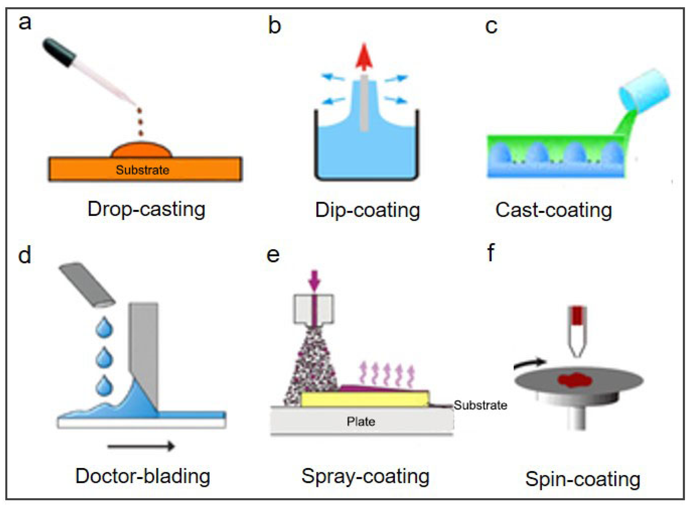
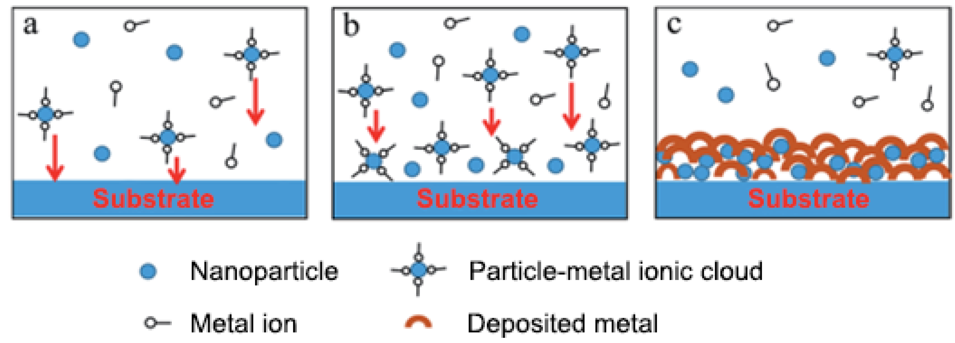
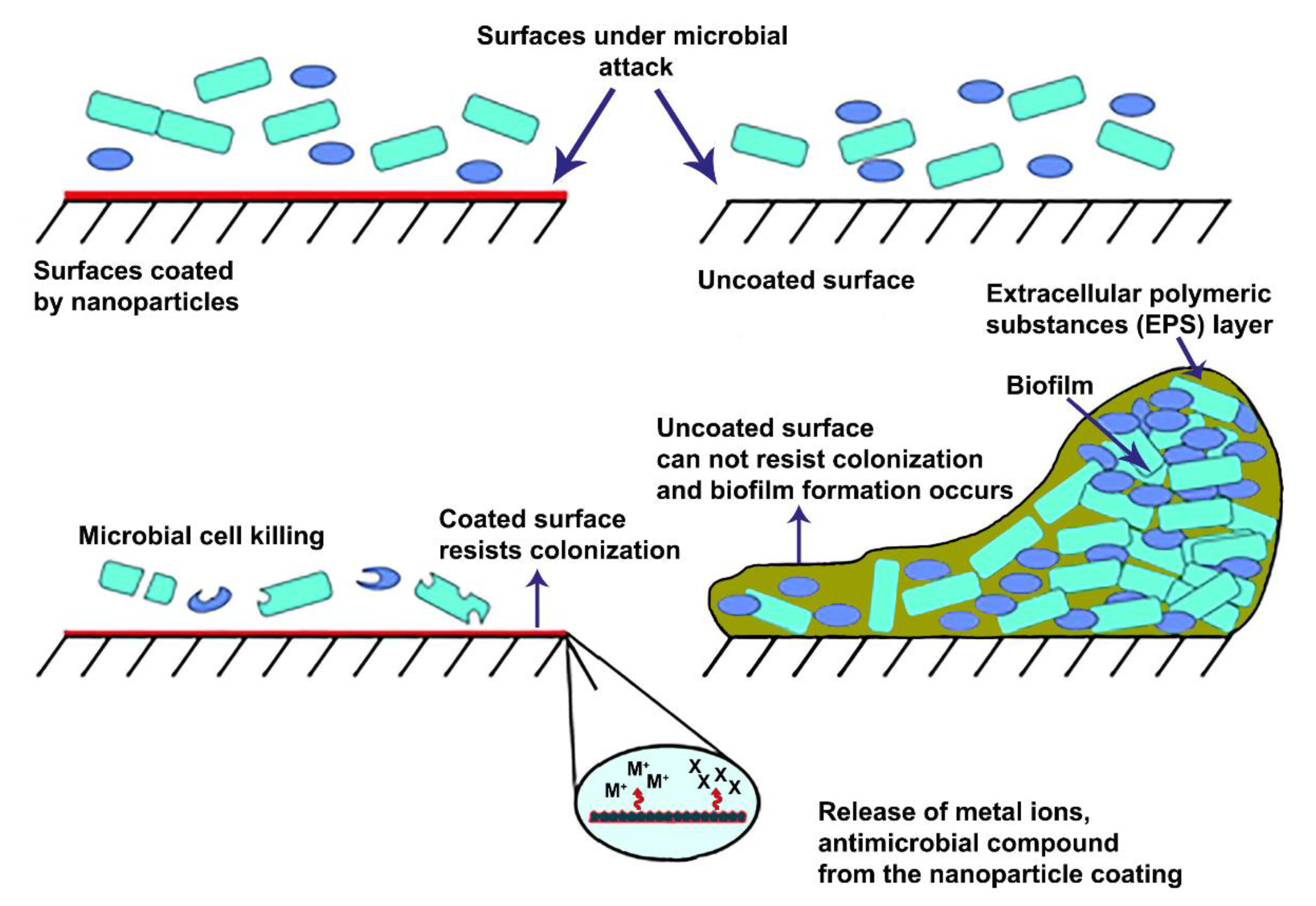
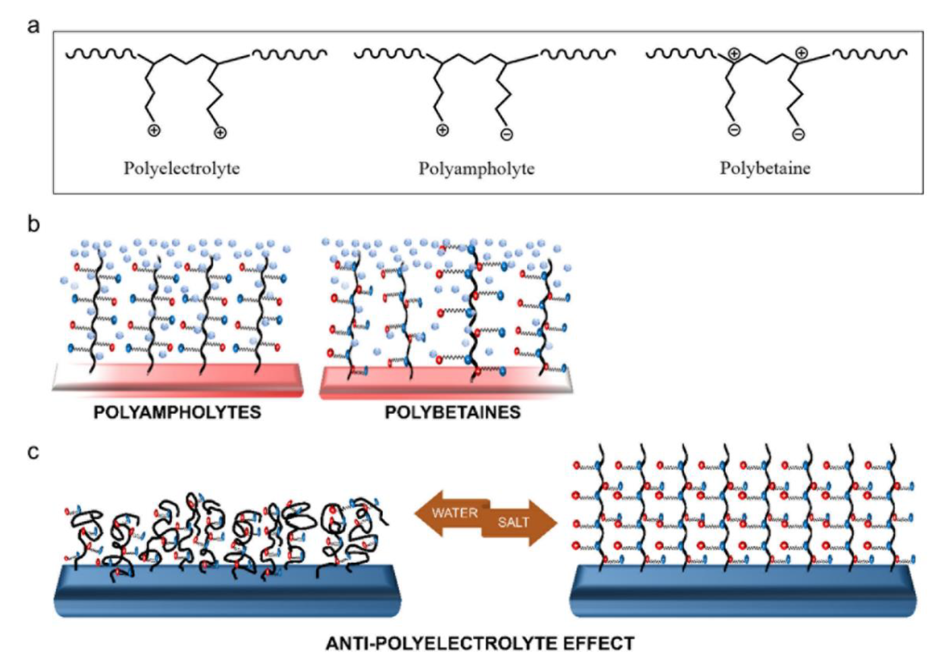
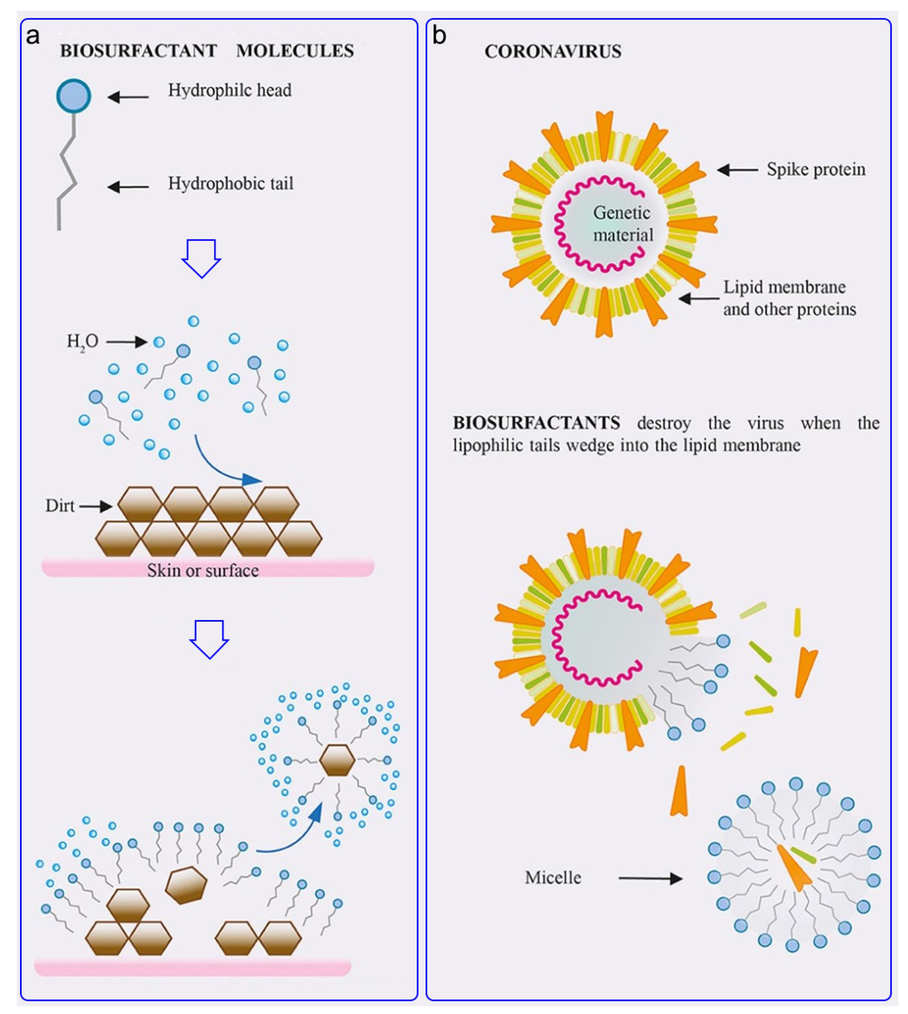
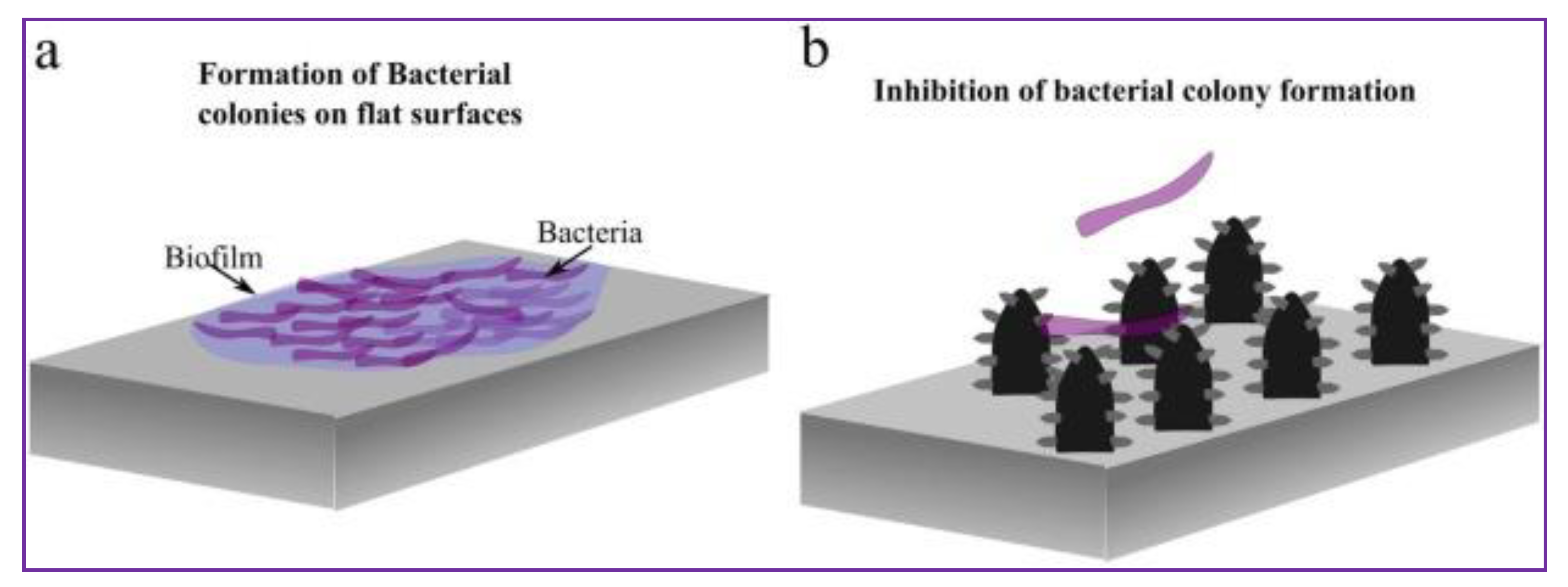
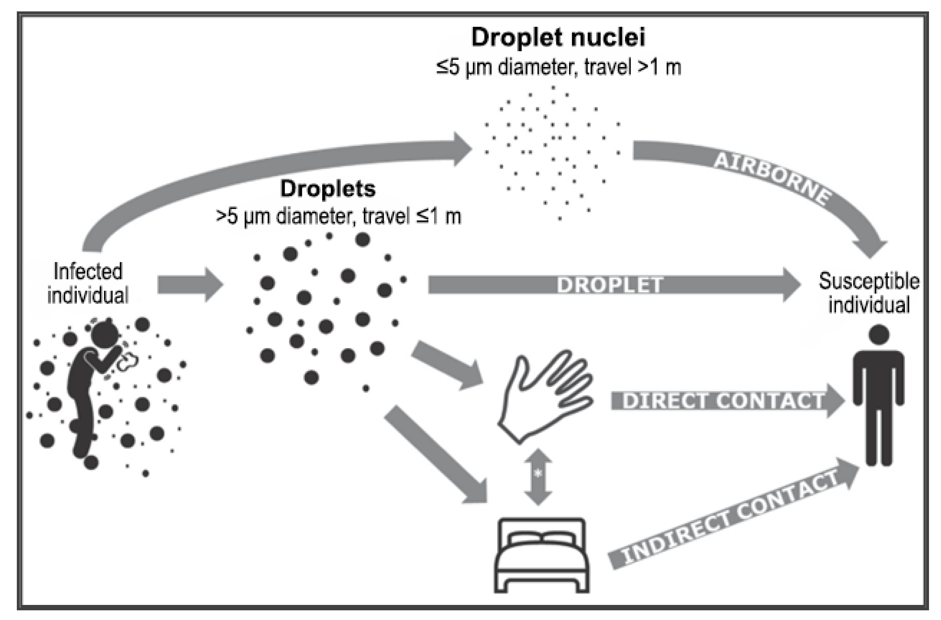
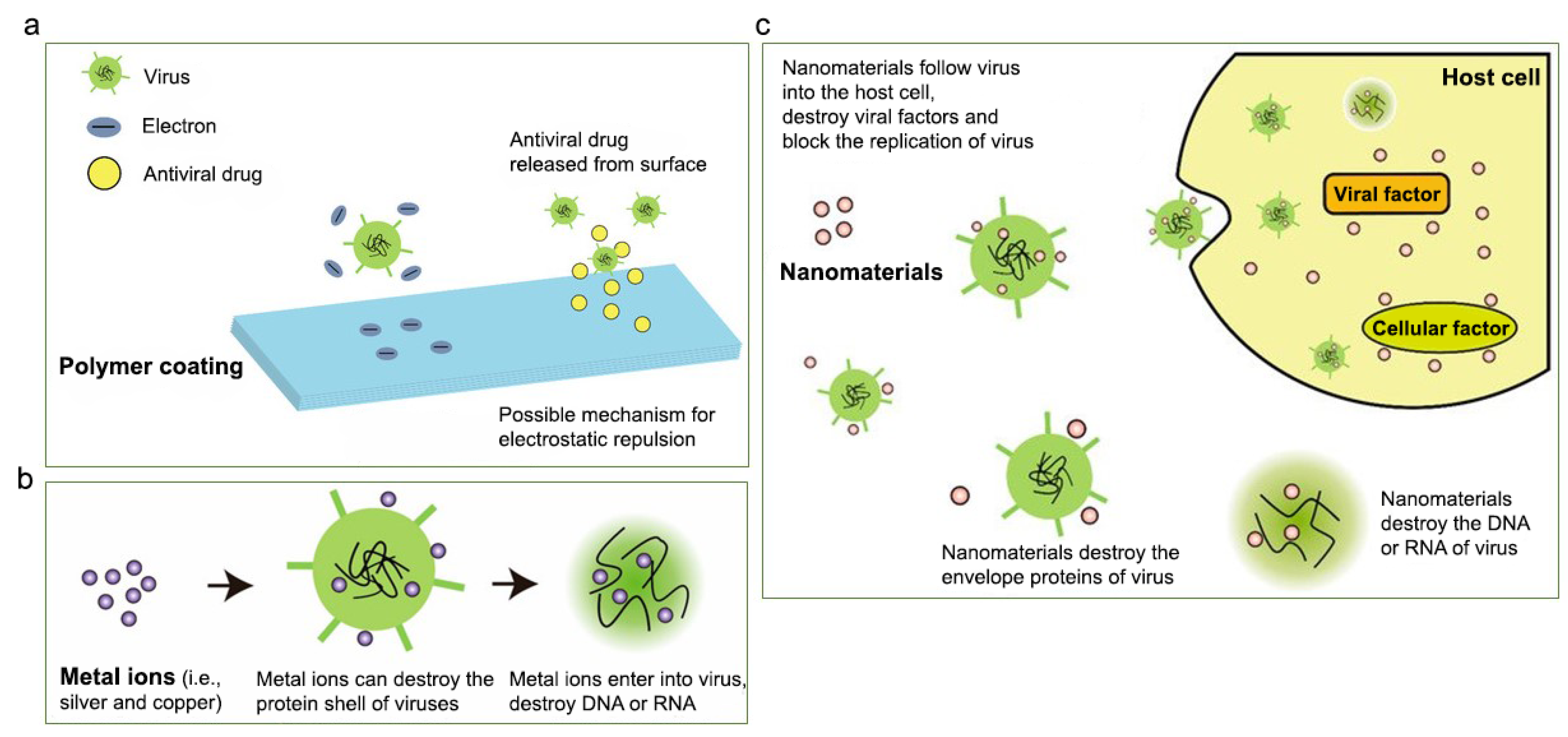
Table 1.
Applications of natural and synthetic cationic polymers.
| Type | Polymer | Application | Species | References |
|---|---|---|---|---|
| Natural | Chitosan | 1 Edible films 2 Virus purification 3 Plasmid DNA-loaded biotinylated chitosan nanoparticles for severe acute respiratory syndrome coronavirus (SARS-CoV) immunization | 1 Murine norovirus, Listeria innocua and Escherichia coli (E. coli) K12 2 Human coronavirus NL63 (HCoV-NL63), human coronavirus OC43 (HCoV-OC43) and mouse hepatitis virus (MHV) 3 SARS-CoV | 1 Amankwaah et al. [45] 2 Ciejka et al. [46] 3 Raghuwanshi et al. [47] |
| Chitosan-based coatings functionalized with methacrylate-based polymer brushes | Implantable sensor | Antifouling, leukocytes, and platelet rich plasma | Buzzacchera et al. [48] | |
| Polypyrrole/chitosan composites | Surface protective and in vitro biocompatible 316L stainless steel implant coating | MG-63 human osteoblast cell | Kumar et al. [49] | |
| Gelatin | 1 Antibacterial agent in the form of hydrogel 2 Injectable hydrogel wound dressing | E. coli and S. aureus 1 S. aureus, E. coli, MRSA 2 | 1 Yi et al. [50] 2 Zheng et al. [51] | |
| Gelatin/chlorhexidine acetate (CHA) | Self-healing hydrogel for wound healing | S. aureus, E. coli | Chen et al. [52] | |
| Cellulose | 1 Superporous hydrogel dressing 2 Natural antimicrobial material | 1S. aureus, E. coli 2E. coli, S. aureus, Proteus microbilis, Proteus vulgari, P. aeruginosa, Enterobacter aerogenes, Bacillus thuringiensis, Salmonella enterica serotype typhmurium, Streptococcus mutans | 1 Wu et al. [53] 2 Demircan et al. [54] | |
| Dextran | 1 Antibacterial hydrogel 2 Hydrogels for biocide release | 1S. aureus, S. Epidermidis, E. coli, P. Aeruginosa 2S. aureus, E. coli, methicillin-resistant S. aureus (MRSA) | 1 Dai et al. [55] 2 Hoque et al. [56] | |
| Cyclodextrin | 1 Polyelectrolyte to microcapsules with antibacterial effect 2 Antibacterial and antiviral agent | 1S. aureus, E. Coli 2E. coli and adenovirus (ADV) | 1 Belbekhouche et al. [57] 2 Pan et al. [58] | |
| Synthetic | 2-(4-methylthiazol-5-yl) ethyl methacrylate (MTA) and N-(3,4-dihydroxyphenethyl) methacrylamide (DOMA) copolymers | Adhesive bacterial coating | E. coli and S. aureus | Chiloeches et al. [59] |
| Poly(dimethylaminoethylmethacrylate)functionalizedgraphene oxide (GO–QPDMAEMA) | Antibacterial and antifouling effects | E. coli and S. aureus | Tu et al. [60] | |
| N-(3,4-dihydroxyphenethyl) methacrylamide (DOMA) and 2-(4-methylthiazol-5-yl) ethyl methacrylate (MTA) quaternized with methyl iodide | Antibacterial effects | S. aureus, S. Epidermidis, E. coli and P. aeruginosa | Plachá et al. [61] | |
| PEG brush surfaces- PLL coils composites and cationically functionalized gold nanoparticles | Antibacterial surface including cationic nanoparticles | S. aureus | Fang et al. [62] | |
| Poly-L-lysine (PLL) and hyaluronic acid (HA) denoted PLL30, PLL90, and PLL400 were used | Antibacterial coating | S. aureus, MRSA, P. aeruginosa and E. coli | Alkekhia et al. [63] | |
| Cationic acrylate-based copolymers (PAMs) by 3-(methacryloylamino) propyltrimethyl ammonium chloride (MPAC) and acrylates (BA, MMA) | Copolymers and their films were sued as surface coatings | E. coli and S.aureus | Wang et al. [64] | |
| Multilayers of Polyethylenimine (PEI) and styrene maleic anhydride copolymer (SMA) | Coating on polypyrrole (PP)-based substrates | E. coli | Bastarrachea et al. [65] | |
| Quaternized poly(4-vinylpyridine-co-N-vinylpyrrolidone) (P(4VP-co-NVP)) copolymers | Antibacterial activity test | E. coli and S. aureus | Gokkaya et al. [66] | |
| Poly-arginine (PAR) | Multifunctional coating polyelectrolyte layers | S. aureus | Özçelik et al. [67] | |
| PEI | 1 Bactericidal agent 2 Antimicrobial agent | 1Pseudomonas strains 2 S. aureus and E. Coli | 1 Khalil et al. [68] 2 Gibney et al. [42] | |
| PEI and PEI-based nanoparticles | Antibacterial agent on polyurethane based medical catheters | S. epidermidis, Acinetobacter baumannii (A. baumannii), S. aureus and Candida albicans | Azevedo et al. [44] | |
| For multiple examples of a polymer, corresponding study, application, and species are shown as superscript numbers. | ||||
Publisher’s Note: MDPI stays neutral with regard to jurisdictional claims in published maps and institutional affiliations. |
© 2021 by the authors. Licensee MDPI, Basel, Switzerland. This article is an open access article distributed under the terms and conditions of the Creative Commons Attribution (CC BY) license (http://creativecommons.org/licenses/by/4.0/).
Share and Cite
MDPI and ACS Style
Erkoc, P.; Ulucan-Karnak, F. Nanotechnology-Based Antimicrobial and Antiviral Surface Coating Strategies. Prosthesis 2021, 3, 25-52. https://0-doi-org.brum.beds.ac.uk/10.3390/prosthesis3010005
AMA Style
Erkoc P, Ulucan-Karnak F. Nanotechnology-Based Antimicrobial and Antiviral Surface Coating Strategies. Prosthesis. 2021; 3(1):25-52. https://0-doi-org.brum.beds.ac.uk/10.3390/prosthesis3010005
Chicago/Turabian StyleErkoc, Pelin, and Fulden Ulucan-Karnak. 2021. "Nanotechnology-Based Antimicrobial and Antiviral Surface Coating Strategies" Prosthesis 3, no. 1: 25-52. https://0-doi-org.brum.beds.ac.uk/10.3390/prosthesis3010005