Anti-Asthmatic Effects of Saffron Extract and Salbutamol in an Ovalbumin-Induced Airway Model of Allergic Asthma

Department of Pharmacology, SVKM’s Dr. Bhanuben Nanavati College of Pharmacy, Vile Parle (W), Mumbai 400056, India
*
Author to whom correspondence should be addressed.
Sinusitis 2021, 5(1), 17-31; https://0-doi-org.brum.beds.ac.uk/10.3390/sinusitis5010003
Submission received: 26 October 2020
/
Revised: 29 December 2020
/
Accepted: 14 January 2021
/
Published: 24 January 2021
(This article belongs to the Special Issue Allergic Rhinosinusitis and Airway Diseases)
Abstract
:Introduction: Asthma is a chronic inflammatory disorder of the airways often characterized by airway remodeling and influx of inflammatory cells into the airways. Saffron (C. sativus) has been reported to possess anti-inflammatory, anti-allergic and immunomodulatory properties. Salbutamol is known to relax airway smooth muscles. Objective: To investigate the combined anti-asthmatic effect of C. sativus extract (CSE) and salbutamol in an ovalbumin (OVA)-induced asthma in rats. Materials and methods: Airway hyperresponsiveness (AHR) was induced in male Sprague-Dawley rats by OVA challenge and treated with CSE (30 mg/kg and 60 mg/kg i.p.) and salbutamol (0.5 mg/kg p.o) for 28 days. After the induction period, various hematological, biochemical, molecular (ELISA) and histological analyses were performed. Results: OVA-induced alterations observed in hematological parameters (total and differential cell counts observed in Bronchoalveolar Lavage Fluid (BALF) were significantly attenuated (p < 0.01) by CSE (30 mg/kg and 60 mg/kg) and salbutamol (0.5 mg/kg). The treatment combination also significantly decreased (p < 0.01) the levels of total protein and albumin in serum, BALF and lung tissues. Treatment with CSE and salbutamol significantly attenuated (p < 0.01) increase in OVA induced Th2 cytokine levels (TNF-α, IL-1β, IL-4, IL-13). Histopathological analysis of lung tissue showed that combined effect of CSE and salbutamol treatment ameliorated OVA-induced inflammatory influx and ultrastructural aberrations. Conclusion: The results obtained from this study show that the combined effect of CSE and salbutamol exhibited anti-asthmatic properties via its anti-inflammatory effect and by alleviating Th2 mediated immune response. Thus, this treatment combination could be considered as a new therapeutic strategy for management of asthma.
1. Introduction
Amongst chronic respiratory diseases, chronic obstructive pulmonary disease (COPD) and asthma are the leading cause of mortality worldwide [1]. Asthma is a complex heterogeneous chronic disease that affects people of diverse age groups. It is characterized by three main features: (1) Chronic inflammation of the airways (2) Variable airflow obstruction (3) Airway hyperresponsiveness (AHR). Other characteristic features include airway constriction, increased mucus production, reversible bronchospasm, multicellular inflammation and airway remodeling.
Inflammatory mediators such as eosinophils, neutrophils, mast cells, T lymphocytes, and dendritic cells play a major role in the development of asthma [2]. Acute lung inflammation followed by lung injury leads to pulmonary fibrosis, which in turn impairs gas exchange. Influx of eosinophils into the airways can cause epithelial damage, exudation of plasma wall, airway wall and basement membrane thickening [3,4].
Various treatment strategies have been devised for effective management and treatment of asthma. These include inhaled Bronchoprovocation challenges, corticosteroids, mast cell stabilizers, β2 adrenergic agonists, anti cholinergics, antihistamines, leukotriene modifiers etc. [5] Corticosteroids along with bronchodilators are the treatment of choice for severe asthmatics. Various studies involving dexamethasone has failed to show significant improvement in respiratory clinical score or hospital admission rate, whereas according to a meta-analysis, beta2-agonists failed to improve clinical symptoms when compared with a placebo [6,7]. In another study, no improvement was observed when dexamethasone, salbutamol, epinephrine was given alone in a similar set of population [8]. However, combination therapy involving dexamethasone and salbutamol has reported an improvement in hospital admission rate or respiratory clinical score when compared with placebo. Although the exact mechanism of action for this combination therapy is unknown, synergism between these two classes of drugs is well documented. Reports suggest that this combination of drugs showed a synergistic effect in children with bronchiolitis [6,8]. Patients experience different comorbidities due to prolong use of different corticosteroids. Hence it is imperative to find a substitute that can replace or to certain extent diminish the side effects associated with corticosteroids.
However, the current treatment regimen largely aims to decrease the symptoms and improve lung function by decreasing airway inflammation and restoring normal lung functions. Despite this, only a fraction of patients receives symptomatic relief. Side effects associated with these treatment strategies are a major concern. Thus, there is a need to develop medications that are not only efficacious but are also safe at the same time [5].
Saffron (C. Sativus) along with salbutamol could be considered as a potential drug to substitute corticosteroids because of its immunomodulatory, anti-inflammatory and airway smooth muscle relaxing effects. The present study aims to investigate the combined effect of saffron extract and Salbutamol in an ovalbumin-induced rat model of asthma.
2. Material and methods
2.1. Preparation of C. sativus Extract (CSE)
C. sativus extract (CSE) was prepared according to the method previously described [9]. An ultrasound-assisted extraction was performed in order to obtain the CSE. Saffron petals were crushed into powder and suspended in distilled water containing 0.3% Tween-80. This suspension was sonicated for 10 min in an ultrasound water bath (water temperature at 25 °C) and was passed through a 0.45 µm filter. The resulting solution (CSE) was then administered intraperitoneally (i.p.) in a volume of 0.5 mL per rat.
2.2. Animals
Healthy Sprague Dawley rats with a weight range of 180–200 g were procured from National Institute of Biosciences. They were housed in Perspex cages and maintained under standard laboratory conditions (12 h: 12 h dark––light cycle, a temperature of (24 ± 2 °C and 45–55% humidity. The animals were fed on standard food pellets and drinking water ad libitum. Before starting the experiment, animals were acclimatized for a period of one week. The entire experimental protocol was reviewed and approved by an Institutional Animal Ethics Committee registered under “Committee for the Purpose of Control and Supervision of Experiment on Laboratory Animals” (CPCSEA), Ministry of Environment and Forests, Government of India. Approval number—CPCSEA/IAEC/P-34/2018.
2.3. Chemicals
Ovalbumin (OVA, egg albumin grade II), and aluminum hydroxide were purchased from Sigma Chemical Co. (St Louis, MO), Salbutamol was purchased from TCI chemicals (Tokyo, Japan), Saffron was purchased from Yucca enterprises (Mumbai, India). Marketed preparation of Dexamethasone sodium phosphate was used (Martin & Brown Biosciences). All other purchases were made from S.D. fine chemicals (Mumbai, India).
2.4. Preparation of Crocus Sativus Extract (CSE):
Different Dosages of CSE Extract Was Freshly Prepared and Administered Intraperitoneally (i.p.) in a Volume of 0.5 mL per Rat. Salbutamol was Freshly Prepared Daily as 1% Aqueous Solution of Saline and Administered Orally at a Dose of 0.5 mg/kg [10]. Dexamethasone Sodium Phosphate (0.1 mg/kg) Was Administered as per Body Weight. Control Animals Received Isovolumetric Amounts (0.5 mL) of 0.3% Tween-80.
2.5. Induction of Asthma
Sensitization procedures and aerosol challenge were performed according to reported procedure with some minor changes [11]. Animals were actively sensitized by an intraperitoneal (i.p.) injection of 0.66 mL of a suspension of 1 mg OVA plus 300 mg of aluminum hydroxide in saline, this was considered as Day 0 of sensitization. Seven days after sensitization, the animals were boosted subcutaneously (s.c.) with an identical injection of aluminum-precipitated OVA in all the mentioned groups except normal control. The aluminum precipitate acts as an adjuvant to promote IgE production. The standard drugs and treatment drugs were administered daily for all 28 days in the standard and treatment groups, respectively. On day 28, intranasal (i.n.) OVA-aerosol challenge was accomplished by placing the rats individually in a polyurethane chamber connected to a nebulizer. This generated an aerosol mist pumped into the exposure chamber with an output volume of 10 mL. Except the Normal Control group, all other groups were exposed to 1% OVA-aerosol challenge for 15 min. Animals in normal control group were given saline aerosol for 15 min. On Day 29, 24 h after final OVA challenge (saline challenge for normal control group), the animals were anesthetized using urethane (1.25 g/kg) for collection of blood and Bronchoalveolar Lavage (BAL) fluid for biochemical evaluation. After that the animals were sacrificed, their lungs were isolated for further analyses. (Figure 1)
Groups:
- Normal Control—0.9% w/v saline
- Negative control—1mg OVA + 300 mg Aluminium hydroxide
- Standard—Dexamethasone (0.1 mg/kg) i.p + Salbutamol (0.5 mg/kg) oral
- Combination 1—Saffron (30 mg/kg) i.p + Salbutamol (0.5 mg/kg) oral
- Combination 2—Saffron (60 mg/kg) i.p + Salbutamol (0.5 mg/kg) oral
2.6. Determination of Cell Count in Blood
Blood (3 mL) was collected in a heparinized tube from each animal under anesthesia (urethane (1.25 mg/kg)) and used for leukocyte count. These samples were collected from retro-orbital plexus of rats. Each sample was centrifuged at 500× g for 10 min at 4 °C; the cells in the pellet were washed in 0.5 mL saline, and total cells were counted using automated cell counter. In order to perform differential analyses, aliquots of the cells were placed onto slides and then stained with Field’s stain to identify eosinophils, lymphocytes, or neutrophils using standard morphologic determinants.
2.7. Collection of Bronchoalveolar Lavage Fluid (BALF)
Ice-cold PBS (0.5 mL) was instilled into the lungs and BALF was obtained by three aspirations (total volume 1.5 mL) via tracheal cannulation [11]. Each BALF was centrifuged, and the supernatant collected was stored at 70 °C. Total inflammatory cell numbers were assessed by counting cells in at least five squares of a hemocytometer, after exclusion of dead cells stained with trypan blue. Cell pellets were suspended in 0.5 mL PBS, and 100 mL of each solution was spun onto a slide.
2.8. Lung Tissue Homogenate Preparation
After collection of BAL fluid, animals were sacrificed. About 1 g of the lung tissue was homogenized in buffer solution (2 mL) of phosphate-buffered saline in a ratio of 1:2 (w/v; 1 g tissue with 2 mL PBS, pH 7.4). Homogenates were centrifuged at 10,000× g for 15 min at 4 °C in a refrigerated centrifuge (Eltrec, India). The supernatants were divided into aliquots, then stored at 20 °C, and used for further evaluation [12].
2.9. Determination of Inflammatory Cytokines Using Enzyme-Linked Immunosorbent Assay (ELISA)
To determine the levels of cytokines in vivo, BALF and lung homogenate samples were collected 24 h after the final OVA challenge. IL-4, IL-13, IL-1 β and Tumor necrosis factor-alpha (TNF-α) were assayed with commercially available ELISA kits (Krishgen Biosystems, India).
2.10. Biochemical Estimation (Total Protein and Albumin)
The activity of albumin in serum, BAL and lungs were measured using commercially available reagent kits (ERBA Diagnostics, India). Blood was centrifuged at 6000 rpm for 10 min. Serum samples were collected and frozen at −80 °C until the time of analysis.
2.11. Enzyme-Linked Immunosorbent Assay (ELISA)
To determine the levels of cytokines in vivo, BALF and lung homogenate, samples were collected 24 h after the final OVA challenge. IL-4, IL-13, IL-1 β and Tumor necrosis factor-alpha (TNF-α) were assayed with commercially available ELISA kits (Krishgen Biosystems, India), according to the manufacturer’s instruction.
2.12. Histopathological Examination
At the end of the study rats from each group were sacrificed by urethane and their lungs were excised, washed and cleaned with 0.9% saline and kept in 10% neutral formalin and given for histopathological examination by staining it with Haematoxylin and Eosin (H & E) [12]. These tissues were trimmed and routinely processed. Tissue processing was done to dehydrate in ascending grades of alcohol, clearing in xylene and embedded in paraffin wax. Paraffin wax embedded tissue blocks were sections at 4–5 μm thickness with the Rotary Microtome. All the slides were stained with Haematoxylin & Eosin (H & E) stain. The prepared slides were examined under microscope and photomicrographs were taken at 40×.
2.13. Statistical Analysis
The statistical evaluation was performed with the assistance of GraphPad prism 5 for 64 bit Windows Version. All the experimental groups were compared to assess the statistical significance using One-way analysis of variance (ANOVA). Each test was followed by the Dunnet’s multiple comparison. The data are represented as mean ± SEM values and p values < 0.05 were considered as statistically significant.
3. Results
3.1. Effect of Treatment Combination on OVA Induced Alterations in Body Weight and Relative Lungs Weight
In the present study, negative control group were observed with reduced body weight and an increased relative lungs weight as compared to normal control group (** p < 0.01). However, Standard group showed significant reduction in body weight as compared to normal control (** p < 0.01). Combination 2 group restored the decrease in body weight as compared to negative control (## p < 0.01) (Table 1)
3.2. Effect of Treatment Combination on OVA-Induced Alteration in Hematological Parameters
Evaluation of different hematological parameters provides better understanding of the diseased state. In the present study, significant changes in hematological parameters of negative control rats were observed as compared to normal control rats. As compared to normal control group, a significant decrease in hemoglobin, red blood cells (RBC) count, PCV%, MCV% was observed, in negative control group (** p < 0.01). Treatment combinations (30 mg/kg CSE + 0.5 mg/kg salbutamol, 60mg/kg CSE + 0.5 mg/kg salbutamol) significantly ameliorated these alterations observed in hematological parameters as compared to negative control rats (## p < 0.01). Standard combination (0.1 mg/kg dexamethasone + 0.5 mg/kg salbutamol) also decreased these aforementioned hematological parameters as compared to negative control group (## p < 0.01). In case of mean platelet count, WBCs, neutrophils and lymphocytes, a significant increase was observed in negative control group as compared to normal control group (** p < 0.01). When compared with normal control rats, Treatment combinations (30 mg/kg CSE + 0.5 mg/kg salbutamol, 60 mg/kg CSE + 0.5 mg/kg salbutamol) and standard group showed significant inhibition in differential WBCs, neutrophil and lymphocyte count (## p < 0.01). (Table 2)
3.3. Inflammatory and Differential Cell Counts in BALF
BALF differential and total cell counts were increased significantly in negative control rats as compared to normal control (*** p < 0.001). However, Treatment combination groups (30 mg/kg CSE + 0.5 mg/kg salbutamol, 60mg/kg CSE + 0.5 mg/kg salbutamol) showed significant decrease in BAL fluid count as compared to negative control group (### p < 0.001). Also in case of Standard (0.1 mg/kg dexamethasone + 0.5 mg/kg salbutamol) group BALF total and differential cell count were significantly decreased as compared to negative control group (## p < 0.01)
3.4. Effect of Treatment Combination on OVA-Induced Alteration in the Levels of Total Protein in Serum, BALF and Lung Tissues of Rats
In the present investigation, Negative control rats showed significant increase in total protein levels in serum (** p < 0.01), BALF (** p < 0.01) and lung tissues (** p < 0.01) as compared to normal control rats. Increase in total protein and albumin was significantly inhibited by treatment combinations (30 mg/kg CSE+ 0.5 mg/kg salbutamol, 60 mg/kg CSE + 0.5 mg/kg salbutamol) in serum, BALF and lung tissues (## p < 0.01) as compared to negative control rats. Standard group (0.1 mg/kg dexamethasone + 0.5 mg/kg salbutamol) significantly decreased total protein levels in serum, BALF and lung tissues of rats (## p < 0.01) as compared to negative control group.
3.5. Effect of Treatment Combination on OVA-Induced Alteration in Albumin Levels in Serum and BALF
An increase in albumin levels in serum and BALF of negative control group was observed as compared to normal control group (** p < 0.01). However standard group (0.1 mg/kg dexamethasone + 0.5 mg/kg salbutamol) and treatment groups (30 mg/kg CSE + 0.5 mg/kg salbutamol, 60 mg/kg CSE + 0.5 mg/kg salbutamol) decreased albumin levels in serum and BALF (## p < 0.01) as compared to negative control group. (Refer Table 3)
3.6. Effect of Treatment Combination on OVA-Induced Alteration in Cytokine Analysis of BALF and Lung Tissues
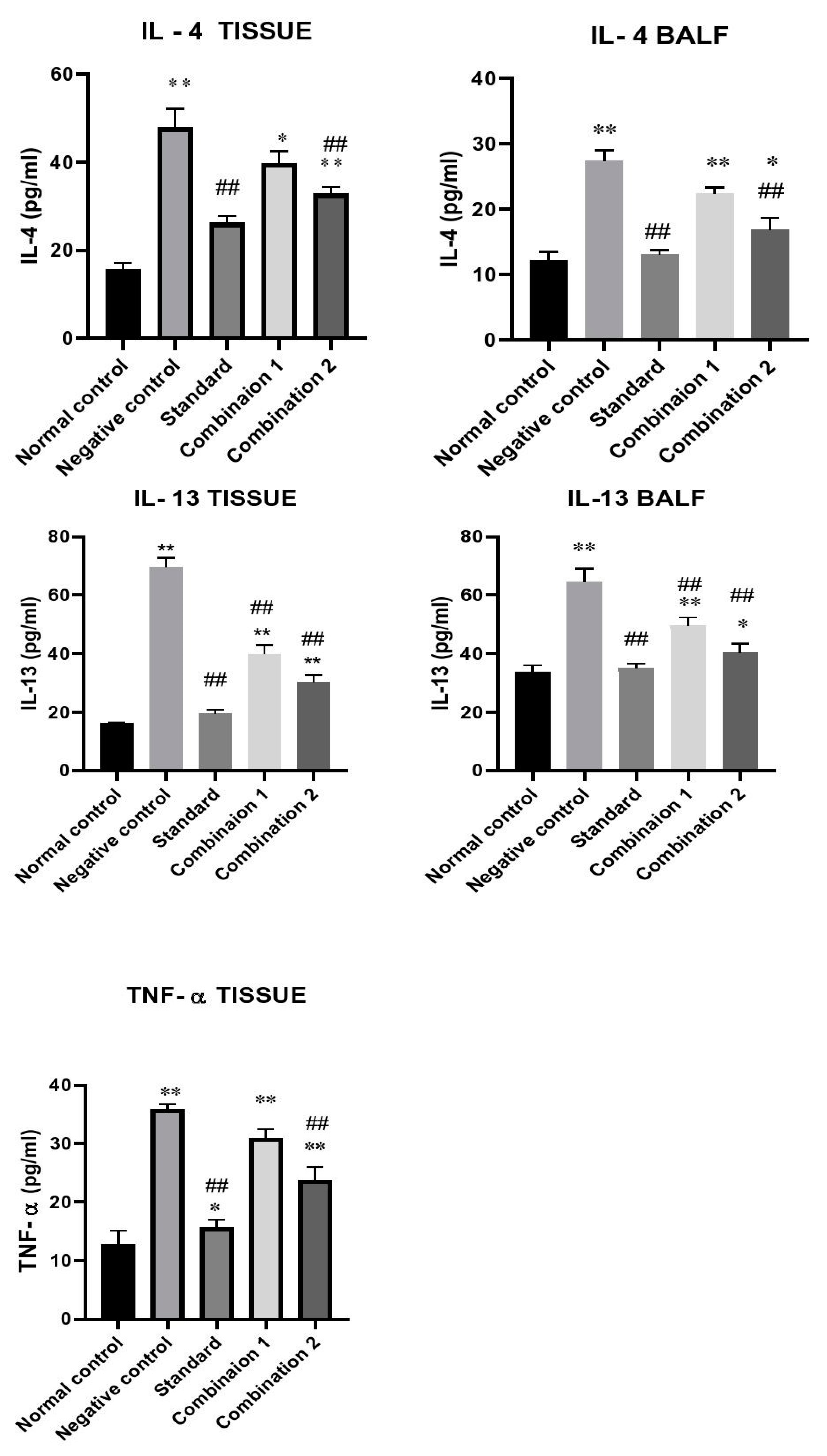
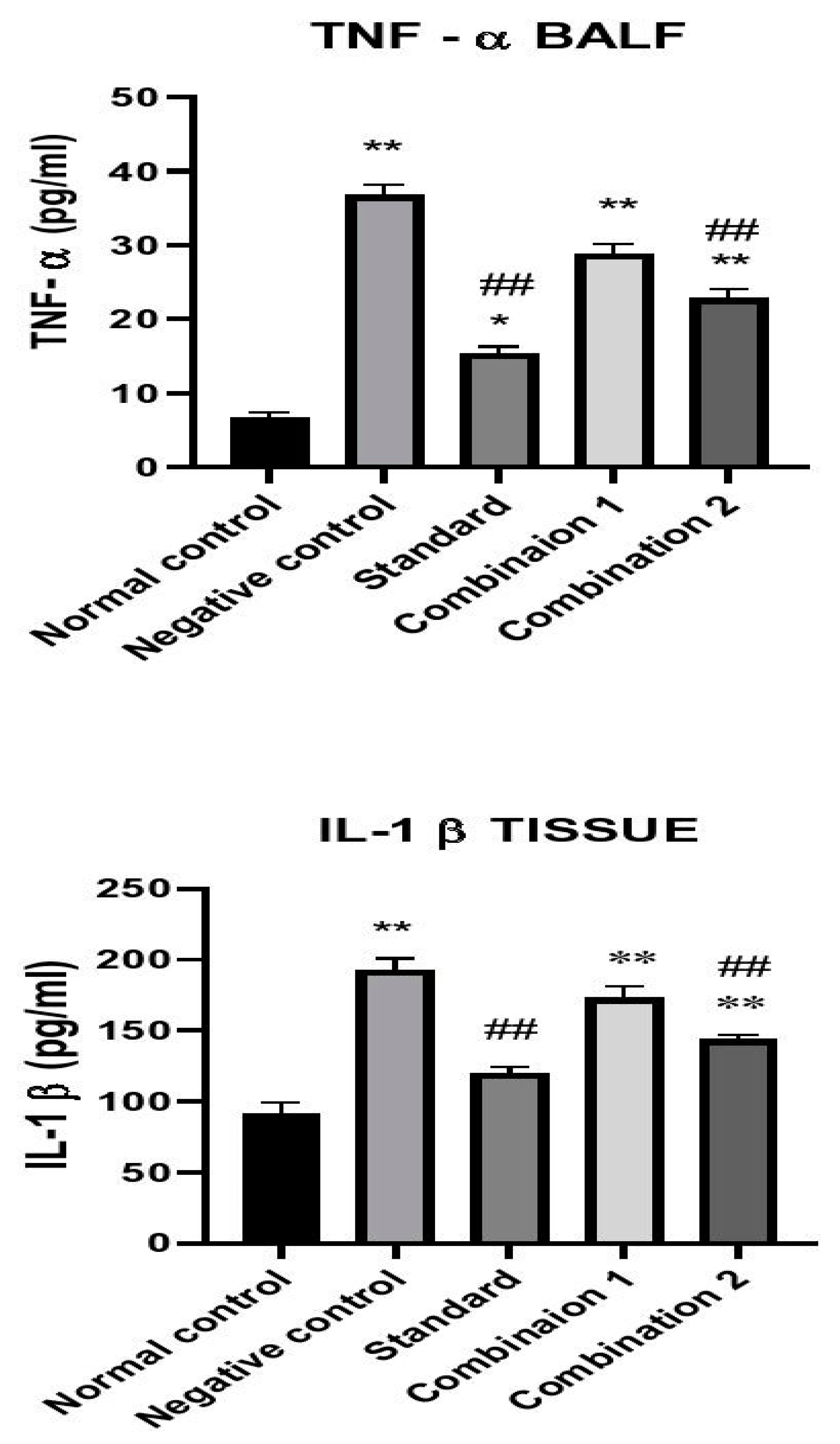
Higher levels of Th2 response related cytokines (TNF-α and IL’s) shows successful induction of asthma. In the current study, a significant up-regulation was observed in cytokine levels (IL-4, IL-13, IL-1 β, TNF-α) (** p < 0.01) of negative control group as compared to normal control group in both lung tissue and BALF. In case of IL-4, combination 2 group showed significant reduction in IL-4 levels as compared to negative control group in lung tissues and BALF of normal control (* p < 0.05) and negative control group (## p < 0.01) respectively. Up-regulation of IL-13 levels were inhibited by standard and treatment groups in lung tissues and BALF as compared to negative control group (## p < 0.01). IL-1β levels in lung tissue of standard and treatment groups were significantly reduced as compared to negative control group (## p < 0.01). Levels of TNF-α was reduced in both standard and treatment groups as compared to negative control group (## p < 0.01). (Figure 2)
3.7. Effect of Treatment Combination on OVA-Induced Histopathological Alteration in Rat Lungs
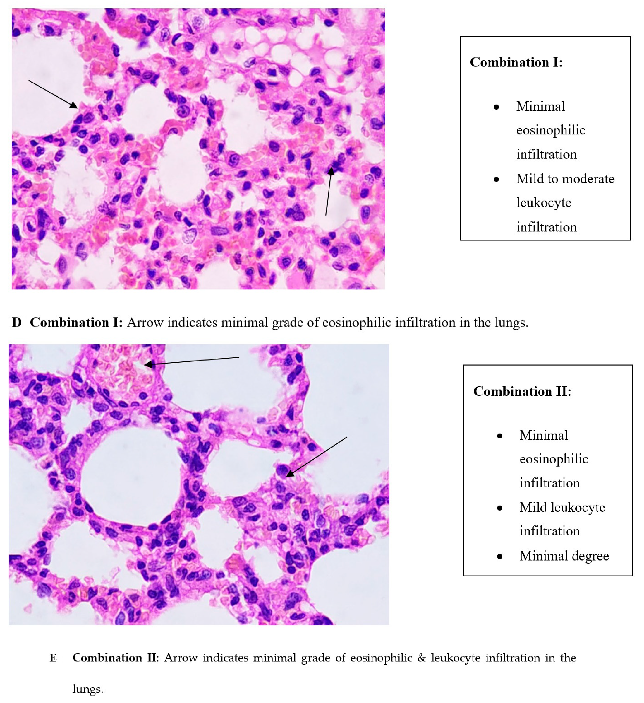
The following pathological changes were observed in the lungs: interstitial infiltration (eosinophil and lymphocyte), grade of infiltration and other notable changes. Normal control group showed no leukocyte or eosinophil infiltration and no notable changes in the lung tissues were observed. However, in the case of the negative control group, mild eosinophilic infiltrations were observed followed by moderate to severe leukocytic infiltration. A minimal degree of hemorrhage was also observed. Standard group showed minimal leukocytic infiltration. As in the case of treatment groups, minimal eosinophilic infiltration was observed and mild to moderate leukocytic infiltrations were observed. However, treatment groups, like negative control group, also showed minimum degree of hemorrhage (Table 4) (Figure 3). Following were the levels of observed grade of infiltration for each group.
Photomicrograph of sections of lungs of:
Figure 3.
Effect of treatment combination on OVA-induced alteration in lung histology of rats.


4. Discussion
Asthma is a chronic, immunoinflammatory disorder of the airways characterized by airway hyperresponsiveness, mucus hypersecretion and bronchial inflammation. It is a well-known fact that asthma leads to chronic inflammation of the tracheo-bronchial tree [13]. Several animal models have given us insights about the probable mechanisms involved in the pathogenesis of asthma. In the present study, we have evaluated the combined effects of saffron extract (C. sativus) along with salbutamol, in OVA—induced model of asthma. Administration of saffron extract (C. sativus) and salbutamol in OVA—challenged rats increased body weights, improved hematological parameters. It inhibited infiltration of BALF cells, improved biochemical parameters (total protein and albumin). The combination also decreased inflammatory cytokine (IL-4, IL-13, TNF-α, IL-1β) levels and improved histopathological parameters of the lung tissue. Thus, these results show the anti-asthmatic activity of saffron extract—salbutamol in an OVA-challenged model of allergic airway inflammation.
A decrease in body weight of rats was observed in standard group as compared to negative control group. This could be attributed to the fact that, daily dose of dexamethasone reduced 5-HT (hydroxytryptamine) levels in the brain. Moreover, dexamethasone also elevated plasma leptin levels in a dose dependent manner [14,15]. Thus, cited literature suggests that leptin level in plasma may play a vital role in dexamethasone-induced anorexia. However, treatment combination groups showed significant improvement in body weights as compared to negative control group. Literature suggests that macrophages play a vital role in innate immune responses [16]. In an inflammatory state, lung tissue cells attract and infiltrate eosinophils, neutrophils, and lymphocytes from the blood. causing bronchial hyperresponsiveness. Thus, increase in the number of eosinophils is a prominent feature of allergic asthma leading to several inflammatory reactions. As a result, these inflammatory reactions play a detrimental role in causing damage to endothelial cells and extracellular matrix (ECM) present in the airways. In line with previous findings [17], results obtained from this study showed alterations in hematological differential cell counts, eosinophil and neutrophil counts, increase in total WBC counts in negative control group after OVA- challenge. Results from current investigation indicate improvement in hematological parameters after administration of saffron extract—salbutamol. Similarly, results obtained from total and differential cell count in BAL fluid of negative control group showed significant aberrations as compared to normal control group, whereas administration of treatment combination inhibited it. Shredding of epithelial cells occurs in the airways and is considered as a hallmark in airway inflammation. The present study also shows increase in epithelial shredding of the airways in negative control group as compared to normal control group. Treatment combination inhibited it to a certain extent.
Increase in protein and albumin levels are considered as a characteristic feature of tissue damage. Thus, the effect of treatment combination on total protein (TP) and albumin levels were studied. Reports from the present study indicate that treatment of OVA sensitized animals with saffron extract and salbutamol prevented increase in TP levels. However, a significant increase in TP levels of negative control group was observed in serum, BALF and lung tissues as compared to normal control group. These results are in line with previous research findings [18]. Similarly, from the present investigation BALF and serum albumin reports suggest that treatment combination has significantly inhibited albumin levels in OVA sensitized rats.
It is well documented that pathogenesis of asthma is due to infiltration of Th2 cytokines (IL-4, IL-13) and they also play an important role in the inflammatory mechanism of allergic asthma [19]. Switching of B lymphocyte to produce IgE and activation of macrophage and dendritic cells requires signals by Th2 cells, mainly IL-4. IL-13 activates signal transducer and transcription factor (STAT 6) via IL-4 mediated signal transduction, leading to AHR in the allergic lung [20,21]. Thus, Th2 cytokines play a significant role in regulating allergic responses. In this study, it was observed that IL-4 and IL-13 levels in lung tissues and BALF were significantly increased in negative control group. Results reveal that administration of saffron extract and salbutamol inhibited the levels of Th2 cytokines, IL-4 and IL-13 in both BALF and lung tissues.
TNF-α and IL-1β are pleiotropic cytokines which drive inflammatory responses causing changes in airway smooth muscle (ASM) responsiveness in asthma. Asthmatics have demonstrated high levels of these pro-inflammatory cytokines in the BAL fluid which are responsible for contraction of ASM [22]. Taking these evidences into consideration, levels of TNF-α and IL-1β in rat lung tissues and BALF were studied. In negative control rats, an upregulation of TNF- α and IL-1β levels were observed after OVA challenge. However, treatment combination inhibited this pro-inflammatory mediator, thus showing its anti-inflammatory effect. Data from the present study indicate that treatment combination of saffron extract and salbutamol decreased the levels of TNF-α and IL-1β in lung tissue as compared to negative control group, thus showing its anti-inflammatory potential against allergen-induced immunoinflammatory disease. The finding of the previous investigator [23] also showed the anti- inflammatory potential of saffron extract via inhibition of TNF-α and IL-1β and our results are also in line with the findings of the investigator.
Inflammatory infiltration in the lungs due to tissue damage is a unique histological feature of OVA challenged allergic asthma model. Histopathological findings illustrated moderate to severe eosinophilic and leukocytic infiltrations along with minimal hemorrhage in the lung tissues of OVA exposed rats of negative control group. However, saffron extract and salbutamol treated rats showed minimal degree of inflammatory infiltration along with minimal hemorrhage in the lungs. Thus, this treatment combination ameliorated histological aberrations caused by OVA inhalation.
5. Conclusions
Currently available treatment regimens for asthma focus on controlling asthmatic exacerbations and are associated with many side effects. Based upon the results obtained from the present study, the combination of C. sativus extract and salbutamol exhibits its anti-asthmatic activity against OVA induced allergic asthma. The treatment combination decreased total and differential cell count in blood and BALF, thereby showing its anti-inflammatory property. The treatment combination also ameliorated total protein, albumin levels, Th2 cytokines (IL-4 and IL-13) and immune-inflammatory responses (TNF-α and IL-1β) in lung tissues and BALF, thus exhibiting its immunomodulatory property. These results suggest that, combined effects of C. sativus extract and salbutamol can be considered as an “add-on therapy” for asthmatics or could be used along with current available anti-asthmatic drugs.
Author Contributions
Conceptualization K.P. and P.N. ; methodology, K.P. and P.N.; software, K.P. and P.N., validation, K.P., formal analysis, K.P., investigation, K.P. and P.N., resources, K.P. and P.N., data curation, K.P. and P.N., writing—original draft preparation, P.N., writing—P.N., visualization, K.P. and P.N., supervision, K.P., project administration, funding acquisition, N/A. All authors have read and agreed to the published version of the manuscript.
Funding
This research received no external funding.
Institutional Review Board Statement
The entire experimental protocol was reviewed and approved by an Institutional Animal Ethics Committee registered under “Committee for the Purpose of Control and Supervision of Experiment on Laboratory Animals” (CPCSEA), Ministry of Environment and Forests, Government of India. Approval number—CPCSEA/IAEC/P-34/2018.
Informed Consent Statement
Not applicable.
Data Availability Statement
The data presented in this study are available on request from the corresponding author.
Conflicts of Interest
The authors declare no conflict of interest.
References
- Symptoms, R. Economic burden of asthma in India. Lung India Off. Organ Indian Chest Soc. 2018, 35, 281. [Google Scholar]
- Nakagome, K.; Matsushita, S.; Nagata, M. Neutrophilic inflammation in severe asthma. Int. Arch. Allergy Immunol. 2012, 158, 96–102. [Google Scholar] [CrossRef] [PubMed]
- Calhoun, W.J. Nocturnal Asthma. Chest 2003, 123, 399–405. [Google Scholar] [CrossRef] [PubMed]
- Access, O.; Keglowich, L.F.; Borger, P. The Three A’s in Asthma—Airway Smooth Muscle. Airw. Remodel. Angiogenesis 2015, 70–80. [Google Scholar] [CrossRef] [Green Version]
- Barnes, P. Drugs for asthma. Br. J. Pharmacol. 2016, 147, S297–S303. [Google Scholar] [CrossRef]
- Tal, A.; Bavilski, C.; Yohai, D.; Bearman, J.E.; Gorodischer, R.; Moses, S.W. Dexamethasone and salbutamol in the treatment of acute wheezing in infants. Pediatrics 1983, 71, 13–18. [Google Scholar]
- Pauwels, R.A.; Löfdahl, C.G.; Postma, D.S.; Tattersfield, A.E.; O’Byrne, P.; Barnes, P.J.; Ullman, A. Effect of inhaled formoterol and budesonide on exacerbations of asthma. Formoterol and Corticosteroids Establishing Therapy (FACET) International Study Group. N. Engl. J. Med. 1997, 337, 1405–1411, Erratum in: N. Engl. J. Med. 1998, 338, 139. [Google Scholar] [CrossRef]
- Plint, A.C.; Johnson, D.W.; Patel, H.; Wiebe, N.; Correll, R.; Brant, R.; Mitton, C.; Gouin, S.; Bhatt, M.; Joubert, G.; et al. Pediatric Emergency Research Canada (PERC). Epinephrine and dexamethasone in children with bronchiolitis. N. Engl. J. Med. 2009, 360, 2079–2089. [Google Scholar] [CrossRef] [Green Version]
- Pitsikas, N.; Sakellaridis, N. Crocus sativus L. extracts antagonize memory impairments in different behavioural tasks in the rat. Behav. Brain Res. 2006, 173, 112–115. [Google Scholar] [CrossRef]
- Salami, E.O.; Ozolua, R.I.; Okpo, S.O.; Eze, G.I.; Uwaya, D.O. Studies on the anti—Asthmatic and antitussive properties of aqueous leaf extract of Bryophyllum pinnatum in rodent species. Asian Pac. J. Trop. Med. 2013, 6, 421–425. [Google Scholar] [CrossRef] [Green Version]
- Mukherjee, A.A.; Kandhare, A.D.; Rojatkar, S.R.; Bodhankar, S.L. Ameliorative effects of Artemisia pallens in a murine model of ovalbumin-induced allergic asthma via modulation of biochemical perturbations. Biomed. Pharmacother. 2017, 94, 880–889. [Google Scholar] [CrossRef] [PubMed]
- Zemmouri, H.; Sekiou, O.; Ammar, S.; El Feki, A.; Bouaziz, M.; Messarah, M.; Boumendjel, A. Urtica dioica attenuates ovalbumin-induced inflammation and lipid peroxidation of lung tissues in rat asthma model. Pharm. Biol. 2017, 55, 1561–1568. [Google Scholar] [CrossRef] [PubMed] [Green Version]
- Ye, W.-J.; Xu, W.-G.; Guo, X.-J.; Han, F.-F.; Peng, J.; Li, X.-M.; Guan, W.-B.; Yu, L.-W.; Sun, J.-Y.; Cui, Z.-L.; et al. Differences in airway remodeling and airway inflammation among moderate-severe asthma clinical phenotypes. J. Thorac. Dis. 2017, 9, 2904–2914. [Google Scholar] [CrossRef] [PubMed] [Green Version]
- Jahng, J.W.; Kim, N.Y.; Ryu, V.; Yoo, S.B.; Kim, B.T.; Kang, D.W.; Lee, J.H. Dexamethasone reduces food intake, weight gain and the hypothalamic 5-HT concentration and increases plasma leptin in rats. Eur. J. Pharmacol. 2008, 581, 64–70. [Google Scholar] [CrossRef] [PubMed]
- Michel, C.; Cabanac, M. Effects of dexamethasone on the body weight set point of rats. Physiol. Behav. 1999, 68, 145–150. [Google Scholar] [CrossRef]
- Parihar, A.; Eubank, D.; Doseff, A.I. Monocytes and Macrophages Regulate Immunity through Dynamic Networks of Survival and Cell Death. J. Innate Immun. 2010, 43220, 204–215. [Google Scholar] [CrossRef] [Green Version]
- Vosooghi, S.; Mahmoudabady, M.; Neamati, A.; Aghababa, H. Preventive effects of hydroalcoholic extract of saffron on hematological parameters of experimental asthmatic rats. Avicenna J. Phytomed. 2013, 3, 279–27987. [Google Scholar]
- Gholamnezhad, Z.; Koushyar, H.; Byrami, G.; Boskabady, M.H. The Extract of Crocus sativus and Its Constituent Safranal, Affect Serum Levels of Endothelin and Total Protein in Sensitized Guinea Pigs. Iran J Basic Med Sci 2013, 16, 1022–1026. [Google Scholar]
- Palmqvist, C.; Wardlaw, A.J.; Bradding, P. Chemokines and their receptors as potential targets for the treatment of asthma. Br. J. Pharmacol. 2007, 151, 725–736. [Google Scholar] [CrossRef]
- Poulsen, L.K.; Hummelshoj, L. Triggers of IgE class switching and allergy development. Ann. Med. 2007, 39, 440–456. [Google Scholar] [CrossRef]
- Muraro, A.; Lemanske, R.F.; Hellings, P.W.; Akdis, C.A.; Bieber, T.; Casale, T.B.; Jutel, M.; Ong, P.Y.; Poulsen, L.K.; Schmid-Grendelmeier, P.; et al. Precision medicine in patients with allergic diseases: Airway diseases and atopic dermatitis—PRACTALL document of the European Academy of Allergy and Clinical Immunology and the American Academy of Allergy, Asthma & Immunology. J. Allergy Clin. Immunol. 2016, 137, 1347–1358. [Google Scholar] [CrossRef] [PubMed] [Green Version]
- Dejager, L.; Dendoncker, K.; Eggermont, M.; Souffriau, J.; Van Hauwermeiren, F.; Willart, M.; Van Wonterghem, E.; Naessens, T.; Ballegeer, M.; Vandevyver, S.; et al. Neutralizing TNFα restores glucocorticoid sensitivity in a mouse model of neutrophilic airway inflammation. Mucosal Immunol. 2015, 8, 1212–1225. [Google Scholar] [CrossRef] [PubMed] [Green Version]
- Xiong, Y.; Wang, J.; Yu, H.; Zhang, X.; Miao, C. Nti-asthma potential of crocin and its effect on MAPK signaling pathway in a murine model of allergic airway disease. Immunopharmacol. Immunotoxicol. 2015, 37, 236–243. [Google Scholar] [CrossRef] [PubMed]
Figure 1.
Timeline of the study.

Figure 2.
Effect of treatment combination on OVA-induced alteration in Cytokine analysis (IL-4, IL-13, TNF-α, IL-1β) of lung tissues and BALF. Data were analyzed by one-way ANOVA followed by Dunnett’s Multiple Comparisons Test. Values are expressed as Mean ± S.E.M. (n = 4). Statistical significance was assessed as * p < 0.05, ** p < 0.01 vs. normal control group and # p < 0.05, ## p < 0.01 vs. negative control group. Standard: 0.1 mg/kg dexamethasone + 0.5 mg/kg salbutamol, Combination 1: 30 mg/kg CSE + 0.5 mg/kg salbutamol, Combination 2: 60 mg/kg CSE + 0.5 mg/kg salbutamol.


Table 1.
Effect of treatment combination on OVA induced alterations in Body Weight and Relative lungs weight.
Table 1.
Effect of treatment combination on OVA induced alterations in Body Weight and Relative lungs weight.
| Parameter | Normal Control | Negative Control | Standard | Combination 1 | Combination 2 |
|---|---|---|---|---|---|
| Body weight (gm) | 273 ± 4.74 | 202 ± 3.94 ** | 193 ± 3.16 ** | 210.2 ± 3.26 ** | 223.2 ± 4.79 **## |
| Relative Lungs weight (gm) | 0.72 ± 4.55 | 1.76 ± 0.20 ** | 0.87 ± 1.47 | 1.02 ± 2.14 ** | 0.95 ± 3.17 * |
Data were analyzed by one-way ANOVA followed by Dunnett’s Multiple Comparisons Test. Values are expressed as Mean ± S.E.M. (n = 6). Statistical significance was assessed as * p < 0.05, ** p < 0.01 vs normal control group and ## p < 0.01 vs. negative control group. Standard: 0.1 mg/kg dexamethasone + 0.5 mg/kg salbutamol, Combination 1: 30 mg/kg CSE + 0.5 mg/kg salbutamol, Combination 2: 60 mg/kg CSE + 0.5 mg/kg salbutamol.
Table 2.
Effect of treatment combination on OVA-induced alteration in hematological parameters and differential cell counts in BAL fluid of rats.
Table 2.
Effect of treatment combination on OVA-induced alteration in hematological parameters and differential cell counts in BAL fluid of rats.
| Parameter | Normal Control | Negative Control | Standard | Treatment 1 | Treatment 2 |
|---|---|---|---|---|---|
| Hemoglobin (gm/dl) | 15.05 ± 0.25 | 11.15 ± 0.11 ** | 14.38 ± 0.35 **## | 12.73 ± 0.11 **## | 13.93 ± 0.24 **## |
| RBC | 8.84 ± 0.2 | 8.18 ± 0.12 | 8.74 ± 0.10 | 8.09 ± 0.16 | 8.76 ± 0.23 |
| PCV% | 43.2 ±1.34 | 35.98 ± 0.86 ** | 44.11 ± 0.83 ## | 37.73 ± 0.43 ** | 41.91 ± 1.51 ## |
| MCV% | 47.93 ± 0.42 | 41.05 ± 0.34 ** | 48.78 ± 1.44 ## | 46.03 ± 1.20 ## | 46.88 ±0.78 ## |
| Mean Platelets | 4.84 ± 0.30 | 8.24 ± 0.67 *** | 5.46 ± 0.19 ## | 7.10 ± 0.69 * | 6.01 ± 0.35 # |
| WBC (× 103/mm3) | 15.83 ± 0.85 | 26.89 ± 1.49 ** | 16.41 ± 0.56 ## | 19.88 ± 1.30 *## | 18.45 ± 1.13 ## |
| N (%) | 28.5 ± 1.05 | 46.16 ± 1.03 ** | 29.83 ± 0.87 ## | 35.5 ± 0.42 **## | 66 ± 0.89 ## |
| L (%) | 62.83 ± 1.40 | 74.33 ± 1.41 ** | 63.66 ± 1.28 ## | 32.66 ± 0.76 *## | 64.83 ± 0.98 ## |
| BALF- Total cell count | 9.63 ± 0.52 | 49.85 ± 2.53 *** | 15.01 ± 1.05 | 27.45 ± 1.24 *** | 21.09 ± 1.79 *** |
| N (× 103) | 2.69 ±0.17 | 15.32 ± 1.66 ** | 4.17± 0.28 ## | 8.35 ± 0.42 *## | 6.18 ± 0.54 **## |
| L (× 103) | 3.113 ± 0.23 | 15.57 ± 1.62 ** | 4.72 ± 0.33 ## | 9.33 ± 0.42 **## | 7.80 ± 0.76 **## |
| E (× 103) | 0.16 ± 0.02 | 2.29 ± 0.22 ** | 0.22 ± 0.04 ## | 0.77 ± 0.04 **## | 0.67 ± 0.09 *## |
| M (× 103) | 0.15 ± 0.17 | 1.39 ± 0.19 ** | 0.26 ± 0.02 ## | 1.09 ± 0.04 ** | 0.53 ± 0.10 *## |
| Epithelial cells (× 103) | 3.507 ± 0.21 | 15.26 ± 0.51 ** | 5.45 ± 0.42 ## | 7.89 ± 0.52 **## | 5.89 ± 0.50 ## |
Data were analyzed by one-way ANOVA followed by Dunnett’s Multiple Comparisons Test. Values are expressed as Mean ± S.E.M. (n = 6). Statistical significance was assessed as * p < 0.05, ** p < 0.01, *** p < 0.001 vs. normal control group and # p < 0.05, ## p < 0.01, vs. negative control group. N—Neutrophils, L—Lymphocytes, E—Eosinophils, M—Monocytes. Standard: 0.1 mg/kg dexamethasone + 0.5 mg/kg salbutamol, Combination 1: 30 mg/kg CSE + 0.5 mg/kg salbutamol, Combination 2: 60 mg/kg CSE + 0.5 mg/kg salbutamol.
Table 3.
Effect of treatment combination on OVA-induced alteration in the levels of total protein and albumin in serum, BALF and lung tissues of rats.
Table 3.
Effect of treatment combination on OVA-induced alteration in the levels of total protein and albumin in serum, BALF and lung tissues of rats.
| Parameter | Normal Control | Negative Control | Standard | Combination 1 | Combination 2 |
|---|---|---|---|---|---|
| Serum total protein (gm/dl) | 7.36 ± 0.20 | 9.43 ± 0.21 ** | 7.66 ± 0.20 ## | 9.43 ± 0.21 ** | 8.36 ± 0.20 *# |
| BALF total protein (gm/dl) | 1.12 ± 0.06 | 2.98 ± 0.04 ** | 1.27 ± 0.05 ## | 1.95 ±0.08 **## | 1.52 ± 0.07 *## |
| Lung total protein (gm/dl) | 0.822 ± 0.11 | 2.26 ± 0.08 ** | 1.17 ± 0.03 *## | 1.37 ± 0.10 **## | 1.83 ± 0.05 *## |
| Serum albumin (gm/dl) | 0.56 ± 0.02 | 1.11 ± 0.04 ** | 0.59 ± 0.05 ## | 0.89 ± 0.07 **# | 0.69 ± 0.08 ## |
| BALF albumin (gm/dl) | 0.084 ± 0.02 | 0.17 ± 0.02 ** | 0.112 ± 0.01 # | 0.11 ± 0.03 ## | 0.098 ± 0.02 ## |
Data were analyzed by one-way ANOVA followed by Dunnett’s Multiple Comparisons Test. Values are expressed as Mean ± S.E.M. (n = 6). Statistical significance was assessed as * p < 0.05, ** p < 0.01 vs. normal control group and # p < 0.05, ## p < 0.01 vs. negative control group. Standard: 0.1 mg/kg dexamethasone + 0.5 mg/kg salbutamol, Combination 1: 30 mg/kg CSE+ 0.5 mg/kg salbutamol, Combination 2: 60 mg/kg CSE + 0.5 mg/kg salbutamol.
Table 4.
Histopathological findings.
| Groups | Eosinophilic Infiltration | Leukocyte Infiltration | Other Changes | Grade of Infiltration |
|---|---|---|---|---|
| Normal control | Not noted | Not noted | Not noted | 00 |
| Negative control | Mild | Moderate to severe | Minimal degree hemorrhage | 3–4 |
| Standard | --- | Minimal | --- | 0–1 |
| Combination 1 | Minimal | Mild to moderate | Minimal degree hemorrhage | 2 |
| Combination 2 | Minimal | Mild | Minimal degree hemorrhage | 1–2 |
Standard: 0.1 mg/kg dexamethasone + 0.5 mg/kg salbutamol, Combination 1: 30 mg/kg CSE + 0.5 mg/kg salbutamol, Combination 2: 60 mg/kg CSE+ 0.5 mg/kg salbutamol.
Publisher’s Note: MDPI stays neutral with regard to jurisdictional claims in published maps and institutional affiliations. |
© 2021 by the authors. Licensee MDPI, Basel, Switzerland. This article is an open access article distributed under the terms and conditions of the Creative Commons Attribution (CC BY) license (http://creativecommons.org/licenses/by/4.0/).
Share and Cite
MDPI and ACS Style
Nair, P.; Prabhavalkar, K. Anti-Asthmatic Effects of Saffron Extract and Salbutamol in an Ovalbumin-Induced Airway Model of Allergic Asthma. Sinusitis 2021, 5, 17-31. https://0-doi-org.brum.beds.ac.uk/10.3390/sinusitis5010003
AMA Style
Nair P, Prabhavalkar K. Anti-Asthmatic Effects of Saffron Extract and Salbutamol in an Ovalbumin-Induced Airway Model of Allergic Asthma. Sinusitis. 2021; 5(1):17-31. https://0-doi-org.brum.beds.ac.uk/10.3390/sinusitis5010003
Chicago/Turabian StyleNair, Pranav, and Kedar Prabhavalkar. 2021. "Anti-Asthmatic Effects of Saffron Extract and Salbutamol in an Ovalbumin-Induced Airway Model of Allergic Asthma" Sinusitis 5, no. 1: 17-31. https://0-doi-org.brum.beds.ac.uk/10.3390/sinusitis5010003