Case Report

Chronic Antibody-Mediated Liver Rejection: More than Meets the Eye

by
Claudia Geraldine Rita
1,*,
Ignacio Iturrieta-Zuazo
1,
Rubén Ballester-González
1,
Nieves Alonso-Alarcón
1,
Esther Moreno-Moreno
2,
José Luis Castañer-Alabau
1 and
Israel Nieto-Gañán
1
1
Immunology Department, Ramón y Cajal University Hopital, Carretera Colmenar Viejo km 9,100, 28034 Madrid, Spain
2
Pathology Department, Ramón y Cajal University Hopital, Carretera Colmenar Viejo km 9,100, 28034 Madrid, Spain
*
Author to whom correspondence should be addressed.
Submission received: 1 December 2020 / Revised: 17 December 2020 / Accepted: 20 December 2020 / Published: 3 January 2021
(This article belongs to the Collection Progress and Recent Advances in Solid Organ Transplantation)

Abstract

:
Understanding the role of donor-specific antibodies (DSAs) in liver transplantation remains an investigative priority. Acute and chronic rejection associated with DSAs have been described. However, most transplant protocols did not consider the presence of DSAs at the moment of liver transplantation (LTx) or for the follow-up. A 65-year-old man received an ABO-compatible LTx for cirrhosis. Ten years after the LTx, he presented with a progressive elevation of liver enzymes and bilirubin. The single antigen Luminex bead assay showed the presence of DSAs against several DQ2, DQ7, and DQ8 alleles. The patient received several desensitization treatments regarding the persistence of DSAs. The anatomopathological study confirms chronic rejection. Although in this case the immunohistochemical deposits of C4d were negative, the data revealed morphological criteria of chronic graft injury and DSAs’ incompatibilities explained by structural analysis. These data support an antibody-mediated rejection (AMR). It could be reasonable to establish a protocol for human leukocyte antigen (HLA) typing of every LTx donor and recipient as well as a periodic follow-up to assess the presence of DSAs. This will make it possible to carry out studies of donor–recipient incompatibility and to confirm the existence of probable cases of AMR.

Graphical Abstract

1. Introduction

The importance of human leukocyte antigen (HLA) histocompatibility and donor-specific antibodies (DSA) in liver transplantation is still controversial [1,2,3]. Potential associations between the presence of DSA and degrees of liver injury have been demonstrated [1]. Acute and chronic antibody-mediated rejection (AMR) in liver transplantation (LTx) are now recognized as being associated with DSAs [4]. Despite growing evidence of its clinical importance, most transplant programs did not consider the presence of DSAs at transplantation or during the follow-up, partly due to shortage of suitable donor organs and the clinical urgency of many of the cases. However great efforts are being made in the area of risk assessment, in evaluating both memory and primary alloimmune risks in the setting of organ transplantation [5]. Herein, we describe a case report of a patient with chronic antibody-mediated liver rejection, with favorable resolution after retransplantation.

2. Case Report

We describe a clinical case of a 65-year-old man (HLA typing shown in Table 1) who received an ABO-compatible cadaveric LTx (Table 1) for enolic and hepatitis B virus-related liver cirrhosis. Ten years after the LTx, he presented with a progressive elevation of liver enzymes (cytolytic and cholestatic pattern) and bilirubin (Table 2). Toxic, infectious, and autoimmune etiology were excluded, although lower serum levels of tacrolimus were found (4.10 ng/mL normal range 10–20 ng/mL). The immunological assessment showed the presence of DSAs by a single antigen Luminex bead assay. The first determination of anti-HLA antibodies demonstrated DSAs against several DQ2 and DQ7 alleles with over 100.000 standard fluorescence intensity (SFI), against DQ8 with over 40.000 SFI, and DQ9 over 20.000 SFI. Since the mean fluorescence intensity (MFI) is widely used as units, it is convenient to indicate that we consider risk antibodies those with SFIs higher than 40.000 units and antibodies with probable positive crossmatch for both complement-dependent cytotoxicity and flow cytometry those with SFI higher than 100.000 units. Based on our experience, the equivalence between SFI-MFI units for anti-HLA antibody assays and its correlation with probable crossmatch result is summarized in Table 3.
By structural analysis (HLA-matchmaker software), HLA-class I and HLA-class II incompatibilities between the donor and the receptor were consistent with the DSAs generated, which allowed us to deduce the presence of an AMR. The patient received desensitization treatments which included five sessions of plasmapheresis and five infusions of rituximab (375 mg/m2); firstly, every two weeks, and the last three infusions were scheduled every week. These treatments have shown a decrease in bilirubin as well as the intensity of fluorescence of anti-HLA antibodies assays, but with persistence of DSAs against DQ2 over 100.000 SFI and DQ7 over 40.000 SFI (Figure 1).
The anatomopathological study showed a liver parenchyma with a distorted architecture due to the presence of biliary-type septal fibrosis. A marked cholestatic sign without the presence of bile thrombi was seen. The portal spaces show slight edema and chronic inflammatory infiltrate (Figure 2A). With the cytokeratin 7 (CK7) technique, a positive staining in 90% of the bile ducts (Figure 2B) was shown. C4d staining by immunohistochemistry was negative twice.
By consensus between the gastroenterologists, pathologists, and immunologists, the patient underwent a new LTx (second donor HLA, Table 1), and at this moment the patient remains asymptomatic with a normally functioning graft. He receives tacrolimus 1.5 mg/day, everolimus 0.75 mg/day and ursodeoxycholic acid 600 mg/day. No new samples were sent to measure DSAs.

3. Discussion

The role of AMR on short- and long-term liver transplant outcomes has been controversial for decades. While the clinical significance of AMR in the LTx was initially unclear, it is now generally accepted that antibodies can mediate clinically significant rejection episodes [2]. In contrast to other solid organ transplants (SOTxs), the liver shows intrinsic immunoregulatory properties with an improved response to immune-mediated injuries [4]; one explanation for this relative resistance to the AMR is that the portal venous blood leads to a constant exposure to several antigenic products which fosters a tolerogenic microenvironment [4]. Furthermore, the liver possesses an unmatched capacity to regenerate, even after a substantial immune-mediated hepatocellular injury [6]. Moreover, liver allografts are able to release soluble class I major histocompatibility complex (MHC) antigens into the recipient circulation that could form immune complexes with anti-class I DSA which would eventually be absorbed and cleared by Küpffer cells [4]. It has been reported that some of these DSA disappeared shortly after their detection, especially those with low MFIs.
AMR is caused by DSAs, mostly anti-HLA antibodies [7]. The susceptibility to AMR is dependent on the antibody class, titer, epitope binding, and target distribution [8]. DSA capable of causing AMR may be either preformed or arise de novo post-transplant [9]. Current data support that preformed DSA increases the risk of early rejection and that de novo DSA is associated with a higher risk of acute or chronic AMR [4,10,11]. Anti-HLA class I antibodies tend to appear earlier, while anti-HLA class II antibodies (particularly anti-HLA-DQ antibodies) develop in the later post-transplant period usually in the context of reduced immunosuppression [12]. Risk factors for chronic AMR include young age at transplant, retransplantation, and low levels of immunosuppression or noncompliance [10].
In our case, the patient had a normally functioning liver for the following 10 years after the transplantation. During the assessment, suboptimal immunosuppression levels were found, as well as several anti-HLA class II antibodies. We could not prove de novo development of DSAs because we did not have any sample prior to transplantation, but according to the behavior of the case, we can deduce that the patient might have not preformed DSA against the donor HLA. Regarding the existence of the anti-DQ2 antibody, we analyzed the allele linkage disequilibrium of the donor between HLA-DRB1 and HLA-DQB1 because of a lack of HLA-DQA1 typing of cadaveric donors (Figure 3). We hypothesized that this DQ2 antibody may be directed against the DQA1 allele instead of DQB1 as soon as both (donor and recipient) were DQB1*02:01 (Table 1).
The clinical and biochemical features of chronic AMR are not well characterized [2]. The Banff Working Group recently proposed the following features as suggestive of chronic AMR: histopathological pattern of injury consistent with chronic AMR, recent circulating DSAs, at least focal microvascular C4d deposition, and exclusion of other insults that might cause a similar pattern of graft injury [2]. Unlike what happens in other SOTxs, where complement fragment 4d (C4d) is a reliable tissue biomarker of AMR, the diagnostic utility and functional significance of C4d immunostaining in the liver allograft are controversial and less clearly formed [7,13]. For instance, there is a lack of agreement on a standardized detection technique with different staining protocols including the type of materials (frozen or formalin-fixed tissue), nature of C4d antibodies, and antigen retrieval, which may lead to different interpretations [13,14,15]. In the case we present here, the C4d staining by immunohistochemistry was negative. It is necessary to take into account the technical issues referred above. In this regard, the Banff Working Group recommends that each laboratory should validate its anti-C4d reactions against positive and negative controls to monitor the effect of fixation times, processing techniques, automation and selection of antibodies before C4d can be utilized as a diagnostic marker for AMR in liver transplantation [2,13]. However, some studies suggest that hepatocyte CK7 expression is frequently noted in chronic rejection, and it would appear to reflect ductopenia, as seen in this case [16].
Chronic AMR is a B-cell-mediated production of antibodies against a transplanted organ. However, until now, there is no standardized treatment for late/chronic AMR. The strategies that can effectively reverse early AMR do not work as well in late episodes [17]. Despite this and based on the pathophysiologic of AMR, rituximab, IVIG, and plasmapheresis have been used as treatments for acute and chronic AMR [18,19,20], although some cases required retransplantation [21]. Del Bello et al. suggest that liver transplant patients who present with liver dysfunction should be screened for DSAs, should undergo a liver biopsy that searches for AMR, and should receive early treatment if necessary [10]. In our patient, rituximab and plasmapheresis therapy reduced the SFI of DSAs, but still showed positivity. In our laboratory, to analyze the variation of fluorescence intensity between samples of the same patient, we employed SFI units instead of MFI units. It should be considered that, since the MFI measurements depend on the instrument employed, an exact equivalence between the two units cannot be given (Table 3). Koch et al. found no association between high-intensity DSAs and long-term graft and patient survival [22], contrary to McCaughan et al. who showed that preformed DSA, whenever present at high cumulative mean fluorescent intensity, regardless of class, was associated with recipient mortality at 1 year [23]. Other studies suggest DSAs in patients with chronic rejection have been shown to be more often of multiple IgG subclasses, including IgG3, suggesting that the IgG subclass carries an increased risk for graft loss by itself, even more relevant than the intensity of DSAs [1,24].
In this case, the association of histological, biochemical, and immunological findings, as well as the satisfactory evolution after liver retransplantation, support the diagnosis of chronic AMR. In spite of the C4d staining being negative, the diagnosis cannot be excluded because its diagnostic value in liver transplantation still needs further investigation [13]. Although a consensus on the specific risks of persistent or de novo DSAs remains to be consolidated, routine collection of antibodies before and after transplantation, as well as the HLA typing of donor and recipient, may be important to guide the management decisions in combination with biopsy findings and to define real antibody-mediated injury (acute rejection, chronic rejection or fibrosis development) in liver graft transplants [25].

4. Conclusions

Although it is a single case and further studies with a greater number of patients are necessary, we believe that it could be reasonable to consider the HLA typing of every LTx donor and recipient in order to better understand the impact of DSAs on short- and long-term outcomes of LTx. In this regard, it should be noted that, to facilitate the correlation between the HLA antibodies generated and the donor–recipient HLA incompatibilities in the context of an AMR, the HLA typing must include at least the A, B, C, DRB1, DQA1, and DQB1 loci, and if possible, as is being carried out in kidney transplantation, the DPA1 and DPB1 loci. This would avoid having to deduce haplotypes due to linkage disequilibrium as in the case presented.
Prospective studies are needed, with the aim of correlating the presence of DSAs (before and after transplantation), clinical outcome, and graft biopsy, to be able to recommend the creation of new protocols for the follow-up of these patients. For this, as recommended by other authors, it seems useful to establish the procedures for the extraction of pretransplantation serum periodically after the LTx for an improved follow-up of the patient’s immunological evolution through anti-HLA antibody assays. Finally, conducting additional studies such as the C1q assay to search for complement-fixing antibodies would be especially valuable to assess the existence of humoral rejection.

Author Contributions

Conceptualization, C.G.R., N.A.-A. and I.N.-G.; methodology, I.N.-G. and C.G.R.; software, R.B.-G.; data curation R.B.-G., I.I.-Z. and E.M.-M.; writing—original draft preparation, C.G.R., I.I.-Z. and I.N.-G.; writing—review and editing, C.G.R. and J.L.C.-A.; supervision, I.N.-G. All authors have read and agreed to the published version of the manuscript.

Funding

This research received no external funding.

Institutional Review Board

The study was conducted according to the guidelines of the Declaration of Helsinki, and approved by the Ethics Committee of Hospital Universitario Ramón y Cajal (protocol code 225-20; date of approval 8 June 2020).

Informed Consent

Informed consent was obtained from all subjects involved in the study.

Data Availability Statement

No new data were created or analyzed in this study. Data sharing is not applicable to this article.

Conflicts of Interest

The authors declare no conflict of interest.

References

  1. Wesson, R.N.; Etchill, E.W.; Garonzik-wang, J. Application and interpretation of histocompatibility data in liver transplantation. Curr. Opin. Organ Transplant. 2017, 22, 499–504. [Google Scholar] [CrossRef]
  2. Demetris, A.J.; Bellamy, C.; Hübscher, S.G.; O’Leary, J.; Randhawa, P.S.; Feng, S.; Neil, D.; Colvin, R.B.; McCaughan, G.; Fung, J.J.; et al. 2016 comprehensive update of the Banff working group on liver allograft pathology: Introduction of antibody-mediated rejection. Am. J. Transplant. 2016, 16, 2816–2835. [Google Scholar] [CrossRef]
  3. Muro, M. The endless history or search for the true role of alloantibodies in liver transplantation. Clin. Res. Hepatol. Gastroenterol. 2020. [Google Scholar] [CrossRef]
  4. Vionnet, J.; Sempoux, C.; Pascual, M.; Sánchez-Fueyo, A.; Colmenero, J. Donor-specific antibodies in liver transplantation. Gastroenterol. Hepatol. 2020, 43, 34–45. [Google Scholar] [CrossRef]
  5. Tambur, A.R.; Campbell, P.; Chong, A.S.; Feng, S.; Ford, M.L.; Gebel, H.; Gill, R.G.; Kelsoe, G.; Kosmoliaptsis, V.; Mannon, R.B.; et al. Sensitization in transplantation: Assessment of risk (STAR) 2019 Working Group Meeting Report. Am. J. Transplant. 2020, 20, 2652–2668. [Google Scholar] [CrossRef]
  6. Taner, T.; Stegall, M.D.; Heimbach, J.K. Antibody-mediated rejection in liver transplantation: Current controversies and future directions. Liver Transplant. 2014, 20, 514–527. [Google Scholar] [CrossRef] [PubMed]
  7. Sun, Q.; Yang, Y. Late and chronic antibody-mediated rejection: Main barrier to long term graft survival. Clin. Dev. Immunol. 2013, 2013, 1–7. [Google Scholar] [CrossRef] [PubMed] [Green Version]
  8. Demetris, A.J.; Murase, N.; Nakamura, K.; Iwaki, Y.; Yagihashi, A.; Valdivia, L.; Todo, S.; Iwatsuki, S.; Takaya, S.; Fung, J.J. Immunopathology of antibodies as effectors of orthotopic liver allograft rejection. Semin. Liver Dis. 1992, 12, 51–59. [Google Scholar] [CrossRef] [PubMed]
  9. Ronca, V.; Wootton, G.; Milani, C.; Cain, O. The Immunological Basis of Liver Allograft Rejection. Front. Immunol. 2020, 11, 1–17. [Google Scholar] [CrossRef] [PubMed]
  10. Del Bello, A.; Congy-jolivet, N.; Danjoux, M.; Muscari, F.; Esposito, L.; Cardeau-desangles, I.; Lavayssie, L.; Milongo, D.; Suc, B.; Duffas, J.P.; et al. De novo donor-specific anti-HLA antibodies mediated rejection in liver-transplant patients. Transpl. Int. 2015, 28, 1371–1382. [Google Scholar] [CrossRef]
  11. Legaz, I.; Boix, F.; López, M.; Alfaro, R.; Galián, J.A.; Llorente, S.; Campillo, J.A.; Botella, C.; Ramírez, P.; Sánchez-Bueno, F.; et al. Influence of Preformed Antibodies in Liver Transplantation. J. Clin. Med. 2020, 9, 708. [Google Scholar] [CrossRef] [PubMed] [Green Version]
  12. Valenzuela, N.M.; McNamara, J.T.; Reed, E.F. Antibody-mediated graft injury: Complement-dependent and complement-independent mechanisms. Curr. Opin. Organ Transplant. 2014, 19, 33–40. [Google Scholar] [CrossRef] [PubMed] [Green Version]
  13. Chen, L.; Himmelfarb, E.A.; Sun, M.; Choi, E.K.; Fan, L.; Lai, J.; Kim, C.J.; Xu, H.; Wang, H.L. Immunostaining Patterns of Posttransplant Liver Biopsies Using 2 Anti-C4d Antibodies. Appl. Immunohistochem. Mol. Morphol. 2020, 28, 146–153. [Google Scholar] [CrossRef] [PubMed]
  14. Sakashita, H.; Haga, H.; Ashihara, E.; Wen, M.; Tsuji, H.; Miyagawa-hayashino, A.; Egawa, H.; Takada, Y.; Maekawa, T. Significance of C4d staining in ABO-identical / compatible liver transplantation. Mod. Pathol. 2007, 20, 676–684. [Google Scholar] [CrossRef] [PubMed] [Green Version]
  15. Fayek, S.A. The value of C4d deposit in post liver transplant liver biopsies. Transpl. Immunol. 2020, 27, 166–170. [Google Scholar] [CrossRef] [PubMed]
  16. Bellizzi, A.M.; LeGallo, R.D.; Boyd, J.C.; Iezzoni, J.C. Hepatocyte cytokeratin 7 expression in chronic allograft rejection. Am. J. Clin. Pathol. 2011, 135, 238–244. [Google Scholar] [CrossRef] [Green Version]
  17. Neil, D.A.H.; Hubscher, S.G. Histologic and Biochemical Changes During the Evolution of Chronic Rejection of Liver Allografts. Hepatology. 2002, 35, 639–651. [Google Scholar] [CrossRef]
  18. Salazar, E.; Cohen, D.; Ipe, T.; Leveque, C. Therapeutic Plasma Exchange for Potential Human Leukocyte Antigen Antibody-Mediated Rejection in Liver Transplant Patients. Am. J. Clin. Pathol. 2017, 147, S163. [Google Scholar] [CrossRef]
  19. Kamar, N.; Lavayssière, L.; Muscari, F.; Selves, J.; Guilbeau-Frugier, C.; Cardeau, I.; Esposito, L.; Cointault, O.; Nogier, M.B.; Peron, J.M.; et al. Early plasmapheresis and rituximab for acute humoral rejection after ABO-compatible liver transplantation. World J. Gastroenterol. 2009, 15, 3426–3430. [Google Scholar] [CrossRef]
  20. Oguz, E.G.; Yildirim, T.; Merhametsiz, O.; Haspulat, A.; Ercan, Z.; Gursoy, K.; Bulut, C.; Akoglu, H.; Akdag, I.; Canbakan, B.; et al. Plasmapheresis in Chronic Active Antibody Mediated Rejection. Transplantation 2014, 98, 677. [Google Scholar] [CrossRef]
  21. Vuille-Lessard, É.; Bilodeau, M.; Willems, B.; Marleau, D.; Vincent, C.; Giard, J.; Villeneuve, J.; Soucy, G.; Nguyen, B.; Huard, G. A67 Antibody-mediated rejection after liver transplantation: A case series. J. Can. Assoc. Gastroenterol. 2019, 2, 134–135. [Google Scholar] [CrossRef]
  22. Koch, M.; Marget, M.; Sterneck, M.; Fischer, L.; Thude, H.; Nashan, B. Limited impact of pre-existing donor specific HLA-antibodies (DSA) on long term allograft survival after first adult liver transplantation. Hum. Immunol. 2018, 79, 545–549. [Google Scholar] [CrossRef] [PubMed]
  23. McCaughan, J.A.; Robertson, V.; Falconer, S.J.; Cryer, C.; Turner, D.M.; Oniscu, G.C. Preformed donor-specific HLA antibodies are associated with increased risk of early mortality after liver transplantation. Clin. Transplant. 2016, 30, 1538–1544. [Google Scholar] [CrossRef] [PubMed]
  24. Kaneku, H.; Leary, J.G.O.; Taniguchi, M.; Susskind, B.M. Donor-Specific Human Leukocyte Antigen Antibodies of the Immunoglobulin G3 Subclass Are Associated With Chronic Rejection and Graft Loss After Liver Transplantation. Liver Transpl. 2012, 18, 984–992. [Google Scholar] [CrossRef] [PubMed]
  25. Muro, M.; Moya-Quiles, M.R.; Mrowiec, A. Humoral Response in Liver Allograft Transplantation: A Review of the Role of Anti-Human Leukocyte Antigen (HLA) Antibodies. Curr. Protein Pept. Sci. 2016, 17, 776–784. [Google Scholar] [CrossRef]
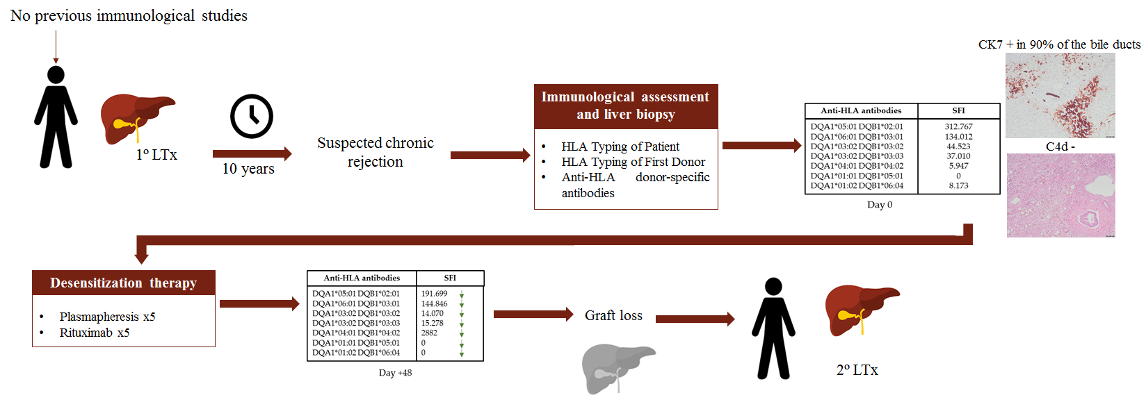
Figure 1. Evolution of anti-HLA antibodies over 40.000 SFI in some of the sera studied. Day 0 refers to the date of the first serum studied.
Transplantology 02 00001 g001
Figure 2. (A) Portal and interstitial fibrosis, with chronic inflammatory infiltrate and cholestatic signs (Hematoxylin-eosin); (B) Bile duct in the portal space with cytokeratin 7 (CK7).
Transplantology 02 00001 g002
Figure 3. Evolution of anti-HLA antibodies against DQ2 antigens of different alleles. Day 0 refers to the date of the first serum studied.
Transplantology 02 00001 g003
Table 1. Human leukocyte antigen (HLA) typing of the patient and the donors.
HLA Typing of PatientA*01:01 A*30:02 B*49:01 B*50:02 C*06:02 C*07:01 DRB1*07:01 DRB1*08:01 DQA*02:01 DQA1*04:02 DQB1*02:01 DQB1*04:02
HLA Typing of First DonorA*30:02 A*26:01 B*18:01 B*38:01 C*05:01 C*12:03 DRB1*03:01 DRB1*14:01 DQB1*02:01 DQB1*05:03 (DQA1*01:01 DQA1*05:01)
HLA Typing of Second DonorA*02:01 A*30:01 B*08:01 B*51:01 C*07:01 C*02:02 C*07:01 DRB1*03:01 DQB1*02:01 (DQA1*05:01)
In brackets, we show the donor DQA1 locus deduced by linkage disequilibrium.
Table 2. Biochemical parameters of liver function.
Biochemical Parameters of Liver FunctionMean (Min–Max)
Aspartate transaminase (AST)146.9 U/L (76–261)
Alanine transaminase (ALT)87.6 U/L (59–114)
Bilirubin15.7 mg/dL (6.6–24.9)
Alkaline phosphatase (ALP)259 U/L (138–352)
Gamma-glutamyltransferase (GGT)62.1 (33–90)
Means were calculated with the determinations within the last 3 months before first donor-specific antibody (DSA) determination. Aspartate transaminase (AST) normal value: 4–50 U/L; Alanine transaminase (ALT) normal value 5–40 U/L; Bilirubin normal value 0.2–1.2 mg/dL; Alkaline phosphatase (ALP) normal value 42–128 U/L; Gamma-glutamyltransferase (GGT) normal value 7–10 U/L.
Table 3. Equivalence standard fluorescence intensity (SFI)-mean fluorescence intensity (MFI) units of anti-HLA antibodies and probable crossmatch results.
SFI UnitsMFI UnitsDonor AntigensCDC XMFC XM
>100.0004ForbiddenProbably positivePositive
100.000–40.0004.000–1.500RiskProbably negativeProbably positive
40.000–20.0001.500–800UndeterminedNegativeProbably negative
<20.000<800No RiskNegativeNegative
CDC: complement-dependent cytotoxicity; FC XM: Flow cytometry crossmatching.
Publisher’s Note: MDPI stays neutral with regard to jurisdictional claims in published maps and institutional affiliations.

