Article

Relationship between the Superior Attachment of the Uncinate Process and Pneumatization of the Middle Turbinate—A Radiological Study

by
Christian Calvo-Henriquez
1,2,*,
Miguel Mayo-Yañez
1,3,
Carlos M. Chiesa-Estomba
1,4,
Gabriel Martinez-Capoccioni
2 and
Carlos Martin-Martin
2,5
1
Young-Otolaryngologists of the International Federations of Oto-Rhino-Laryngological Societies (YO-IFOS) Study Group, 13005 Marseille, France
2
Service of Otolaryngology, Hospital Complex of Santiago de Compostela, Travesía de Choupana s/n., 15706 Santiago de Compostela, Spain
3
Service of Otolaryngology, Hospital Complex of La Coruña. As Juvias 84, 15006 A Coruña, Spain
4
Service of Otolaryngology, Donostia University Hospital, Paseo Dr. Begiristain s/n., 20014 San Sebastian, Spain
5
Department of Otolaryngology, Medicine College, University of Santiago de Compostela, Rua de San Francisco s/n., 15782 Santiago de Compostela, Spain
*
Author to whom correspondence should be addressed.
Submission received: 25 December 2021 / Revised: 17 April 2022 / Accepted: 5 May 2022 / Published: 24 May 2022

Abstract

:
Objective: the superior attachment of the uncinate process (SAUP) is highly variable. Lateral types of SAUP are associated with frontal rhinosinusitis. SAUP in the middle turbinate is more common when the middle turbinate is aerated. We aim to refine these findings by studying the relationship between the SAUP type and the different subtypes of pneumatization of the middle turbinate. Methods: 200 CT sinonasal scans were analyzed by an otolaryngologist and a radiologist. All CT scans were analyzed to study the type of the middle turbinate concha bullosa and lamellar cell and the type of SAUP. Results: 379 sides were analyzed. The prevalence of the various types of SAUP were: Type I: 43.80%, Type II: 22.16%, Type III: 16.36%, Type IV: 3.69%, Type V: 9.76%, and Type VI: 4.22%. When studying the pneumatization of the middle turbinate and the SAUP types, we only found a statistically significant association between the lateral attachment of the uncinate process and the presence of a lamellar cell (p = 0.029). Conclusion: lateral types of SAUP are more likely in the presence of a lamellar cell. This finding, therefore, renders checking for lateral SAUP types in the presence of a lamellar cell even more relevant than when studying the pre-surgical CT scan. In those cases, it is advisable to perform a careful and complete uncinectomy.

1. Introduction

The uncinate process (UP) is a thin bone of the lateral wall of the nose1, medial to the ethmoid infundibulum, being the medial wall of the ostiomeatal complex. There it plays a crucial role in mucociliary activity [1]. Uncinectomy is a mandatory step in functional endoscopic sinus surgery (FESS), necessary to expose the hiatus semilunaris, maxillary ostium, and nasofrontal recess [2,3].
The posterior edge and the posterior segment of the inferior border of the UP are free. On the other hand, the superior attachment of the uncinate process (SAUP) is highly variable. It has been studied by Landsberg and Friedman [4], who classified it into six types (Figure 1): Type I: insertion to the lamina papyracea (52%); Type II: insertion to the posteromedial wall of the agger nasi cell (18.5%); Type III: insertion to both the lamina papyracea and the junction of the middle turbinate with the cribriform plate (17.5%); Type IV: insertion to the junction of the middle turbinate with the cribriform plate (7%); Type V: insertion to the skull base (3.6%); and Type VI: insertion to the middle turbinate (1.4%). Other authors have suggested new subtypes, but they are not standardized yet [5,6,7].
Preoperatory analysis of the SAUP is important for three reasons. First, knowing the insertion type enables the surgeon to perform a safe uncinectomy. Secondly, the SAUP determines the frontal sinus drainage pathway, which is associated with frontal rhinosinusitis. Finally, incomplete uncinectomy has been linked to a higher risk of surgical failure [8].
An aggressive uncinectomy, performed by pulling the UP out in patients with a SAUP in the cranial base (Types III, IV, or V), is a risk factor in developing a cerebrospinal fluid leak [9]. In these cases, it is recommended to avoid traction of the uncinate process.
Frontal rhinosinusitis is more common when the frontal drainage is medial to the uncinate process [5,10]. In those cases, an incomplete uncinectomy is related to the persistence of rhinosinusitis [5]. In fact, an incomplete uncinectomy was found in 37% of patients undergoing revision for FESS [8].
Cheng et al. describe that the SAUP in the middle turbinate (Types III, IV, and VI) is more common when the middle turbinate is aerated [11]. This is called “concha bullosa”. An aerated middle turbinate creates a space compromise, making it difficult to perform a superior uncinectomy.
The pneumatization of the middle turbinate may affect different areas of the middle turbinate. It can be divided into bulbous, affecting the body of the middle turbinate, or lamellar cell, affecting the vertical aspect of the middle turbinate (Figure 2). Both types of middle turbinate aeration have recently been studied and classified by our research team. We have classified the bulbous concha bullosa into five categories [12], and the lamellar cell concha bullosa into four categories [13].
In this work, we aim to refine the findings of Cheng et al. by studying the relationship between the SAUP type and the different subtypes of pneumatization of the middle turbinate.

2. Methods

2.1. Data Source

The file of the latest 200 CT sinonasal scans performed at a tertiary university hospital was obtained independently of the diagnosis.
Inclusion criteria were to have a sinonasal CT scan of less than 1 mm slice resolution. There was no restriction by age or illness. We reviewed the medical records of all patients to obtain diagnosis and history of previous sinonasal surgery. In patients scheduled for sinonasal surgery, the pre-surgery CT scan was also obtained.
Exclusion criterion was presenting a CT scan with a slice resolution higher than 1 mm, as this does not enable the adequate study of the superior attachment of the uncinate process. CT scans not including SAUP were also excluded.

2.2. Image Study

CT scans were analyzed by an otolaryngologist and a radiologist with the DICOM viewer Sectra®. The 3D reconstruction planes were made from the axial images.
All CT scans were analyzed to study the absence or presence (and type) of the middle turbinate concha bullosa and lamellar cell. We examined coronal views to find pneumatization of the middle turbinate. In case of pneumatization, a thorough exam was performed using a modified axial image through the bulla.
All CT scans were analyzed in order to study the type of SAUP using 3D reconstruction planes, which allowed us to study the SAUP in the axial, coronal, and sagittal planes concomitantly.

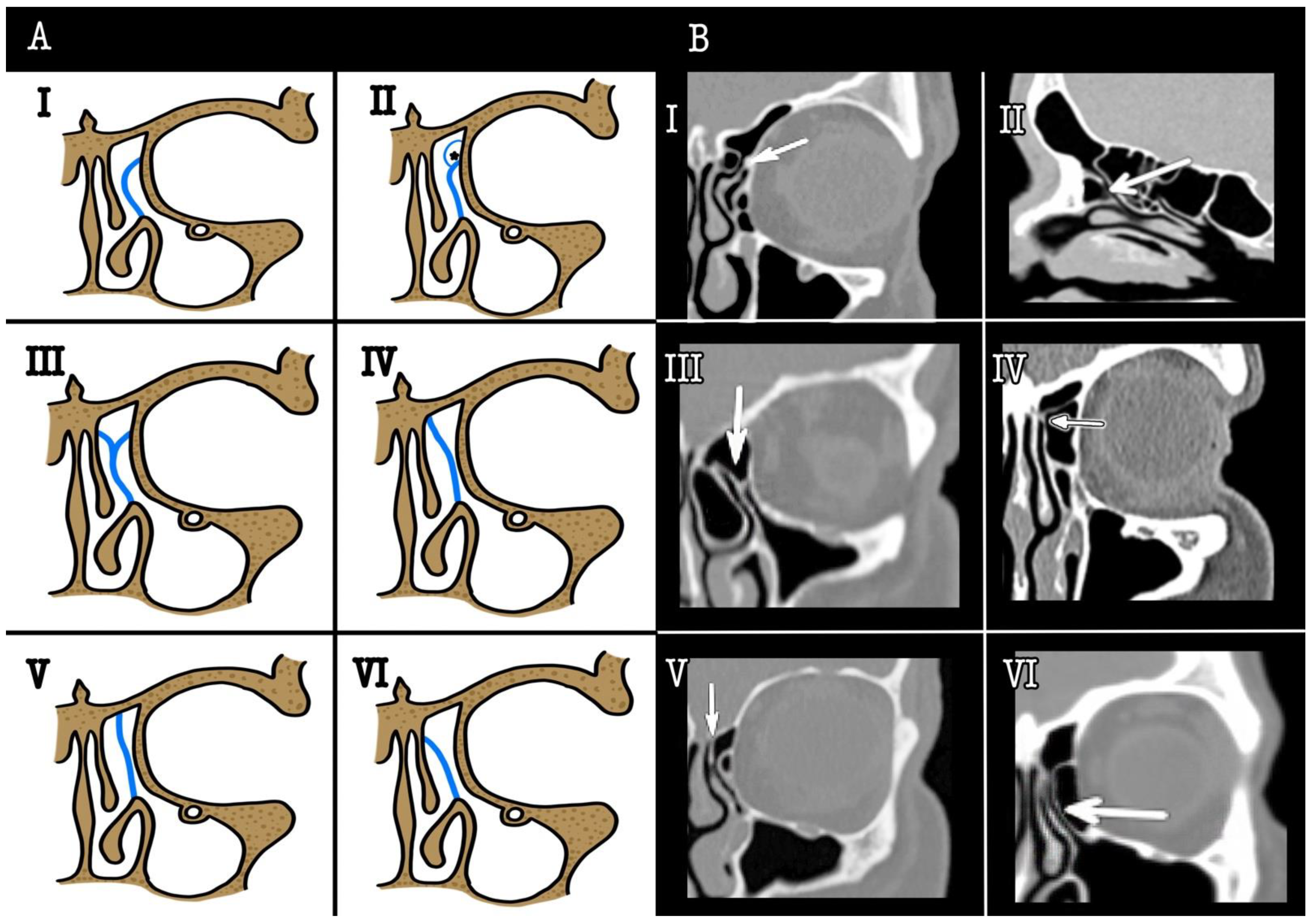
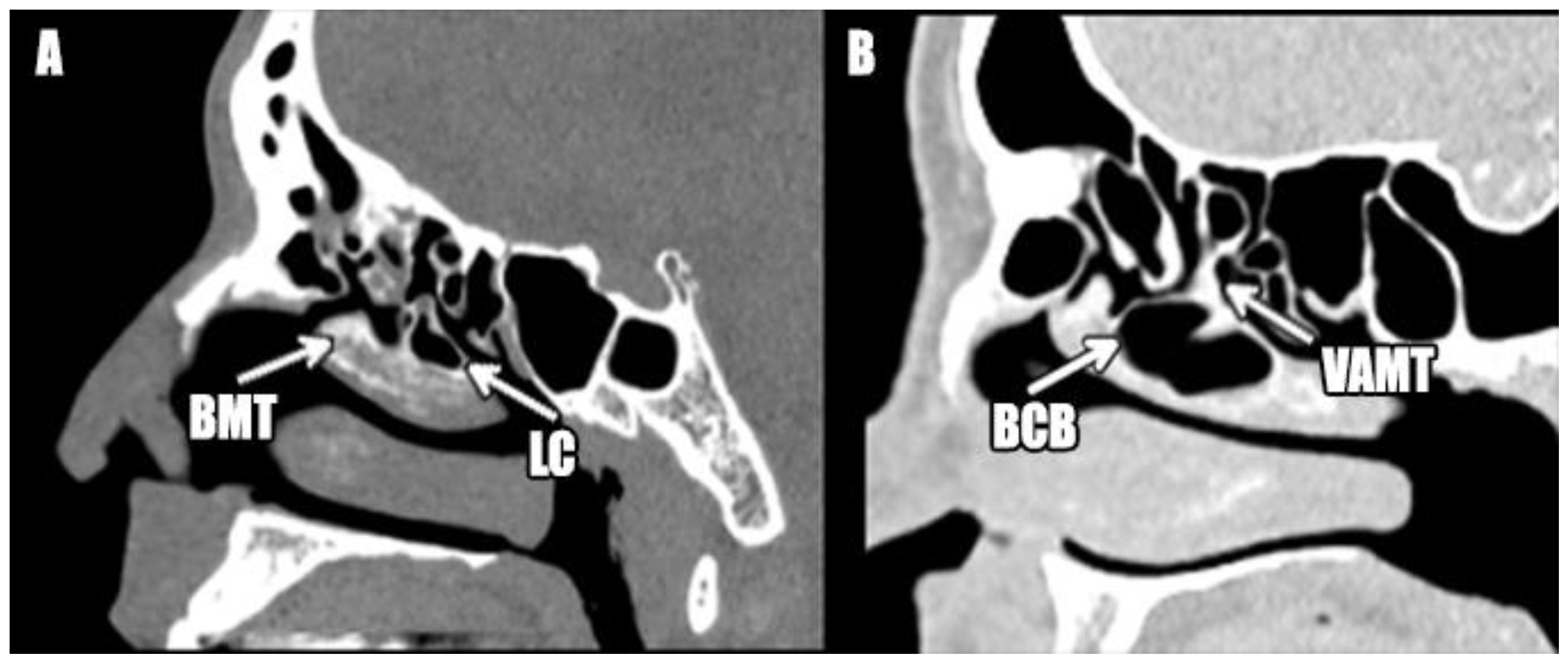
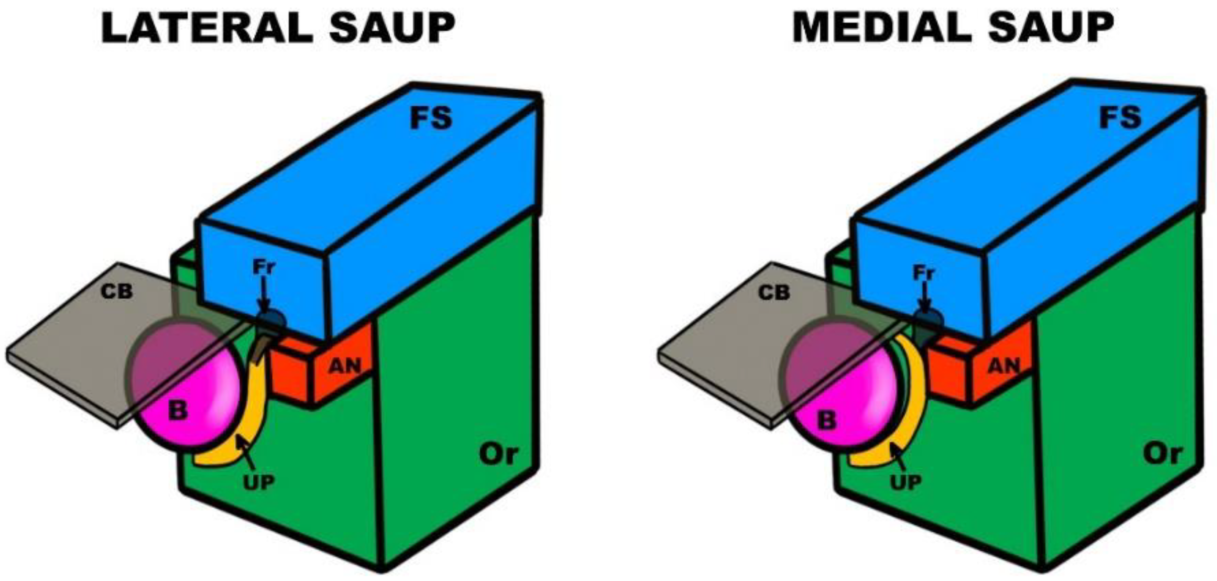
2.3. Concha Bullosa Classification

The concha bullosa was classified into 6 categories according to the degree of pneumatization in the axial plane of the body of the middle turbinate, following the classification previously described by our team [12]: Type 0: no aeration; Type I: aeration of 1/3 anterior; Type II: aeration of 2/3 anterior, or 1/3 in the middle; Type III: complete anterior-posterior aeration; Type IV: aeration of 2/3 posterior, and Type V: aeration of 1/3 posterior.

2.4. Lamellar Cell Classification

The lamellar cell was classified into 5 categories according to the degree of pneumatization of the vertical lamella in the middle turbinate, likewise following the classification previously established by our research team [13] (see Figure 1): Type 0: no cell; Type I: pneumatization of ½ the anterior vertical lamella of the middle turbinate; Type II: pneumatization of ½ the posterior vertical lamella and ground lamella of the middle turbinate; Type III: a combination of Types I and II; Type IV: Type III continued with a concha bullosa.

2.5. Statistical Analysis

Statistical analysis was performed with SPSS for Macintosh v. 23.0 (Chicago, IL, USA, IBM Corp.). We used chi-square analysis. Statistical significance was initially determined at p < 0.05.

2.6. IRB Approval

The study was performed in accordance with the ethical standards laid down in the Declaration of Helsinki. The study protocol was approved by the Research and Ethics Committee of the Hospital Complex of Santiago de Compostela-Spain, register code 2017/038.

3. Results

3.1. Participants

Of the 200 initially selected CT sinonasal scans, 5 were completely excluded: 3 because the middle turbinate could not be studied due to nasal polyps, 1 because the image resolution was poor, and 1 because the superior attachment of the uncinate process was not shown in the CT scan.
We studied each side as a different unit. In 11, cases the uncinate process could not be studied in 1 of the sides due to the mucosal disease that made the bony septa indistinct. Finally, we examined 379 sides.
There were 51.19% females and 48.81% males. Mean age was 54.17 years, median 54, range 6–100, percentile 25–75 (40–68).

3.2. SAUP Type Prevalencez

The prevalence of the SAUP in our sample was Type I: 43.80%, Type II 22.16%, Type III 16.36%, Type IV 3.69%, Type V 9.76% and Type VI 4.22%.
SAUP was the same in both sides at 66.67% (95% CI 59.71–72.96).

3.3. SAUP and Lamellar Cell

Results are shown in Table 1. We found a statistically significant association between the lateral attachment of the uncinate process and the presence of a lamellar cell (p = 0.029). There was no relationship between the presence of a lamellar cell and the SAUP in the middle turbinate (p = 0.944).
Furthermore, there was no statistically significant relationship between the anterior types of lamellar cell and SAUP in the middle turbinate (p = 0.237), nor in the lateral SAUP (p = 0.057).

3.4. SAUP and Concha Bullosa

Results are shown in Table 1. We did not find any significant association between the lateral attachment of the uncinate process and the presence of concha bullosa (p = 0.944), even when considering only the anterior types of concha bullosa (p = 0.884).
Also, there was neither a significant correlation between the middle turbinate SAUP and the presence of concha bullosa (p = 0.782), nor with the anterior types of concha bullosa (p = 0.665).

4. Discussion

Frontal rhinosinusitis is one of the most important challenges for a rhinology specialist, given its frequency, as well as its tendency to relapse.
The anatomy of the frontal recess is intricate. It is limited by neighboring structures, such as the ethmoid bulla, uncinate process, and agger nasi cell. Anatomic variations in those structures are proposed etiologic factors of frontal rhinosinusitis [14], with SAUP being the most relevant [3]. Variations in the SAUP affect the anatomy of the frontal sinus drainage [15] and are a recognized cause of frontal rhinosinusitis [10], as well a cause of failure in surgery [8]. Therefore, knowledge of the anatomic variations in the SAUP is paramount to understanding and properly treating frontal sinus illnesses.
Understanding the 3D anatomy of the frontal recess is complex. The nasofrontal recess is located superior and posterior to the agger nasi cell. The inferior portion of the posterior wall of the agger nasi cell is the uncinate process [7]. When the SAUP is into the lamina papyracea, the ethmoidal infundibulum and the frontal recess are separated from each other so that the ethmoidal infundibulum is closed superiorly to form a blind pouch called a “terminal recess” [16]. Thus, in those cases, the frontal recess opens directly into the middle meatus, medial to the uncinate process. When the SAUP extends superiorly into the skull base or the middle turbinate, the frontal recess drains into the middle meatus via the ethmoidal infundibulum, lateral to the uncinate process (Figure 3). In summary, if the SAUP is lateral (Types I, II, and III), the frontal drainage will be medial to the uncinate process. In the opposite scenario, if the SAUP is medial (Types IV, V, and VI), the drainage will be lateral to the uncinate process.
In our research we found a statistically significant association between the lateral types of SAUP (Types I, II, and III) and the presence of a lamellar cell (p = 0.029). Interestingly, we did not find any significant relationship between the lateral SAUP in the middle turbinate and the lamellar cell, anterior lamellar cell, concha bullosa, or anterior concha bullosa. However, these results should be managed carefully because, if the p value is adjusted for multiple comparisons (eight in this study), the final p value, in order to reject the null hypothesis, is 0.006, which has not been reached in our study.
These results are different from those obtained by Cheng et al., who recently described an increased prevalence of SAUP in the middle turbinate in the presence of a pneumatized middle turbinate [11].
Our findings are crucial from a clinical perspective. A lateral SAUP has been associated with frontal rhinosinusitis [5,10]. When a patient presents a lamellar cell, a lateral SAUP is more likely. Therefore, in these cases, it is paramount to thoroughly study SAUP. Furthermore, in the case of a lamellar cell, a complete uncinectomy could be more difficult to perform due to the lack of space, and an incomplete uncinectomy has been linked to relapse of frontal rhinosinusitis.
It is important to highlight that we have differentiated between bulbous concha bullosa and lamellar cell concha bullosa, and we only found a connection with the lamellar cell. The classical definition of concha bullosa includes a lamellar cell [17]; therefore, in the research of Cheng et al., some patients classified as presenting a concha bullosa would probably be patients with a lamellar cell. This finding is important since concha bullosa could not be linked to chronic rhinosinusitis [18,19]. Future studies should focus on the lamellar cell and frontal rhinosinusitis, given their probable relationship.
Regarding the prevalence of SAUP, our results are similar to those obtained by other authors; a summary is shown in Table 2. Type I is the most common SAUP type for all authors, with the exception of Mahmutoğlu [5], and the less frequent Types are IV, V, and VI. Notice that we have used the Landsberg and Friedman classification [4], while other authors have used other classifications or their own adaptations [5,6,7,20,21,22,23], and therefore we cannot compare our results with theirs. Furthermore, three authors have combined Types I and II in their research [5,10,24]. It is also noteworthy that the work of Turgut et al. [24] was not included in the table since they used the same sample as Ercan et al. [24]. We highlight the research of Al-Qudah and Mardini, who studied SAUP in pediatric patients. They used a different classification in three types, but it remains interesting since pediatric patients have a similar prevalence of SAUP to that of adults [20].
We found a 66.67% prevalence of identical SAUP types on both sides, similarly to Netto et al. [25] (59.9%) and Turgut et al. (65%) [10], but clearly lower than that of Landsberg and Friedman [4] (93%).

5. Conclusions

In conclusion, lateral types of SAUP are more likely in the presence of a lamellar cell. This finding, therefore, renders checking for lateral SAUP types in the presence of a lamellar cell even more relevant than it was when studying the pre-surgical CT scan. In those cases, it is advisable to perform a careful and complete uncinectomy.

Author Contributions

Conceptualization, C.C.-H. and C.M.-M.; methodology, C.C.-H., M.M.-Y., C.M.C.-E., G.M.-C. and C.M.-M.; software, C.C.-H.; validation, C.C.-H., M.M.-Y., C.M.C.-E., G.M.-C. and C.M.-M.; formal analysis, C.C.-H., M.M.-Y., C.M.C.-E., G.M.-C. and C.M.-M.; data curation, C.C.-H., M.M.-Y. and C.M.C.-E.; writing—original draft preparation, C.M.C.-E.; writing—review and editing, M.M.-Y., C.M.C.-E., G.M.-C. and C.M.-M.; supervision, C.M.-M. All authors have read and agreed to the published version of the manuscript.

Funding

This research received no external funding.

Institutional Review Board Statement

The study was conducted in accordance with the Declaration of Helsinki and approved by the Institutional Review Board (or Ethics Committee) of the Hospital Complex of Santiago de Compostela–Spain, register code 2017/038.

Informed Consent Statement

Informed consent was obtained from all subjects involved in the study.

Data Availability Statement

Not applicable.

Conflicts of Interest

The authors declare that there are no conflicts of interest.

References

  1. Nayak, D.R.; Balakrishnan, R.; Murty, K.D. Functional anatomy of the uncinate process and its role in endoscopic sinus surgery. Indian J. Otolaryngol. Head Neck Surg. Off. Publ. Assoc. Otolaryngol. India 2001, 53, 27–31. [Google Scholar] [CrossRef] [PubMed] [Green Version]
  2. Fombeur, J.P.; Koubbi, G.; Seguin, D.; Ebbo, D.; Lecomte, F.; Laurier, J.N. Indications, technics and results of middle meatotomy. Apropos of 94 cases. Ann. Oto-Laryngol. Chir. Cervico Faciale Bull. Soc. Oto-Laryngol. Hopitaux Paris 1989, 106, 515–517. [Google Scholar]
  3. Friedman, M.; Landsberg, R.; Schults, R.A.; Tanyeri, H.; Caldarelli, D.D. Frontal sinus surgery: Endoscopic technique and preliminary results. Am. J. Rhinol. 2000, 14, 393–403. [Google Scholar] [CrossRef] [PubMed]
  4. Landsberg, R.; Friedman, M. A computer-assisted anatomical study of the nasofrontal region. Laryngoscope 2001, 111, 2125–2130. [Google Scholar] [CrossRef] [PubMed] [Green Version]
  5. Mahmutoğlu, A.S.; Çelebi, I.; Akdana, B.; Bankaoğlu, M.; Çakmakçi, E.; Çelikoyar, M.M.; Başak, M. Computed tomographic analysis of frontal sinus drainage pathway variations and frontal rhinosinusitis. J. Craniofac. Surg. 2015, 26, 87–90. [Google Scholar] [CrossRef] [PubMed]
  6. Liu, S.-C.; Wang, C.-H.; Wang, H.-W. Prevalence of the uncinate process, agger nasi cell and their relationship in a Taiwanese population. Rhinology 2010, 48, 239–244. [Google Scholar] [CrossRef]
  7. Zhang, L.; Han, D.; Ge, W.; Xian, J.; Zhou, B.; Fan, E.; Liu, Z.; He, F. Anatomical and computed tomographic analysis of the interaction between the uncinate process and the agger nasi cell. Acta Otolaryngol. 2006, 126, 845–852. [Google Scholar] [CrossRef]
  8. Orlandi, R.R.; Kennedy, D.W. Revision endoscopic frontal sinus surgery. Otolaryngol. Clin. N. Am. 2001, 34, 77–90. [Google Scholar] [CrossRef]
  9. Kirtane, M.V.; Gautham, K.; Upadhyaya, S.R. Endoscopic CSF rhinorrhea closure: Our experience in 267 cases. Otolaryngol. Head Neck Surg. Off. J. Am. Acad. Otolaryngol.-Head Neck Surg. 2005, 132, 208–212. [Google Scholar] [CrossRef]
  10. Turgut, S.; Ercan, I.; Sayin, I.; Başak, M. The relationship between frontal sinusitis and localization of the frontal sinus outflow tract: A computer-assisted anatomical and clinical study. Arch. Otolaryngol. Head Neck Surg. 2005, 131, 518–522. [Google Scholar] [CrossRef] [Green Version]
  11. Cheng, S.-Y.; Yang, C.-J.; Lee, C.-H.; Liu, S.-C.; Kuo, C.-Y.; Lee, J.-C.; Shih, C.-P. The association of superior attachment of uncinate process with pneumatization of middle turbinate: A computed tomographic analysis. Eur. Arch. Oto-Rhino-Laryngol Off. J. Eur. Fed. Oto-Rhino-Laryngol Soc. EUFOS Affil. Ger. Soc. Oto-Rhino-Laryngol-Head Neck Surg. 2017, 274, 1905–1910. [Google Scholar] [CrossRef] [PubMed]
  12. Calvo-Henríquez, C.; Mota-Rojas, X.; Ruano-Ravina, A.; Martinez-Capoccioni, G.; Lattomus, K.; Martin-Martin, C. Concha bullosa. A radiological study and a new classification. Acta Otorrinolaringol. Esp. 2019, 70, 145–150. [Google Scholar] [CrossRef] [PubMed]
  13. Calvo-Henríquez, C.; Ruano-Ravina, A.; Martinez-Capoccioni, G.; Huaranca, M.; Lattomus, K.; Martin-Martin, C. The lamellar cell: A radiological study and a new classification proposal. Eur. Arch. Oto-Rhino-Laryngol Off. J. Eur. Fed. Oto-Rhino-Laryngol Soc. EUFOS Affil. Ger. Soc. Oto-Rhino-Laryngol-Head Neck Surg. 2018, 275, 2713–2717. [Google Scholar] [CrossRef] [PubMed]
  14. Close, L.G.; Stewart, M.G. Looking around the corner: A review of the past 100 years of frontal sinusitis treatment. Laryngoscope 2009, 119, 2293–2298. [Google Scholar] [CrossRef]
  15. Daniels, D.L.; Mafee, M.F.; Smith, M.M.; Smith, T.L.; Naidich, T.P.; Brown, W.D.; Bolger, W.E.; Mark, L.P.; Ulmer, J.L.; Hacein-Bey, L.; et al. The frontal sinus drainage pathway and related structures. AJNR Am. J. Neuroradiol. 2003, 24, 1618–1627. [Google Scholar] [PubMed]
  16. McLaughlin, R.B.; Rehl, R.M.; Lanza, D.C. Clinically relevant frontal sinus anatomy and physiology. Otolaryngol. Clin. N. Am. 2001, 34, 1–22. [Google Scholar] [CrossRef]
  17. Lund, V.J.; Stammberger, H.; Fokkens, W.J.; Beale, T.; Bernal-Sprekelsen, M.; Eloy, P.; Georgalas, C.; Gerstenberger, C.; Hellings, P.; Herman, P.; et al. European position paper on the anatomical terminology of the internal nose and paranasal sinuses. Rhinol. Suppl. 2014, 24, 1–34. [Google Scholar]
  18. Hatipoğlu, H.G.; Cetin, M.A.; Yüksel, E. Concha bullosa types: Their relationship with sinusitis, ostiomeatal and frontal recess disease. Diagn. Interv. Radiol. Ank. Turk. 2005, 11, 145–149. [Google Scholar]
  19. Balikci, H.H.; Gurdal, M.M.; Celebi, S.; Ozbay, I.; Karakas, M. Relationships among concha bullosa, nasal septal deviation, and sinusitis: Retrospective analysis of 296 cases. Ear Nose Throat J. 2016, 95, 487–491. [Google Scholar] [CrossRef] [Green Version]
  20. Al-Qudah, M.; Mardini, D. Computed tomographic analysis of frontal recess cells in pediatric patients. Am. J. Rhinol. Allergy 2015, 29, 425–429. [Google Scholar] [CrossRef]
  21. Yang, Q.-T.; Shi, J.-B.; Kang, Z.; Chen, H.-X.; Wang, T.; Lü, J.-T.; Xu, G. Computer-assisted anatomical study of nasofrontal region. Zhonghua Er Bi Yan Hou Ke Za Zhi 2004, 39, 349–352. [Google Scholar] [PubMed]
  22. Han, D.; Zhang, L.; Ge, W.; Tao, J.; Xian, J.; Zhou, B. Multiplanar computed tomographic analysis of the frontal recess region in Chinese subjects without frontal sinus disease symptoms. ORL J. Oto-Rhino-Laryngol Its Relat. Spec. 2008, 70, 104–112. [Google Scholar] [CrossRef] [PubMed]
  23. Srivastava, M.; Tyagi, S. Role of Anatomic variations of Uncinate Process in Frontal Sinusitis. Indian J. Otolaryngol. Head Neck Surg. Off. Publ. Assoc. Otolaryngol. India 2016, 68, 441–444. [Google Scholar] [CrossRef] [PubMed] [Green Version]
  24. Ercan, I.; Cakir, B.O.; Sayin, I.; Başak, M.; Turgut, S. Relationship between the superior attachment type of uncinate process and presence of agger nasi cell: A computer-assisted anatomic study. Otolaryngol. Head Neck Surg Off. J. Am. Acad Otolaryngol.-Head Neck Surg. 2006, 134, 1010–1014. [Google Scholar] [CrossRef]
  25. Netto, B.; Piltcher, O.B.; Meotti, C.D.; Lemieszek, J.; Isolan, G.R. Computed tomography imaging study of the superior attachment of the uncinate process. Rhinology 2015, 53, 187–191. [Google Scholar] [CrossRef]
Figure 1. SAUP types (modified from Landsberg and Friedman (2001). (A): graphical representation. Abbreviations: uncinate process in blue; asterisk (agger nasi cell). (B): CT scans. A white arrow shows the uncinate process superior attachment.
Surgeries 03 00015 g001
Figure 2. Bulbous and lamellar concha bullosa: (A): Lamellar concha bullosa. Note that the cell is only lamellar cell (LC) and is not affecting the body of the middle turbinate (BMT). (B): Bulbous concha bullosa (BCB). Note that the cell is not affecting the vertical aspect of the middle turbinate (VAMT).
Surgeries 03 00015 g002
Figure 3. Schematic representation of the frontal recess (Fr). Abbreviations: Frontal sinus (FS), Agger nasi cell (AN), Orbit (Or), Cranial base (CB), Uncinate process (UP), Ethmoidal bulla (B), Lateral SAUP: representation of Type I SAUP (lamina papyracea), Medial SAUP: representation of Type V (cranial base). Note that the Fr has been simplified as a duct for an educational objective.
Surgeries 03 00015 g003
Table 1. p values for concha bullosa, lamellar cell and SAUP. (bold if statistically significant p < 0.05).
Concha BullosaLamellar Cell
All TypesAnterior TypesAll TypesAnterior Types
Middle turbinate SAUPp = 0.782p = 0.665p = 0.235p = 0.237
Lateral SAUPp = 0.944p = 0.884p = 0.029p = 0.057
Table 2. Prevalence of SAUP types.
Author/YearSampleSAUP Type (%)
IIIIIIIVVVIOthers
Calvo-Henríquez (this study)37943.8022.1616.363.699.764.22-
Mahmutoğlu (2015) [5]91928.236.14.10.90.717.4
Netto B (2015) [25]29263.59.59.56.36.30.9
Ercan (2006) [24]37162.63.111.614.48.3-
Landsberg (2001) [4]1735218.517.573.61.4-
Publisher’s Note: MDPI stays neutral with regard to jurisdictional claims in published maps and institutional affiliations.

