Article

A Comparison of Microbial Communities of Mango and Orange Residues for Bioprospecting of Biosurfactant Producers

by
Fernanda de Paula
1,
Natália Vama Vieira
1,
Gabriela Fiori da Silva
1,
Tiago Palladino Delforno
2 and
Iolanda Cristina Silveira Duarte
1,*
1
Department of Biology, Laboratory of Applied Microbiology, Federal University of São Carlos, Rodovia João Leme dos Santos km 110, Itinga, São Carlos 18052-780, SP, Brazil
2
Innovation Institute for Biotechnology (SENAI-Biotech), Rua Anhaia, 1321, Bom Retiro, São Paulo 01130-000, SP, Brazil
*
Author to whom correspondence should be addressed.
Submission received: 7 April 2022 / Revised: 14 May 2022 / Accepted: 17 May 2022 / Published: 20 May 2022
(This article belongs to the Special Issue Feature Papers of Ecologies 2022)

Abstract

:
Plants and their derivatives, such as fruits, can be inhabited by different bacteria. However, this microbiota is still poorly studied. Among the wide variety of metabolites that bacteria produce, biosurfactants have been identified as potential molecules in the development of bioprocesses for various industrial sectors. In this work, we analyzed and compared the microbiota of fruit residues (mango and orange), in order to compare two possible sources of bioprospecting. For this, a bioinformatics approach was used to perform the taxonomic analysis and the prediction of the functional profile of the microbiota present in the samples. The results showed that the microbiota present in both fruit residues have the potential in biotechnological applications to produce biosurfactants, as these microbiota have genes related to the biosynthesis of these compounds. The common core of the microbiota present in the samples—Stenotrophomonas, Klebsiella, Serratia and Citrobacter—proved, according to the literature, to be composed of biosurfactant producers, showing the biosurfactant potential of the bacteria isolated from orange and mango residues.

1. Introduction

Surfactants are amphiphilic molecules with hydrophilic and hydrophobic portions, which have properties to decrease the surface or interfacial tension of two liquid phases. They are compounds that play a wide and significant role in several market areas and can also be used in bioremediation processes. However, synthetic surfactants come from the petrochemical industry, and their use has caused a great deal of environmental damage. Therefore, biosurfactants, which are produced by microorganisms, are an environmentally favorable alternative. Biosurfactants are synthesized by microorganisms, such as bacteria, yeasts and filamentous fungi. The production of these compounds occurs through secondary metabolic pathways in a microbial fermentation, which starts during the stationary phase, often related to the lack of nutrients, and continues until the phase of death [1,2,3,4,5]. Biosurfactants are effective in oil spill recovery and removal, wastewater treatment, agribusiness, pharmaceutical and cosmetic industries, and also have the ability to remove potentially toxic metals from contaminated soil and water [6].
Biosurfactant-producing microorganisms are present in various aquatic and terrestrial habitats, including extreme environments, seawater and bilge water [7,8,9]. Some biosurfactant-producing microorganisms can be isolated from high-sugar environments, such as fruits and other parts of plants. Using agro-industrial residues to produce biosurfactants is an environmentally favorable and low-cost alternative. Therefore, knowledge of the diversity of the microbiota present in fruit residues is needed to verify the functional potential of these microorganisms and their applicability to various industrial sectors [7,8,9].
In this scenario, Brazil is the world’s largest orange producer, accounting for approximately 50% of the world’s orange juice production and holds 85% of the world’s exports of the product [10]. However, after extracting the orange juice, approximately 50% of the fruit is discarded in the form of bagasse (peel, seed and pulp). Part of this waste is used to supplement cattle and pig herds, but much of it is wasted [11]. Another prominent tropical fruit in production is mangoes. Brazil is a large producer and exporter of mangoes. In 2020, the mango export to the American and European markets reached the level of 243.2 thousand tons of mangoes, totaling the value of $246.9 million USD. Similar to the case in oranges, the large amount of mango production generates a lot of residue and waste from the pulp and juice industries [12].
In our study, we were interested in evaluating the microbiota present in fruit samples of the Mangifera indica L. (mangoes) and Citrus sinensis L. (oranges) after enriched fermentation. In order to compare these possible sources of bioprospecting from the sequencing of the 16S rRNA gene, the microbiota present in these enriched fruit residues were taxonomically assigned. As the functional profiles cannot be directly identified using 16S rRNA gene sequence data, the prediction of the functional profiles associated with the microbiota present in the fruit samples was performed using PICRUSt2, which is a software for predicting functional abundances based only on marker gene sequences. In this research, we specifically focused only on the analysis of the genes related to biosurfactant production.

2. Materials and Methods

2.1. Sampling

Mangoes (1.2 kg) and oranges (1.2 kg) unfit for consumption were collected at street fairs in the city of Sorocaba, SP, Brazil. The orange bagasse was obtained from peeled and squeezed oranges after juice removal. To obtain the mango pulp, the peel and kernel were discarded. The orange bagasse and mango pulp were ground separately using a domestic blender, homogenized in distilled water in a 1:1 (w/v) ratio and stored in airtight plastic bags at 4 °C until use.

2.2. Microbial Enrichments

Each homogenized sample of fruit was enriched separately in duplicate, and 25 g L−1 of total solids were added to 250 mL of Luria–Bertani (LB) medium in an Erlenmeyer flask (500 mL). The total incubation period for the assays was 19 days, at a temperature of 34 °C and agitation of 150 rpm [13]. After 5 days of incubation, a 2.5 mL aliquot was transferred to a 250 mL flask with a new medium containing 1% glycerol and kept in a rotary shaker (150 rpm) for 7 days. After 7 days, 2.5 mL of the fermentative broth was removed and added to a new medium under the same conditions as above [14].

2.3. DNA Isolation and 16S rRNA Sequencing

After fermentation, microbial genomic DNA was extracted from the enriched fruit samples using a PowerSoil DNA isolation kit (MOBIO Laboratories, Carlsbad, CA, USA), following the manufacturer´s instructions. Two replicates from each enriched fermentation were sequenced, resulting in a total of 4 sequencing data sets. Evaluation of the DNA quality was performed by visual comparison by electrophoresis on a 0.8% agarose gel. The assessment of the taxonomic profile of the microorganisms occurred utilizing the approach of analyzing the 16S ribosomal RNA gene (16S rRNA) on a large scale. The 16S rRNA sequencing was performed using the Illumina MiSeq platform 2 × 250 bp [15]. DNA amplification was performed using primers 341F (5′-CCT ACG GGR SGC AGC AG-3′) and 806R (5′-GGA CTA C V GGG TWT CTA AT-3′) for the Bacteria and Archaea domain [16].

2.4. Bioinformatics and Statistical Analysis

Raw data quality control was performed using FASTQC v.011.5 software [17]. Low-quality sequences (Phred score ≤ 20) and adapters were filtered using the Trimmomatic v.0.39 tool [18]. The QIIME 2 program (Quantitative Insights Into Microbial Ecology version 2019.4) [19] was used to analyze the results obtained with the Illumina platform and the QIIME 2 plugin tool was used to import quality read filters as an ‘artifact’ file. Afterward, the chimeras were removed and clustered into representative sequences and Amplicon Sequence Variants (ASVs) using the ‘dada2’ plugin [20].
Taxonomic classification was performed in the SILVA ribosomal RNA gene database version 138, and the ASVs were determined using 97% similarity. The predicted functional profiles obtained in the 16S were defined using the phylogenetic method of the PICRUSt2 software (Phylogenetic Investigation of Communities of Unobserved States) [21]. In PiCRUSt2, the metagenome inference step depends on an ASV table obtained from taxonomic analysis and is then used as input for the PICRUSt2 QIIME2 plugin. Prediction results were obtained from the PICRUst2 output against the KEGG Orthology (KO) database [22] to reconstruct the metabolic pathways.
Genes were analyzed using the KOs in STAMP v2.1.3 [23]. Poor or non-specific functional data were eliminated from the analysis and used only to calculate the frequency profiles. The comparison of the functional profile between the two samples of Orange and the two samples of Mango was performed with a two-sided Welch’s t-test combined with Welch’s inverted method for 95% confidence intervals. Moreover, a false discovery rate was applied as a multiple test correction using the FDR method. An extended error bar graph was used to group the different functional profiles (KEGG level 2) by similarity. To do this, specific KEGGs records related to biosurfactant production were selected from articles analyzing microbial metabolites.
Afterward, the ASVs were exported to the R statistical environment for the purpose of performing the statistical analysis and graphical computation of the data. The taxonomic data were imported as a phyloseq object in order to perform a set of custom graphical functions for taxonomic sequencing analysis [24]. The maximum value of the ASVs observed in each sample was determined using the rarefaction curves. To assess the richness and diversity of the samples, an alpha diversity graph was used. From this, three indices were analyzed: the observed, the Chao index and the Shannon index [25]. The dissimilarities between the samples (Orange and Mango) were evaluated from the beta diversity analysis using Principal Coordinate Analysis (PCoA). Thus, the main coordinate analysis of the Bray–Curtis and Jaccard dissimilarity was performed with the two replicas of Orange and Mango [24]. The Bray–Curtis index can be expressed as a proportion of similarity or dissimilarity in species abundance. The Jaccard index, on the other hand, indicates the proportion of species shared between two samples in relation to the total number of species [26]. The DESeq2 package was used to perform the differential analysis of genera between the samples. The sequencing reads were submitted to the European Nucleotide Archive under project accession PRJEB36395 and sample accessions ERS4265892 (Mango-1), ERS4265891 (Mango-2), ERS4265890 (Orange-1), ERS4265889 (Orange-2).

3. Results

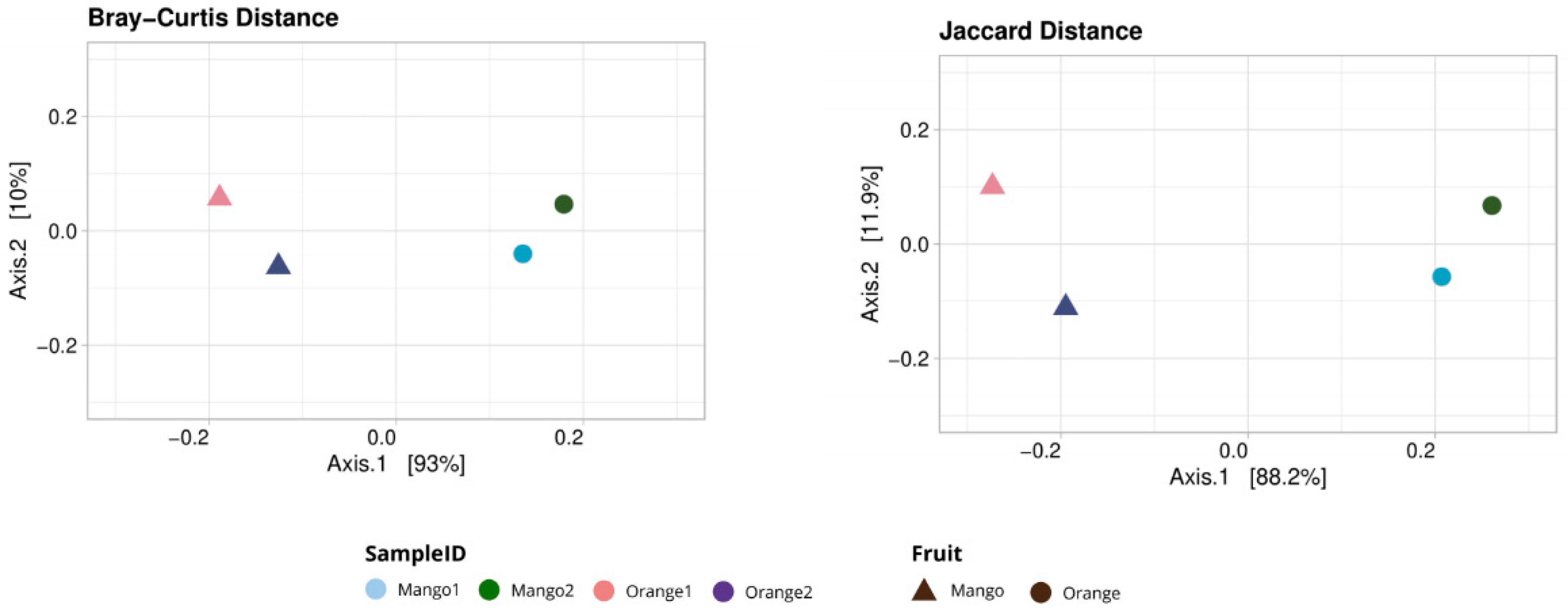
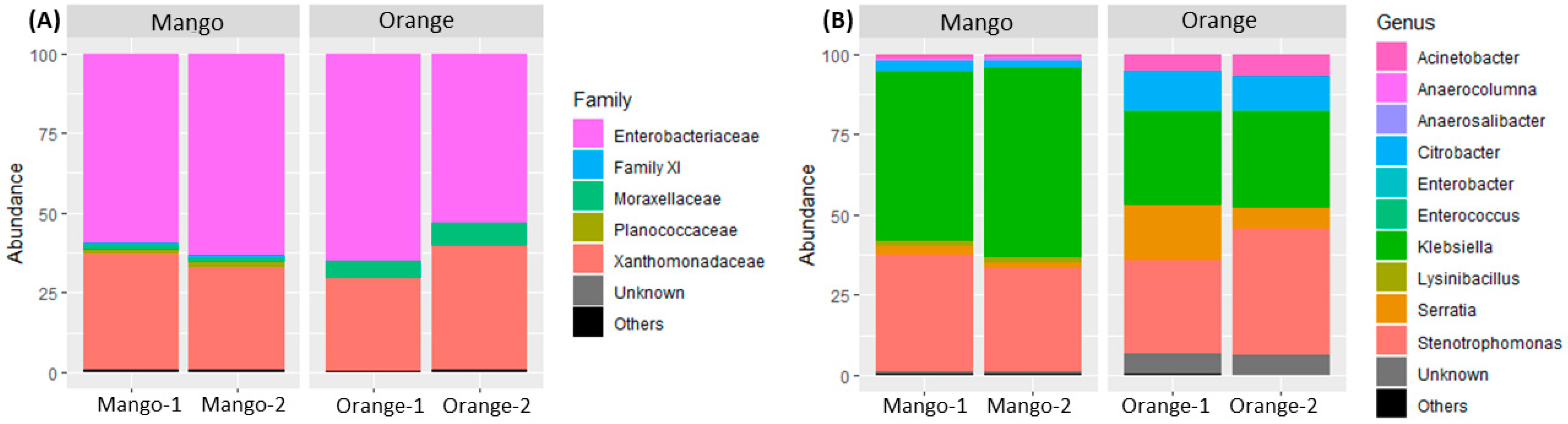
3.1. Taxonomy Analysis

Two samples (Orange and Mango) with two replicates for each fruit were sequenced, resulting in four data sets (Orange-1, Orange-2, Mango-1 and Mango-2). The microbiota present in the data was normalized for comparative analysis based on the sample with the lowest number of reads.
The rarefaction curves reached the plateau, showing that the depth of the samples was satisfactory to assess the microbial diversity. The richness estimator (Chao1) showed a higher value for the Mango (23.5 ± 0.5) than the Orange (13.0) sample. It was observed that the richness of the samples in the present work was achieved since Chao1 was similar to the observed richness. Moreover, the Shannon diversity index showed a higher value for the Orange (1.61 ± 0.05) than the Mango (1.11 ± 0.07) sample. An analysis of variation (ANOVA test, p < 0.05) was performed with the Shannon index and the Chao1 index results, showing that there was a significant difference between the samples. Thus, the microbial diversity based on the Shannon index showed that Orange was higher when compared to Mango (Figure S1, Supplementary Materials).
The Principal Coordinate Analysis (PCoA) based on the Bray–Curtis and Jaccard index (Figure 1) allowed us to evaluate the similarities and differences in the composition of the microbiota between the samples and their replicates. The points from two replicates of each sample were grouped, indicating a high correlation between the replicates. The different samples were relatively ungrouped, indicating lower correlations between samples than the replicates.
A taxonomic analyses revealed that all the sequences were dominated by the Bacteria domain (100%). The five most representative families among the Orange and Mango samples (Figure 2) were: Enterobacteriaceae, Family XI, Moraxellaceae, Planococcaceae and Xanthomonadaceae. Enterobacteriaceae and Xanthomonadaceae are the families with the highest relative frequency in each of the samples, based on the classification of the representative sequences of the ASVs.
By analyzing the taxonomic composition at the genus level, the four most frequent genera were observed in the two enriched Orange samples: Stenotrophomonas (Orange-1 = 28.86% and Orange-2 = 39.18%), Klebsiella (Orange-1 = 29.2% and Orange-2 = 29.8%), Serratia (Orange-1 = 16.9% and Orange-2 = 6.6%) and Citrobacter (Orange-1 = 12.2% and Orange-2 = 10.3%). Regarding the enriched Mango samples, there was a greater predominance of Klebsiella (Mango-1 = 52.5% and Mango-2 = 59%), Stenotrophomonas (Mango-1 = 36.6% and Mango-2 = 32, 3%), Serratia (Mango-1 = 2.5% and Mango-2 = 1.62%) and Citrobacter (Mango-1 = 0.22% and Mango-2 = 0.17%).
The common microbial nucleus between the Orange and Mango sample fruits comprised mainly the genera Stenotrophomonas, Klebsiella, Serratia and Citrobacter, which form the common core of the microbiota present in the samples. The results of the DESeq2 showed that a total of nine ASVs were statistically different (Figure S2, Supplementary Materials), represented by the genera Lysinibacillus (ASV_13), Anaerosalibacter (ASV_16), Anaerocolumna (ASV_17), Klebsiella (ASV_24), Micrococcales (ASV_3), Lactococcus (ASV_15), Corynebacterium (ASV_2), Paenibacillus (ASV_12) and Stenotrophomonas (ASV_29) and are present in a greater abundance in Mango samples.

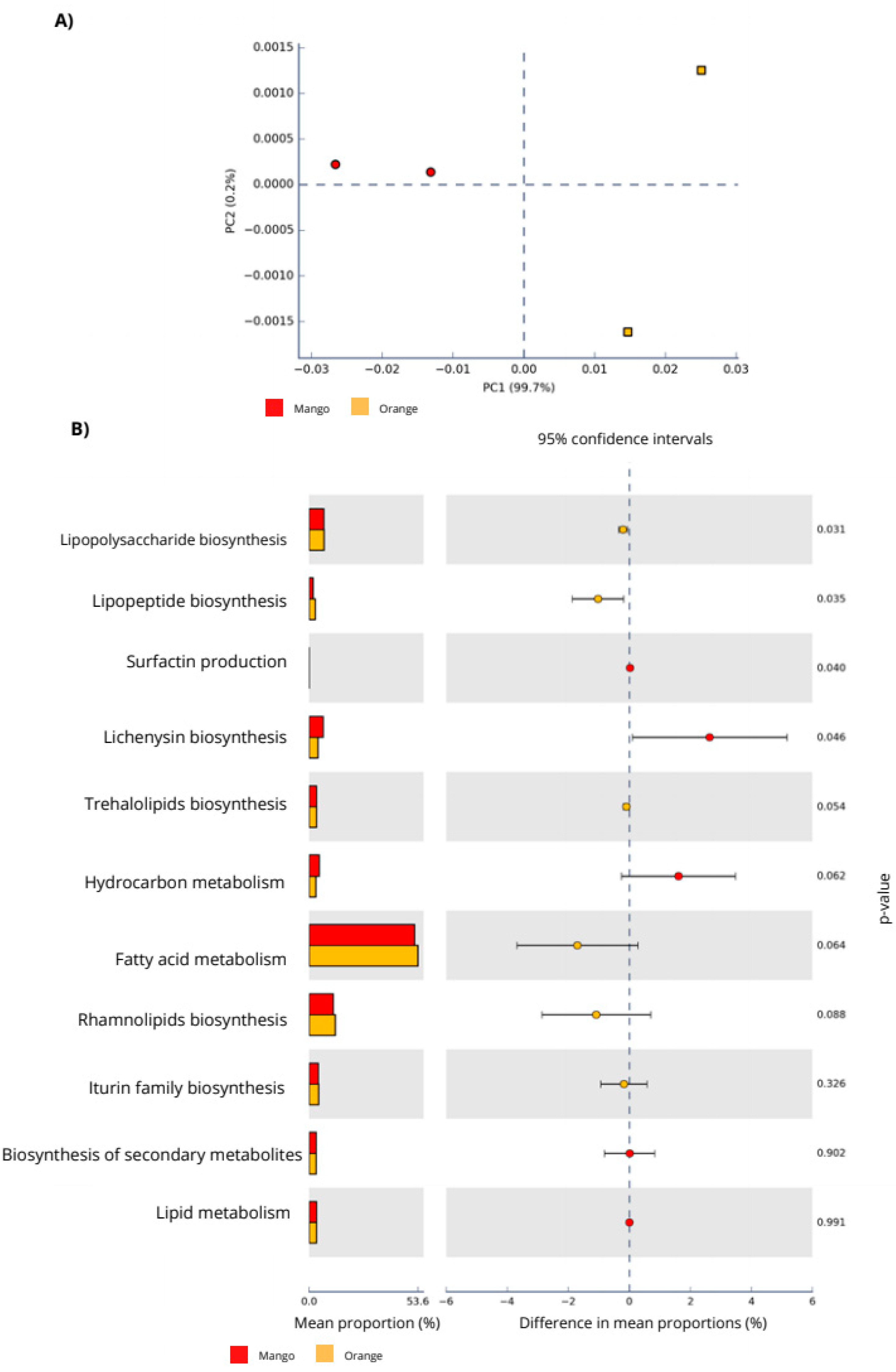
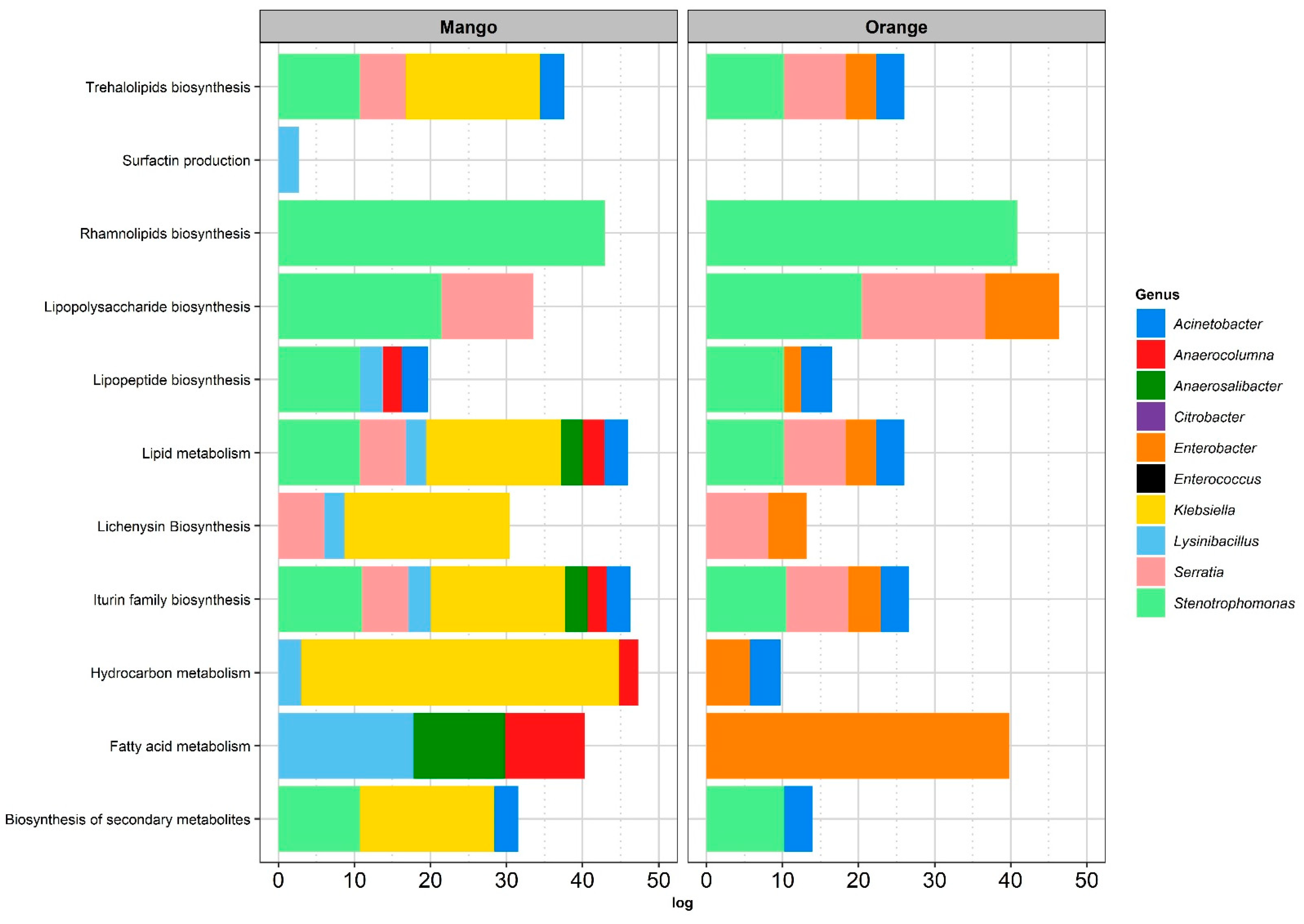
3.2. Functional Analysis

The functional prediction information of the 16S rRNA sequences was analyzed in STAMP and the functional profiles related to biosurfactant biosynthesis were evaluated. From the PCA analysis (Figure 3A), it can be observed that the pathways related to biosurfactant biosynthesis predicted in the Orange (Orange-1 and Orange-2) and Mango replicates (Mango-1 and Mango-2) were more associated with each other and were more heterogeneous between the groups, as observed in the taxonomic analysis. In the PCA graph (Figure 3A), the X and Y axes indicate that Principal 1 and 2 explain 99.7% and 0.2% of the total variation in the predicted functions, respectively.
Among the predicted genes (Table S1, Supplementary Materials), some were found that were related to fatty acid metabolism (fabB, fabH, fabG, fabI, fabV, fadB, fadL, alkM, echA and fadJ), rhamnolipids biosynthesis (rmlB, rmlC, rmlD, rmlA, rfbF), hydrocarbon metabolism (adh, ldh and hpaD), trehalolipids biosynthesis (otsA), lipopolysaccharide biosynthesis (lpxK, waaA, waaS, rfbN and lpxK), lipopeptide biosynthesis (sfp), lichenysin biosynthesis (licA3) and surfactin production (srfATE and srfAD).
In order to predict the functional profile of the samples from the 16S rRNA marker gene, the genes of interest that were related both directly and indirectly to the production of biosurfactants previously described in the literature were grouped into metabolic classes. In both fruit samples, the prediction of the functional profile related to biosurfactant production showed the highest abundance in fatty acid metabolism, followed by rhamnolipid, lipopolysaccharide and lichenysin biosynthesis (Table 1).
The genes involved in surfactin biosynthesis were the least abundant in the samples and were found only in the microbiota present in the Mango samples. Lipopeptide and lipopolysaccharide biosynthesis metabolisms were significantly more abundant in the Orange samples, whereas lichenysin biosynthesis was significantly more abundant in the Mango samples (Figure 3B). In general, the specific metabolisms of KEGG Orthology were found to be related to the production of different types of biosurfactants.
The functional genes predicted in the samples were taxonomically assigned. Among the 10 most abundant genera in the samples, Stenotrophomonas was the common genus between Orange and Mango that was related to most metabolisms and was the only genus related to rhamnolipid biosynthesis in all the samples. In the Mango samples, the genus Klebsiella was related to most metabolisms (six metabolisms of interest), while in the Orange samples, the genus Enterobacter was related to eight metabolisms of interest (Figure 4).

4. Discussion

The results of the Alpha diversity analysis showed that the bacterial diversity was significantly higher in the Mango samples than in the Orange samples (p < 0.05). The Chao1 richness estimator showed that the Mango samples had a higher species richness when compared to the Orange samples, indicating a higher total amount of species in the Mango. In addition, the results of the beta diversity analysis showed that the bacterial composition between the Mango and Orange samples was more homogeneous among themselves, which means that there were more species in common between the samples of the same fruit, indicating that the replicates were satisfactory.
According to Shannon’s index, which considers species richness and its uniformity, the Orange samples showed significantly greater richness and, therefore, greater evenness. That is, the species were more homogeneously distributed within the samples. The enrichment of the samples with glycerol may have influenced the diversity, explaining the low values obtained in the Shannon index and especially in the Orange samples. This enrichment may have further restricted the diversity of the microbiota, since the orange fruit already contains inhibitory compounds, such as limonene, which promotes the natural selectivity of microorganisms. On the other hand, enrichment favored the growth of similar taxonomic groups that were shared in both fruit samples.
Our alpha and beta diversity analysis results were used to better understand the microbial composition of the fruit samples. The taxonomic analysis showed that Oranges and Mangoes shared the same most abundant families (Enterobacteriaceae and Xanthomonadaceae), which represent almost all the bacterial diversity observed based on the classification of the representative sequences of the ASVs. The Enterobacteriaceae and Xanthomonadaceae families were mentioned in several studies on the production of biosurfactants from hydrocarbons [27,28,29].
At the genus level, we observed the predominance of Klebsiella (Enterobacteriaceae), Stenotrophomonas (Xanthomonadaceae), Serratia (Enterobacteriaceae) and Citrobacter (Enterobacteriaceae) in all the samples. Several works describe these genera as having great biosurfactant potential. A strain identified as Stenotrophomonas maltophilia was isolated from water contaminated with hydrocarbons and characterized as a biosurfactant producer with emulsifying activity levels above 70% [28].
The Serratia genus has been highlighted in biotechnological studies, as the biosurfactants produced by this genus are shown to be a promising source of antimicrobial, anti-fouling and antitumor compounds that have emulsification and surface activity [30]. Araújo et al. (2019) obtained a biosurfactant by Serratia marcescens with a CMC (critical micellar concentration) of 1.5% and a surface tension of 25.92 mN/m [31].
Another genus that is present in both samples and that has biosurfactant potential is Citrobacter. A strain of Citrobacter isolated from capable engine oil-contaminated soil was able to produce biosurfactants, which are of interest in many environmental and industrial applications, such as bioremediation processes and improved oil recovery [32]. Furthermore, Anaukwu et al. (2021) [33] observed that the capacity of Citrobacter murliniae to produce biosurfactants using Olive Oil as a carbon source obtained an emulsification index (E24) of 66.67% and an oil displacement diameter of 1.8 cm.
The genus Acinetobacter of the Moraxellacea family was also frequent in the Orange samples. Acinetobacter has several industrial applications, such as the bioremediation of wastewater and effluents, degradation of petrochemical products, production of biopolymers and biosurfactants [34]. Ohadi et al. (2017), who optimized the production of biosurfactants by an Acinetobacter junii, found a surface tension reduction to 45 mN/m after 48 h of incubation and 51% emulsification [35].
The taxonomic analysis showed that the enriched samples of Orange and Mango share the most abundant families and genera in common, which shows the similarity in the microbiota of the fruits. Moreover, the microbiota described in the present study was reported previously in the literature as associated with biosurfactant production. Thus, we can consider these enriched fruit residues as promising sources for the isolation of microorganisms.
In general, the specific metabolisms of the KEGG Orthology were found to be related to the production of different types of biosurfactants. Ten genes related to fatty acid metabolism were predicted that participate in one of the metabolic pathways involved in the synthesis of precursors for the production of biosurfactants, which is a precursor for the synthesis of lipids. The enzyme Enoyl-CoA hydratase is essential for fatty acid metabolism, in beta oxidationto produce both acetyl-CoA and energy in the form of ATP. Some prokaryotes are able to incorporate fatty acid directly in order to produce a biosurfactant [36,37].
Rhamnolipid biosynthesis was the second most common metabolism among the samples in this study, and Stenotrophomonas was the only genus assigned to this group of genes (Figure 4). Ryan et al. (2009) highlight that the Stenotrophomonas species have genes rmlB, rmlA, rmlC and rmlD, which encode the enzymes involved in lipopolysaccharide and exopolysaccharide biosynthesis, and which are bacteria with the potential to be bioremediation agents [38].
Three genes related to hydrocarbon metabolism (adh, ldh and hpaD) were predicted to be more associated with Klebsiella (Mango) and Enterobacter (Orange). Alcohol Dehydrogenase (adh), Lactate Dehydrogenase (ldh) and 2,3-dioxygenase (hpaD) are enzymes that play an important role in the biodegradation of hydrocarbons. Another gene that was predicted in our study was the Sfp (Phoshopantetheinyl transferase), which is fundamental for the biosynthesis of the lipopeptide biosurfactant [39]. In addition, we found licA3, a more specific gene related to the production of a type of biosurfactant lipopeptide (lichenysin). Coronel et al. (2017) [40] showed that lichenysins produced by Bacillus licheniformis are anionic lipopeptide biosurfactants with cytotoxic, antimicrobial and hemolytic activities that have enormous potential for chemical and biological applications [37,38,39,40,41].
In addition, the functional genes for trehalolipid biosynthesis were also predicted in smaller proportions, but they are present in several genera of the samples. Trehalolipids are excellent emulsifying compounds with applications in microbial oil recovery and oil spill treatment and are mainly produced by strains of the genus Rhodococcus [42,43]. In our study, we found genes for the biosynthesis of trehalolipids in five genera present in the samples: Stenotrophomonas, Serratia, Klebsiella, Enterobacter and Acinetobacter.

5. Conclusions

The taxonomic analysis and functional prediction provided in our samples were described in several studies that isolated biosurfactant-producing bacteria in already known environments, showing the potential of the bacteria isolated from fruits for biotechnological use. In addition, several specific genes for the biosynthesis of biosurfactants were predicted, showing the potential of the genera found in the enriched Mango and enriched Orange samples. The microbiota associated with the genes predicted in this work are known and described in the literature as good producers of biosurfactants, emphasizing that this study on the microbial diversity of enriched orange and mango residues is essential for future research that seeks to isolate bacteria with biosurfactant potential.

Supplementary Materials

The following are available online at https://0-www-mdpi-com.brum.beds.ac.uk/article/10.3390/ecologies3020010/s1, Figure S1: Shannon index, Chao1 richness and observed richness of the samples (Orange-1, Orange-2, Mango-1 and Mango-2), Figure S2: DESEq2 analysis of fold-change bacterial genera between Orange and Mango samples (p < 0.005), Table S1: Genes related to the production of functional biosurfactants used for pre-production analysis of Mango and Orange samples.

Author Contributions

F.d.P., I.C.S.D. and G.F.d.S. conceived and planned the experiments. F.d.P. and N.V.V. carried out the fermentation assays; F.d.P., G.F.d.S. and T.P.D. conceived and carried out the bioinformatic analysis. F.d.P. wrote the manuscript; I.C.S.D., G.F.d.S., T.P.D. and N.V.V. reviewed the manuscript. All authors provided critical feedback and helped shape the research, analysis and manuscript. All authors have read and agreed to the published version of the manuscript.

Funding

This research was funded by FAPESP, grant number 2020/06431-7.

Data Availability Statement

The four sequences are available from the European Nucleotide Archive under project accession PRJEB36395 and sample accessions ERS4265892 (Mango-1), ERS4265891 (Mango-2), ERS4265890 (Orange-1), ERS4265889 (Orange-2).

Conflicts of Interest

The authors declare no conflict of interest.

References

  1. Benincasa, M.; Abalos, A.; Oliveira, I.; Manresa, A. Chemical structure, surface properties and biological activities of the biosurfactant produced by Pseudomonas aeruginosa LBI from soapstock. Antonie Van Leeuwenhoek 2004, 85, 1–8. [Google Scholar] [CrossRef] [PubMed]
  2. Domingues, R.R.; Trugilho, P.F.; Silva, C.A.; De Melo, I.C.N.A.; Melo, L.C.A.; Magriotis, Z.M.; Sánchez-Monedero, M.A. Properties of biochar derived from wood and high-nutrient biomasses with the aim of agronomic and environmental benefits. PLoS ONE 2017, 12, e0176884. [Google Scholar] [CrossRef] [PubMed] [Green Version]
  3. Singh, P.; Patil, Y.; Rale, V. Biosurfactant production: Emerging trends and promising strategies. J. Appl. Microbiol. 2018, 126, 2–13. [Google Scholar] [CrossRef] [PubMed] [Green Version]
  4. Fariq, A.; Yasmin, A. Production, characterization and bioactivities of biosurfactants from newly isolated strictly halophilic bacteria. Process Biochem. 2020, 98, 1–10. [Google Scholar] [CrossRef]
  5. Ribeiro, B.G.; Guerra, J.M.C.; Sarubbo, L.A. Biosurfactants: Production and application prospects in the food industry. Biotechnol. Prog. 2020, 36, e3030. [Google Scholar] [CrossRef]
  6. Fracchia, L.; Ceresa, C.; Franzetti, A.; Cavallo, M.; Gandolfi, I.; Van Hamme, J.; Gkorezis, P.; Marchant, R.; Banat, I.M. Industrial applications of biosurfactants. In Biosurfactants: Production and Utilization—Processes, Technologies, and Economics; CRC Press: Boca Raton, FL, USA, 2014; pp. 245–260. [Google Scholar]
  7. Rizzo, C.; Caldarone, B.; De Luca, M.; De Domenico, E.; Giudice, A.L. Native bilge water bacteria as biosurfactant producers and implications in hydrocarbon-enriched wastewater treatment. J. Water Process Eng. 2021, 43, 102271. [Google Scholar] [CrossRef]
  8. Ben Belgacem, Z.; Bijttebier, S.; Verreth, C.; Voorspoels, S.; Van De Voorde, I.; Aerts, G.; Willems, K.A.; Jacquemyn, H.; Ruyters, S.; Lievens, B. Biosurfactant production by Pseudomonas strains isolated from floral nectar. J. Appl. Microbiol. 2015, 118, 1370–1384. [Google Scholar] [CrossRef]
  9. Kumar, A.P.; Janardhan, A.; Viswanath, B.; Monika, K.; Jung, J.-Y.; Narasimha, G. Evaluation of orange peel for biosurfactant production by Bacillus licheniformis and their ability to degrade naphthalene and crude oil. 3 Biotech. 2016, 6, 43. [Google Scholar] [CrossRef] [Green Version]
  10. FAO. FAOSTAT; Food and Agriculture Organization of the United Nations: Rome, Italy, 2018. [Google Scholar]
  11. Cypriano, D.Z.; Da Silva, L.L.; Bohórquez, M.A.M.; Tasic, L. Orange Biomass By-products. Rev. Virtual Quím. 2017, 9, 176–191. [Google Scholar] [CrossRef]
  12. Empresa Brasileiro de Pesquisa Agropecuária. Exportação de manga brasileira bate recorde em 2020, totalizando US$ 246 milhões. Available online: https://www.embrapa.br/busca-de-noticias/-/noticia/60585117/exportacao-de-manga-brasileira-bate-recorde-em-2020-totalizando-us-246-milhoes (accessed on 21 August 2021).
  13. Mehmood, N.; Iqbal, M.; Asgher, M. Production and Optimization of Biosurfactants from Bacillus subtilis. EC Microbiol. 2018, 5, 211–224. [Google Scholar]
  14. Napp, A.P.; Pereira, J.E.S.; Oliveira, J.S.; Silva-Portela, R.C.; Agnez-Lima, L.F.; Peralba, M.C.; Bento, F.M.; Passaglia, L.M.; Thompson, C.E.; Vainstein, M.H.; et al. Comparative metagenomics reveals different hydrocarbon degradative abilities from enriched oil-drilling waste. Chemosphere 2018, 209, 7–16. [Google Scholar] [CrossRef] [PubMed]
  15. Lang, K. Projeto Primer para NGS curtas com amplicon. Imunol. Hum. 2013, 74, 8. [Google Scholar] [CrossRef]
  16. Caporaso, J.G.; Lauber, C.L.; Walters, W.A.; Berg-Lyons, D.; Huntley, J.; Fierer, N.; Owens, S.M.; Betley, J.; Fraser, L.; Bauer, M.; et al. Ultra-high-throughput microbial community analysis on the Illumina HiSeq and MiSeq platforms. ISME J. 2012, 6, 1621–1624. [Google Scholar] [CrossRef] [PubMed] [Green Version]
  17. Babraham Bioinformatics. FastQC: A Quality Control Tool for High Throughput Sequence Data. Available online: https://www.bioinformatics.babraham.ac.uk/projects/fastqc/ (accessed on 25 March 2022).
  18. Bolger, A.M.; Lohse, M.; Usadel, B. Trimmomatic: A flexible trimmer for Illumina sequence data. Bioinformatics 2014, 30, 2114–2120. [Google Scholar] [CrossRef] [Green Version]
  19. Bolyen, E.; Rideout, J.R.; Dillon, M.R.; Bokulich, N.A.; Abnet, C.C.; Al-Ghalith, G.A.; Alexander, H.; Alm, E.J.; Arumugam, M.; Asnicar, F.; et al. Reproducible, Interactive, Scalable and Extensible Microbiome Data Science using QIIME. Nat. Biotechnol. 2019, 37, 852–857. [Google Scholar] [CrossRef]
  20. Callahan, B.J.; Mcmurdie, P.J.; Rosen, M.J.; Han, A.W.; Johnson, A.J.A.; Holmes, S.P. DADA2: High-resolution sample inference from Illumina amplicon data. Nat. Methods 2016, 13, 581–583. [Google Scholar] [CrossRef] [Green Version]
  21. Langille, M.G.I.; Zaneveld, J.; Caporaso, J.G.; McDonald, D.; Knights, D.; Reyes, J.A.; Clemente, J.C.; Burkepile, D.E.; Vega Thurber, R.L.; Knight, R.; et al. Predictive functional profiling of microbial communities using 16S rRNA marker gene sequences. Nat. Biotechnol. 2013, 31, 814–821. [Google Scholar] [CrossRef]
  22. Kanehisa, M.; Sato, Y.; Kawashima, M.; Furumichi, M.; Tanabe, M. KEGG as a reference resource for gene and protein annotation. Nucleic Acids Res. 2016, 44, D457–D462. [Google Scholar] [CrossRef] [Green Version]
  23. Parks, D.H.; Tyson, G.W.; Hugenholtz, P.; Beiko, R.G. STAMP: Statistical analysis of taxonomic and functional profiles. Bioinformatics 2014, 30, 3123–3124. [Google Scholar] [CrossRef] [Green Version]
  24. McMurdie, P.J.; Holmes, S. phyloseq: An R package for reproducible interactive analysis and graphics of microbiome census data. PLoS ONE 2013, 8, e61217. [Google Scholar] [CrossRef] [Green Version]
  25. Ferraz, A.C.P.; Gadelha, B.D.Q.; Aguiar-Coelho, V.M. Análise faunística de Calliphoridae (Diptera) da Reserva Biológica do Tinguá, Nova Iguaçu, Rio de Janeiro. Rev. Bras. Èntomol. 2009, 53, 620–628. [Google Scholar] [CrossRef] [Green Version]
  26. Albuquerque, M.A.; Silva, E.L.; Barros, K.N.N.O.; Junior, S.F.A.X. Comparação entre coeficientes similaridade uma aplicação em ciências florestais. Matemática Estatística Foco 2016, 4, 102–114. [Google Scholar]
  27. Nwaguma, I.V.; Chikere, C.B.; Okpokwasili, G.C. Isolation, characterization, and application of biosurfactant by Klebsiella pneumoniae strain IVN51 isolated from hydrocarbon-polluted soil in Ogoniland, Nigeria. Bioresour. Bioprocess. 2016, 3, 40. [Google Scholar] [CrossRef] [Green Version]
  28. Gargouri, B.; Contreras, M.D.M.; Ammar, S.; Segura-Carretero, A.; Bouaziz, M. Biosurfactant production by the crude oil degrading Stenotrophomonas sp. B-2: Chemical characterization, biological activities and environmental applications. Environ. Sci. Pollut. Res. 2017, 24, 3769–3779. [Google Scholar] [CrossRef] [PubMed]
  29. Adeyemi, J.O.; Onajob, I.B.; Adebajo, L.O.; Orji, F.A.; Ilusany, O.A.F.; Samson, O.J. Biossurfactant production potentials of some bacteria recovered from agro-industrial wastes. Trends Sci. Technol. J. 2019, 4, 870–873. [Google Scholar]
  30. Clements, T.; Ndlovu, T.; Khan, W. Broad-spectrum antimicrobial activity of secondary metabolites produced by Serratia marcescens strains. Microbiol. Res. 2019, 229, 126329. [Google Scholar] [CrossRef]
  31. Araújo, H.W.C.; Andrade, R.F.S.; Montero-Rodríguez, D.; Rubio-Ribeaux, D.; Da Silva, C.A.A.; Campos-Takaki, G.M. Sustainable biosurfactant produced by Serratia marcescens UCP 1549 and its suitability for agricultural and marine bioremediation applications. Microb. Cell Fact. 2019, 18, 2. [Google Scholar] [CrossRef]
  32. Ibrahim, H.M. Characterization of biosurfactants produced by novel strains of Ochrobactrum anthropi HM-1 and Citrobacter freundii HM-2 from used engine oil-contaminated soil. Egypt. J. Pet. 2018, 27, 21–29. [Google Scholar] [CrossRef]
  33. Anaukwu, C.; Ekwealor, A.; Ezemba, C.; Anakwenze, V.; Okafor, U.; Archibong, E. Pseudomonas monteilii and Citrobacter murliniae, Biosurfactant-Producing Bacteria Isolated from Nigerian Soil. Br. Microbiol. Res. J. 2015, 10, 1–9. [Google Scholar] [CrossRef]
  34. Teixeira, L.M.; Merquior, V.L.C. The family moraxellaceae. In The Prokaryotes—Gammaproteobacteria, Rosenberg, E., DeLong, E.F., Lory, S., Stackebrandt, E., Thompson, F., Eds.; Springer: Berlin/Heidelberg, Germany, 2014; Volume 22, pp. 443–476. [Google Scholar]
  35. Ohadi, M.; Dehghannoudeh, G.; Shakibaie, M.; Banat, I.M.; Pournamdari, M.; Forootanfar, H. Isolation, characterization, and optimization of biosurfactant production by an oil-degrading Acinetobacter junii B6 isolated from an Iranian oil excavation site. Biocatal. Agric. Biotechnol. 2017, 12, 1–9. [Google Scholar] [CrossRef]
  36. Fontes, G.C.; Amaral, P.F.F.; Coelho, M.A.Z. Biosurfactants production by yeasts. Química Nova 2008, 31, 2091–2099. [Google Scholar] [CrossRef]
  37. Jimoh, A.A.; Lin, J. Biosurfactant: A new frontier for greener technology and environmental sustainability. Ecotoxicol. Environ. Saf. 2019, 184, 109607. [Google Scholar] [CrossRef] [PubMed]
  38. Phetcharat, T.; Dawkrajai, P.; Chitov, T.; Mhuantong, W.; Champreda, V.; Bovonsombut, S. Biosurfactant-Producing Capability and Prediction of Functional Genes Potentially Beneficial to Microbial Enhanced Oil Recovery in Indigenous Bacterial Communities of an Onshore Oil Reservoir. Curr. Microbiol. 2019, 76, 382–391. [Google Scholar] [CrossRef]
  39. Ryan, R.P.; Monchy, S.; Cardinale, M.; Taghavi, S.; Crossman, L.; Avison, M.B.; Berg, G.; Van Der Lelie, D.; Dow, J.M. The versatility and adaptation of bacteria from the genus Stenotrophomonas. Nat. Rev. Microbiol. 2009, 7, 514–525. [Google Scholar] [CrossRef] [PubMed]
  40. Coronel, J.R.; Marqués, A.; Manresa, Á.; Aranda, F.J.; Teruel, J.A.; Ortiz, A. Interaction of the Lipopeptide Biosurfactant Lichenysin with Phosphatidylcholine Model Membranes. Langmuir 2017, 33, 9997–10005. [Google Scholar] [CrossRef]
  41. Parthipan, P.; Preetham, E.; Machuca, L.L.; Rahman, P.; Murugan, K.; Rajasekar, A. Biosurfactant and Degradative Enzymes Mediated Crude Oil Degradation by Bacterium Bacillus subtilis A1. Front. Microbiol. 2017, 8, 193. [Google Scholar] [CrossRef] [Green Version]
  42. Pacheco, G.J.; Ciapina, E.M.P.; Gomes, E.D.B.; Junior, N.P. Biosurfactant production by Rhodococcus erythropolis and its application to oil removal. Braz. J. Microbiol. 2010, 41, 685–693. [Google Scholar] [CrossRef] [Green Version]
  43. Liu, C.-W.; Liu, H.-S. Rhodococcus erythropolis strain NTU-1 efficiently degrades and traps diesel and crude oil in batch and fed-batch bioreactors. Process Biochem. 2011, 46, 202–209. [Google Scholar] [CrossRef]
Figure 1. Principal Coordinate Analysis (PCoA) of beta diversity using a Bray–Curtis and Jaccard distance from microbial communities of fruits (Mango and Orange) and their technical replicates, represented in a non-metric multidimensional scaling (NMDS). The blue and green dots refer to the Mangoes and their replicates. The pink and purple triangles refer to the Oranges and their replicates.
Ecologies 03 00010 g001
Figure 2. Taxonomic profile of the microbiota present in the enriched Mango and Orange samples analyzed by 16S rRNA gene sequencing by relative abundance (%): (A) family level and (B) genus level. Data were normalized from the sample with the lowest number of reads.
Ecologies 03 00010 g002
Figure 3. Graphics generated using STAMP software. (A) Principal Component Analysis (PCA) between Orange and Mango predicted functional profiles and (B) General pathways related to biosurfactant biosynthesis predicted in the microbiota present in enriched Mango and Orange samples.
Ecologies 03 00010 g003
Figure 4. Metabolic activities from specific pathways related to the production of biosurfactants associated with the 10 most abundant genera.
Ecologies 03 00010 g004
Table 1. Abundance (%) of predicted gene clusters in enriched fruit samples related to the production of biosurfactants.
Biosurfactant Relation MetabolismMango Abundance (%)Orange Abundance (%)
Fatty acid metabolism51.9453.64
Rhamnolipids biosynthesis11.8512.92
Lipopolysaccharide biosynthesis7.177.37
Lichenysin Biosynthesis6.924.29
Hydrocarbon metabolism4.963.34
Iturin family biosynthesis4.464.63
Lipid metabolism3.653.65
Trehalolipids biosynthesis3.533.63
Biosynthesis of secondary metabolites3.503.50
Lipopeptide biosynthesis2.003.00
Surfactin production0.020.00
Publisher’s Note: MDPI stays neutral with regard to jurisdictional claims in published maps and institutional affiliations.

