Review

Are Structurally Modified Galactomannan Derivatives Biologically Active?

by
Maurycyo Silva Geronço
1,
Igor Frederico da Silveira Ramos
1,
Edson Cavalcanti da Silva Filho
1,
Márcia dos Santos Rizzo
1,
Alessandra Braga Ribeiro
2,* and
Marcilia Pinheiro da Costa
1
1
Graduate Program in Material Science and Engineering, Federal University of Piauí, 64049-550 Teresina, Piauí, Brazil
2
Centre of Biotechnology and Fine Chemistry, Faculty of Biotechnology, Catholic University of Portugal, 4169-005 Porto, Portugal
*
Author to whom correspondence should be addressed.
Submission received: 29 November 2020 / Revised: 25 December 2020 / Accepted: 5 January 2021 / Published: 8 January 2021
(This article belongs to the Collection Bioactive Polysaccharides)

Abstract

:
Galactomannans are versatile macromolecules with broad industrial potential. The influence of changes in the chemical structures and respective bioactivities of these polysaccharides have been extensively studied. The derivatives obtained by sulfation, complexation, and phosphorylation are the most studied biological properties in galactomannans. The derivatives obtained have shown several pharmacological activities such as antiviral, antimicrobial, anticoagulant, fibrinolytic, chemopreventive, anticancer, antioxidant, chondroprotective, analgesic, immunomodulatory, and antileishmanial. Considering the relevance of these studies, we aim to provide an overview of studies that apply galactomannan modification or derivatization strategies to improve their properties for applications in the biomedical area. We identified the success of most modified galactomannans for pharmacological purposes. However, some studies found loss of bioactivity of the original polysaccharide after chemical changes to its original structures.

1. Introduction

The development of new biomaterials and “functionalized polymers,” especially from renewable sources, has practical potential for applications in biomedicine and industry [1]. In this direction, the growing interest in the chemical modification or derivatization of natural polysaccharides to create derivatives with properties adapted to the desired applications is remarkable [2,3]. Molecular modification and improvement of the biologically active properties of polysaccharides have sparked interest about the development of biomedical materials. Therefore, it is important to find the correlation between the chemical structure and the functional activity of the polysaccharides [4,5,6].
Galactomannans are polysaccharides which can be obtained from microorganisms, including fungi and yeasts, but the vast majority originate from plants [7]. Among the plant-derived polysaccharides, galactomannans have received much attention, since these biopolymers can be obtained in their pure form, with a high yield, by aqueous extraction from the endosperm of the seeds of leguminous plants [6,8]. Furthermore, since galactomannans are an abundant plant resource, their production on an industrial scale has been widely targeted [5,9], especially in the food, pharmaceutical, and cosmetic industries [10].
Galactomannans have a heterogeneous polymeric structure, consisting of a main chain of mannose units linked by β-1,4-glycosidic bonds and galactose side units linked to mannose by α-1,6-glycosidic bonds (Figure 1) [7,11]. Galactomannans from different sources differ from each other in the ratio of mannose/galactose units, molecular weight, and the distribution of individual galactose branches in the main chain, which significantly influences their biochemical properties [12,13].
In addition, the structural peculiarities of galactomannans make them soluble in water at different temperatures, versatile in their applications, and chemically and biochemically reactive [1]. These factors, combined with the absence of ionic charge in the structure of these polysaccharides, make galactomannans more susceptible to molecular changes than many polysaccharides found in nature. Alterations can be made in order to inherit the characteristics of the native polymer, such as biocompatibility, to expand its technological functionality, improve its intrinsic biological properties, and/or create new functional properties, opening a range of application possibilities in the biomedical areas [12,14,15].
The galactomannans are suitable for used in chemical modification methods, as they have a simple structure. Their derivatives can be easily characterized chemically. Sulfation is an effective, versatile, and simple modification method applied to galactomannans to improve their biological activity. However, other methods can also be used to change the structure of galactomannans [16,17,18]. Thus, Figure 1 provides a schematic representation of the main chemical changes in galactomannans and the various biological activities evaluated.
In view of the aforementioned, this work aimed to provide an overview of the studies that applied methods for structural modification of galactomannans to evaluate or improve their properties for applications in the biomedical area. This review focuses on the study of the intrinsic pharmacological activities of structurally modified galactomannan derivatives. A search for articles referring to the modified galactomannans was conducted. The keywords “modified galactomannans and biological activity” or “galactomannan derivatives and biological activity” were used to procure scientific articles in electronic databases, including PubMed, Scopus, SciELO, and Web of Science. The selection of the articles was based on the following inclusion criteria: full-text articles published containing the keywords above that led to chemical modifications and evaluated the intrinsic pharmacological properties of the modified biopolymer, without restricting the year of publication. Modified galactomannans that had not been evaluated for biological characterization were excluded from the study. Modified galactomannans that were formulated with the addition of active substances were also not considered.

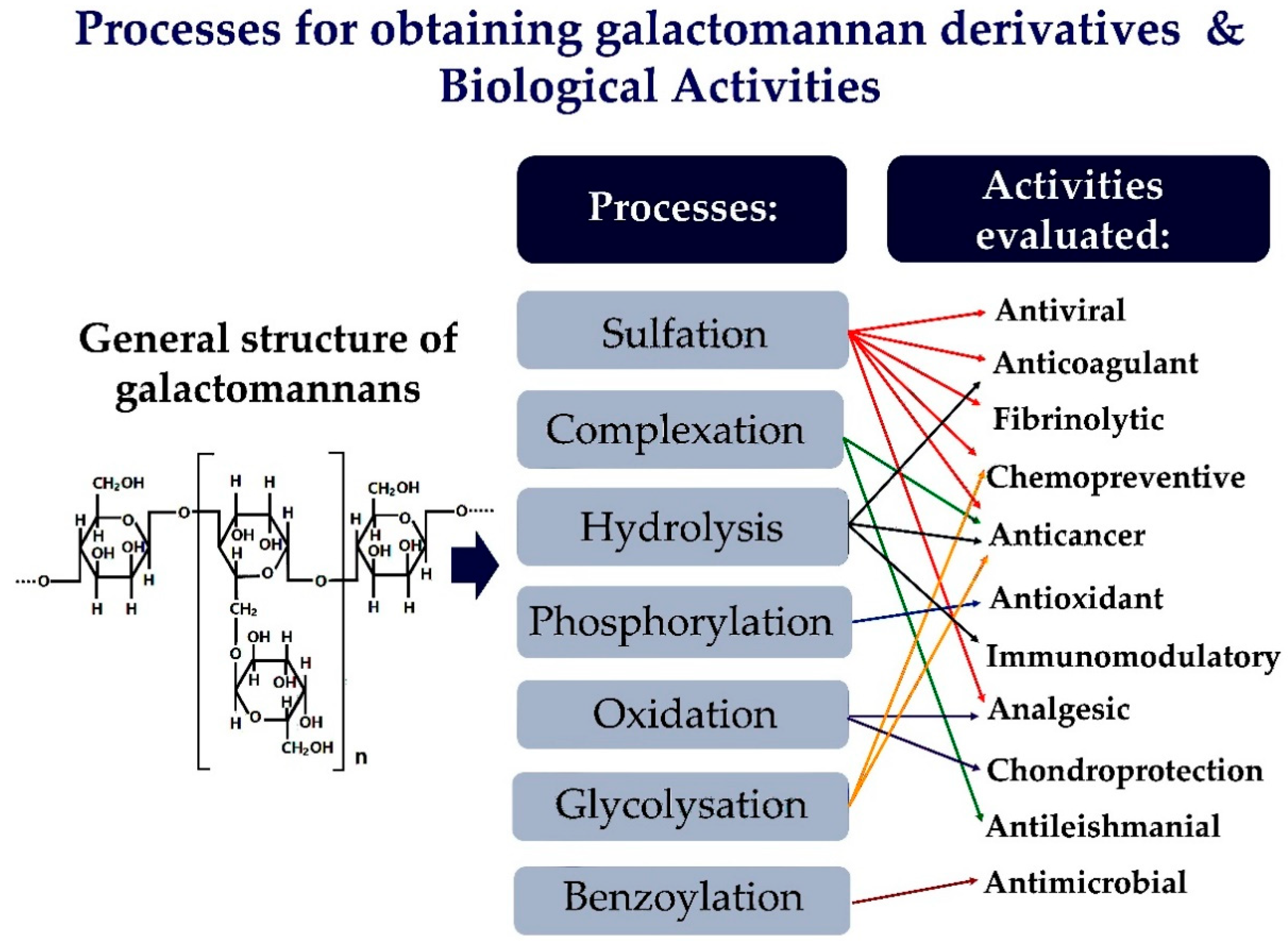
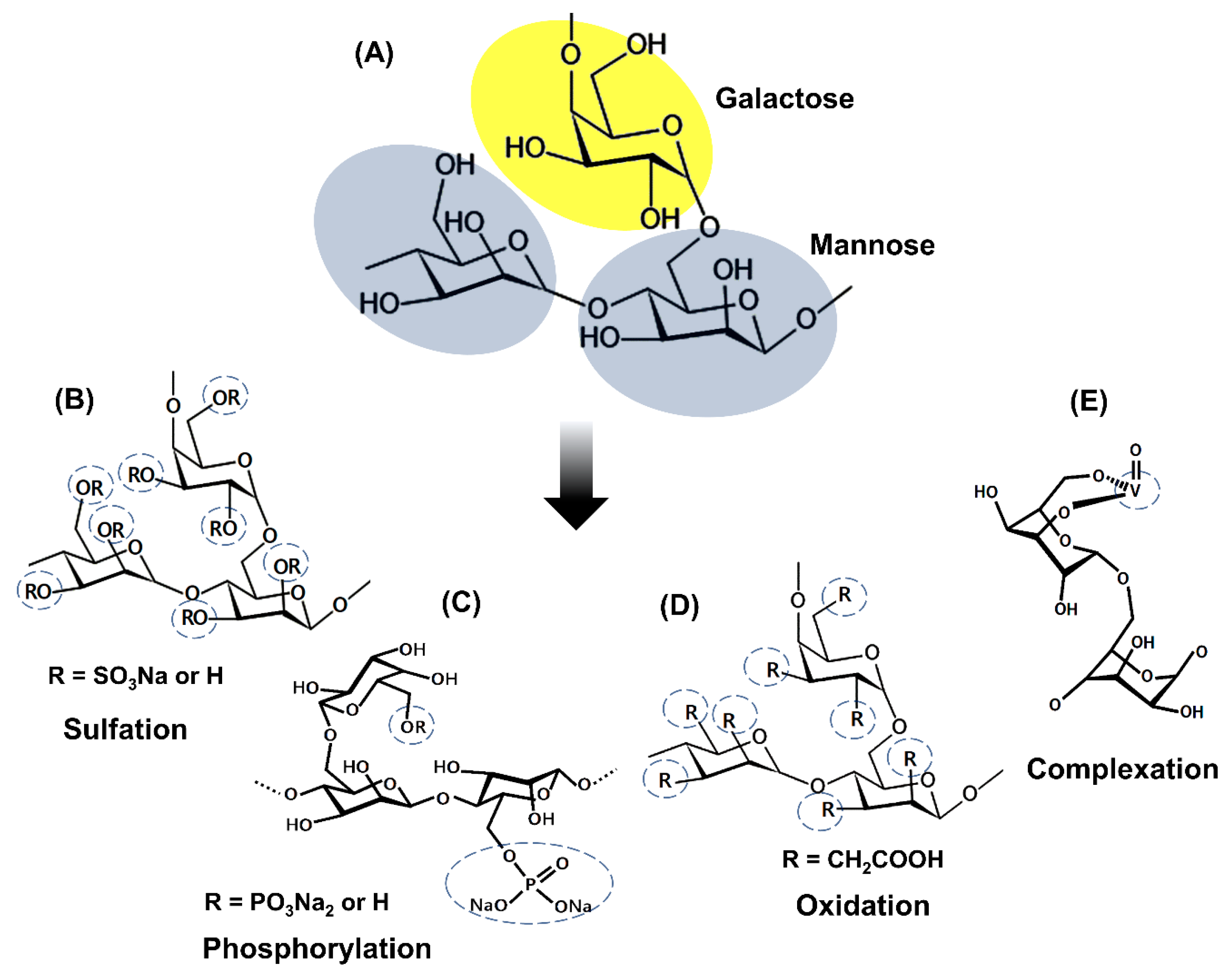
2. Strategies for Obtaining Galactomannan Derivatives

Biological activities of polysaccharides are affected by several factors, among them, monosaccharide composition, molecular weight, molecular structure, functional groups, degree and pattern of substitution, flexibility, and chain configuration [4,19,20]. In addition to the evident biological activity of natural polysaccharides, derivatives obtained by hydrolysis, complexation, or chemical modifications, such as sulfation, phosphorylation, oxidation, and carboxymethylation, were also considered in many studies as successful bioactive derivatives [5,21,22,23].
Among these polysaccharides, galactomannans have been extensively studied for molecular modifications. Galactomannans can be modified mainly by chemical or enzymatic methods. A large number of hydroxyl groups of galactomannans can be altered by etherification, esterification, or oxidation. The changes that add hydrophilic groups to molecules of galactomannans can reduce hydrogen bonds between the molecules and improve solubility and swelling characteristics. Other characteristics that are also improved by the increase in hydrophilicity are the clarity of the gum and the compatibility with electrolytes [24].
The changes in chemical structure and biological activities of galactomannans have been extensively studied. The most studied biological properties in galactomannans are derivatives obtained by complexation or chemical modifications, such as sulfation, oxidation, and phosphorylation. Figure 2 represents the main strategies for obtaining galactomannan derivatives.
Sulfation is the most studied chemical modification in galactomannans. Most studies about sulfation of galactomannans used dimethylformamide as the solvent and SO3 pyridine complex as a sulfating reagent [6,8,9,23,26,27,28,29]. Several studies report preferred sulfation for the 6-CH2 groups due to their higher reactivity with esterifying agents and a degree of sulfating (DS) ranging from 0.4 to 1.85. These sulfated galactomannans are composed of negatively charged sulfate groups that are related to their biological activities [4,5,6,8,9,23,26,27,28,29]. The addition of sulfate groups in the polysaccharide structure improves solubility and enhances the biological properties compared to the non-sulfated polysaccharides. Anticoagulant, antiviral, antioxidant, and antitumor activities are successful examples of sulfated polysaccharides [4,23,28,29].
Polysaccharides can also undergo oxidation in several ways to generate distinct structures. For example, primary alcohol may be oxidized to form the aldehyde or later a carboxylic acid [30]. Protein-free guar gum was oxidized by the method of the TEMPO (2,2,6,6-tetramethylpiperidine-1-oxide radical)/NaBr/NaClO system [25]. This oxidized derivative presents a degree of substitution of 0.36, and the oxidation happened preferentially in the carbon 6 of the mannose residue. FT-IR and 13C NMR spectrum showed the presence of the COOH group in the oxidized molecule.
Phosphorylated polysaccharides exhibit several biological activities, including antioxidant, antitumor, and immunomodulatory properties. Wang et al. [20] phosphorylated the galactomannan chains of Cyamopsis tetragonolobo (guar gum) using POCl3/pyridine and obtained phosphorylated derivatives with the degree of substitution (DS) of 0.35–0.52. They observed an expanded conformation of phosphorylated derivatives with intramolecular hydrogen bonds due to the electrostatic potential of SO3H groups. Experiments with the phosphorylated derivatives with different DS were conducted to evaluate the influence of this parameter on the antioxidant activity.
Hydrolysis is an important strategy for polysaccharide modification. In the enzymatic method, the galactose side chains, or the main mannose chain can be cleaved, and the α-galactosidase and β-mannanase can be used. Compared with the chemical modification method, the enzymatic method is simple to control, and the reaction conditions are milder [24]. The biological activity of the locust bean gum galactomannan hydrolyzed by thermostable β-D-mannanase was evaluated by Chen et al. [31]. In this study, after 24 h of the enzymatic reaction, the weight average molecular weight of galactomannan derivative reduced from 5,580,010 to 3188, and the hydrolysate containing the following manno-oligosaccharides: mannobiose, mannotriose, and mannotetrose.
According to Adriazola et al. [22], complexation with metal ions can make complexes more biocompatible and soluble, less toxic, and endow them with some pharmacological activities, such as antimicrobial and antitumor. Galactomannans can be modified by introducing complexing metals. Vanadium is the metal generally used to complex with galactomannans in order to originate biologically active complexes. Polysaccharides are suitable binders for complexation reactions with the cationic form of vanadium due to the affinity of this metal for hydroxyl groups free of these polyhydroxyl compounds. Complexation of vanadium (IV, V) with monosaccharides is facilitated by the presence of ligands containing vicinal cis-OH groups [19,32]. Anticancer and antileishmanial activities are recognized as examples of the success of galactomannans complexed with vanadium [19,33].
The pharmacological activities of galactomannan derivatives are detailed below.

3. Pharmacological Activities of Galactomannan Derivatives

The intrinsic characteristics of polysaccharides can be changed, for example, by partial hydrolysis and by the removal or introduction of specific chemical groups [34]. When a galactomannan is modified, a biologically inert polymer can be transformed into a biologically active one [35].
Pharmacological activities can be related to changes in the chemical structure of the molecules caused by the type and number of functional groups. Several functional groups, such as -CN, -NO2, -NH2, -OH, -SH, -CHO, -COOH, -COOR, -CONH2, -SO3H, and -PO3H, contribute to drug-receptor interaction. The interaction of a compound with a specific receptor is directly related to a pharmacological effect. Changes in the conformation and flexibility of the molecules can also affect the interaction of molecules with receptors. In addition, several physicochemical properties of a molecule, such as molecular weight, lipophilicity, solubility, permeability, and acid-base ionization, can alter its activity [36,37].
In general, the chemical structure of a molecule determines its physicochemical characteristics and, consequently, determines its absorption, distribution, metabolism, excretion, and toxicity (ADME/Tox) properties and influences the pharmacological effect of the molecule [37].
Table 1 presents the main pharmacological activities of galactomannan derivatives, their source of origin, and the processes for obtaining derivatives.

3.1. Antiviral Activity

Several studies have reported the low cytotoxicity, safety, and therapeutic potential of sulfated polysaccharides [26,29,38]. According to Godoi et al. [38], sulfated polysaccharides are among the most reported pharmacological properties. Moreover, these derivates have a broad spectrum of in vitro antiviral activity.
Ono et al. [26] tested the effects of native and sulfated galactomannans obtained from the seeds of Mimosa scabrella (BR and BRS) and Leucaena leucocephala (LL and LLS) against two flaviviruses using in vitro and in vivo models: the yellow fever virus (YFV) (BeH111 strain) and the dengue 1 virus (DEN-1) (Hawaii strain). In the in vitro tests to determine the quantitative response to antiviral activity, the concentrations that produced a 100-fold reduction in viral titer compared to the positive control were 586 mg·L−1 and 387 mg·L−1 for BRS and LLS against YFV, respectively; and for DEN-1 were 347 mg·L–1 and 37 mg·L−1, respectively. When comparing the results of both viral species, the best response was obtained using LLS. In the in vivo experiment, mice received a simultaneous injection of YFV plus BRS or LLS (49 mg·kg−1, i.p). BRS and LLS promoted protection against death in 87.7% and 96.5% of the animals, respectively. The results reinforced the idea that the antiviral activity of some sulfated polysaccharides is not related only to the mechanisms of inhibition of viral replication, in which the entry of the polysaccharide-virus complex into cells is inhibited. Mice inoculated with viruses and in association with modified galactomannans (YFV + BRS or YFV + LLS) developed viral resistance, while the absence of BRS or LLS at the time of viral inoculation did not show any protective effect. Therefore, in the presence of polysaccharides, there was no development of several diseases, and the immune system of the animals recognized the viral antigens, since animals in the YFV + BRS and YFV + LLS groups developed neutralizing antibodies. Furthermore, the immunomodulatory action of the polysaccharides is considered because the intraperitoneal inoculation, together with viral suspension, seems to activate peritoneal macrophages.
In another study, Chrestani et al. [8] observed that the sulfated galactomannan from Mimosa scabrella seeds (BRS) exerted activity against herpes simplex virus type 1 (HSV-1). However, this effect was not verified when tested with the simian rotavirus A/SA11 (SiRV-A/SA11). BRS had a selective inhibition against HSV-1 during its binding step, with an IC50 of less than 2.5 µg·mL−1, suggesting that BRS has a higher selectivity against an enveloped virus that uses heparan sulfate on the surface of the host cell membrane for its internalization. The antiviral action of sulfated polysaccharides has been attributed to a mechanism for inhibiting viral adsorption by the cell. The lack of response against SiRV-A/SA11 reinforced this possibility since SiRV-A/SA11 is known to have a negative surface charge at neutral pH, not allowing a strong interaction between rotavirus and polyanions, such as sulfated polysaccharides.
Gemin et al. [27] also demonstrated that the sulfated galactomannan from Leucaena leucocephala seeds (LLS-2) affected the early stages of HSV-1 infection, with an EC50 below 2.5 µg·mL−1. The native polysaccharide did not have an antiviral effect, proving that the anti-herpetic activity of LLS-2 depended on the presence of negatively charged groups.
In the study by Lopes et al. [28], the sulfated galactomannan from Caesalpinia ferrea (SPLCf) inhibited several stages of infection by herpes simplex virus type 1 (HSV-1) and Poliovirus type 1 (PV-1) (ATCC, VR-58). The results showed an IC50 of 405 µg·mL−1 and 1.73 µg·mL−1, for HSV-1 and PV-1, respectively. The antiviral activity was mainly attributed to the polyanionic character of SPLCf, which interfered mainly in the virus adsorption stage, as well as in the viral particles and expression of the viral protein. A synergistic effect was found between SPLCf and Acyclovir, a drug used in most treatments against HSV, in which all combinations of different concentrations inhibited 100% of HSV-1 replication. In a study reported by Godoi et al. [38], the sulfated galactomannan derivative from the seed of Adenanthera pavonina (SPLSAp) showed an IC50 of 1.18 µg·mL−1 against poliovirus type 1 (PV-1) (ATCC, VR-58), acting upon different stages of viral replication. This property is probably related to the distinct structural characteristics of the polysaccharide and not only to the high charge density. In addition, SPLSAp could also present polysaccharide components with low molecular weight. The high density of negative charge due to sulfation would also interfere in the electrostatic interactions between the positive charges of the viral proteins and the negative ones of the cellular receptors. This would explain the inhibition of viral protein expression or the post-translational polyprotein cleavage and the inhibition of viral RNA synthesis. Godoi et al. [23] reported that the same derivative (SPLSAp) showed better antiviral activity against HSV-1 (IC50 of 15 µg·mL−1) than the native polysaccharide (IC50 of 744 µg·mL−1). The inhibition of HSV-1 probably occurred because sulphation provides higher charge density in the SPLSAp molecule, interfering with the viral electrostatic interactions of the glycoprotein and with the surface receptor. Moreover, this polysaccharide can be accumulated on the cell surface more efficiently, blocking the virus penetration.
In Marques et al. [29], the inhibition of DENV-2 dengue virus (New Guinea strain) replication with sulfated galactomannans from Caesalpinia ferrea Mart., Dimorphandra gardneriana Tull, and Adenanthera pavonina L. at 25 µg·mL−1 was 96%, 94%, and 77%, respectively. The authors suggested that these polysaccharides bind to the viral surface and act at the beginning of the infection. Muschin et al. [5] performed the sulfation of galactomannans from fenugreek gum, guar gum, tara gum, and locust bean gum, which all showed anti-HIV and anti-dengue activities of 0.04–0.8 and 0.2–1.1 µg·mL−1, respectively. Surface plasmon resonance revealed a strong interaction with poly-l-lysine as a model composed of viral proteins and suggested that specific biological activities may originate from the electrostatic interaction of negatively charged sulfate groups of galactomannan derivative and the positively charged amino groups found in the surface of these viruses.

3.2. Antimicrobial Activity

Few studies have reported the antimicrobial effect of structurally modified galactomannan; nevertheless, some research evidences the success of this activity. Nakamura et al. [39] demonstrated the antimicrobial effect of the lysozyme-galactomannan (guar gum) conjugate (LGC), prepared via Maillard reaction. LGC conjugate exhibited antibacterial activity against Gram-negative (Vibrio parahuemolyticus, Escherichia coli, Aeromonas hydrophila, Proteus mirabilis, and Klebsiella pneumoniae) bacteria. Another study, with the same LGC conjugate, showed that the galactomannan improved the bactericidal effect of lysozyme against Edwardsiella tarda infection in carp [40].
Das et al. [41] synthesized a new guar gum benzamide (GGBA). The benzoylation of this galactomannan was motivated by the safety and biocidal potential of benzoic acid. Antimicrobial activity of GGBA film was investigated by zone inhibition and viable cell count assays against Gram-positive (Staphylococcus aureus and Bacillus subtilis) and Gram-negative (Salmonella enterica and Escherichia coli) bacteria. Qualitative and quantitative biocide activity of GGBA film was established against all bacterial strains tested.

3.3. Anticoagulant and Fibrinolytic Activities

Although heparin is the drug of choice in the clinical prophylaxis of thrombotic events, several studies have sought substitutes with fewer adverse effects. Many heparinoids prepared by polysaccharide sulfation have been studied since the activity of this class of anticoagulants depends on its sulfate content [42]. Hussein et al. [10] sulfated the galactomannans extracted from whole seeds of Leucaena sp., Medicago sativa, and Phoenix dactylifera, and seed husks of Glycine max, and then evaluated the anticoagulant and fibrinolytic activities. The native galactomannans of P. dactylifera and G. max exhibited anticoagulant activities comparable to standard sodium heparin. However, the other two native galactomannans presented lower anticoagulant activity. The anticoagulant activity of sulfated derivatives of the M. sativa and G. max gum was ten times higher than that of the corresponding non-sulfated ones. The sulfated product of Leucaena sp. exhibited twice the anticoagulant activity of its original galactomannan. However, the sulfation of the galactomannan of P. dactylifera did not increase its anticoagulant activity. The sulfated products P. dactylifera and Leucanena sp. provided a high fibrinolytic activity, but the sulfated of M. sativa and G. max exhibited a deficient fibrinolytic activity comparable to their corresponding non-sulfated products. The results indicated a relationship between chemical composition and biological activity, showing that most sulfated galactomannans with a higher proportion of mannose residues were more biologically active than those with a lower mannose/galactose (M/G) index, noting that galactomannan with a higher M/G ratio and a lower degree of polymerization exhibited more significant fibrinolytic activity in this study.
The anticoagulant activity of sulfated galactomannan from Senna macranthera seeds was evaluated by Pires et al. [42] through the assessment of activated partial thromboplastin time (APTT). The synthesized product had anticoagulant activity at a concentration of 45 IU·mg−1, while heparin from porcine intestinal mucosa showed the same activity at a concentration of 183 IU·mg−1. In the same study, the sulfated galactomannan was separated by chromatography on an agarose-antithrombin III (AT-III) column. Two obtained fractions of the sulfated derivative differed in their affinity for AT-III gel. Only a fraction exhibited high affinity for AT-III, as well as heparin. Moreover, this fraction with a high affinity for AT-III was the only one that achieved intense anticoagulant activity.
In another study, the galactomannan from Cyamopsis tetragonoloba (L.) Taub seeds. (guar gum) underwent enzymatic hydrolysis, followed by sulfation of fractions with different molecular weights. All sulfated derivatives exhibited anticoagulant activity [9]. That study also demonstrated that the mechanism of inhibition of blood plasma coagulation factors by galactomannan sulfate differed from the mechanism of inhibition by heparin. This difference in the mechanism of action was because the anti-factor Xa (aXa) activity of galactomannan sulfates is lower than its antithrombin activity (anti-factor IIa—aIIa). In contrast, the aXa activity of heparin is equal or higher than its aIIa activity.
In a study by Muschin et al. [5], the galactomannans of fenugreek gums (Trigonella foenum-graecum), guar gum, tara gum (Caesalpinia spinosa), and locust bean gum (Ceratonia siliqua) were sulfated and evaluated for anticoagulant activity. The derivatives displayed anticoagulant activity in concentrations between 13.4 and 36.6 unit·mg−1. Nevertheless, these values were lower than those of the clinically used heparin, which is 169 unit·mg−1.

3.4. Chemopreventive Activity

Cancer chemoprevention uses drugs to inhibit, delay, or reverse the process of carcinogenesis [43]. The assessment of the potential of chemopreventive substances for cancer was based on tests that evaluate the inhibition of cancerous metabolic activators, for example, the carcinogenic activating enzyme (CYP1A) and induction of carcinogenic detoxification enzymes glutathione-S-transferases (GSTs). The work of Gamal-Eldeen et al. [35] demonstrated the possibility of using guar gum derivatives to prevent cancer when used as a functional food supplement. In this work C-glycosylated (GG) and sulfated C-glycosylated (SGG) derivatives were obtained, the latter formed by sulfation of guar gum. The C-glycosylated derivative of guar gum (GG) had better chemopreventive activity since it inhibited the enzyme CYP1A and induced the GSTs, while the sulfated derivative (SGG) inhibited both enzymes.
Additionally, both GG and SGG showed anticancerogenic properties, as indicated by their antioxidant and anti-inflammatory activities. SGG was a more effective radical scavenger than GG. The two modulated macrophage functions in an anti-inflammatory pattern, and the relatively similar effect of which, indicated that this property appears to be due to the composition of the guar gum itself. Both derivatives strongly inhibited the generation of NO (nitric oxide) and the production of TNF-α (tumor necrosis factor α), which may suggest a vital role as anti-inflammatory agents, since the inhibition of these factors is one of the most important strategies in chemoprevention.

3.5. Anticancer Activity

In the study conducted by Gamal-Eldeen et al. [35], the C-glycosylated (GG) and C-glycosylated derivatives followed by guar gum sulfation (SGG) were evaluated for their cytotoxic activity in human cell lines of lymphoblastic leukemia (1301), hepatocellular carcinoma (HepG2), and breast carcinoma (MCF-7). Both polymers, GG and SGG, exhibited significant antiproliferative activity against the HepG2 line. Neither samples exhibited cytotoxicity against 1301 leukemia cells and only SGG was specifically cytotoxic for MCF-7 cells.
In another study evaluating cytotoxicity of galactomannan, Noleto et al. [19] evaluated the effects on HeLa cells of the complexation with oxovanadium of a galactomannan isolated from Mimosa scabrella seeds (GALMAN-A, native form) and its partially hydrolyzed form (GALMAN-B, form partially degraded by endogenous glycosidases) (IV/V, represented as VO2+/VO3+), forming GALMAN-A: VO2+/VO3+ and GALMAN-B: VO2+/VO3+, respectively. Vanadium is a transition metal that can complex with biomolecules and make them biologically active. Vanadium compounds are proven to be cytotoxic against a variety of cancer cell strains. Thus, since the structural characteristics of carbohydrates give rise to multiple possibilities for complexation with metal ions, the partially hydrolyzed polysaccharide (GALMAN-B) has been worked on in order to favor a greater number of binding sites for vanadium, so that complexes with a higher concentration of vanadium were obtained.
In the in vitro study, the galactomannans GALMAN-A and GALMAN-B did not affect cell viability in HeLa cells. GALMAN-A promoted a slight increase in cell proliferation in the same strain, while GALMAN-B showed a small decrease. However, the complexed forms exhibited reduced cell viability and a significantly decreased in cell proliferation. Among them, GALMAN-B: VO2+/VO3+ was extremely more cytotoxic than GALMAN-A: VO2+/VO3+. Since GALMAN-B consists of polymer fragments, this effect may have been greater in GALMAN-B: VO2+/VO3+ due to the more significant number of sites available for binding with oxovanadium ions and, consequently, more metal in this sample. Many studies have sought to obtain more potent and less toxic vanadium complexes. This study showed that the introduction of vanadium in the polysaccharide structure gave rise to highly cytotoxic compounds since the complexes were more cytotoxic than the non-complexed polymers.
In the study of Cunha de Padua et al. [6], the galactomannan from Schizolobium amazonicum seeds (SAGM) was sulfated, and two derivatives were obtained (SAGMS1 and SAGMS2). The SAGM1 sample had a sulfation degree of 0.4, while the SAGM2 derivative had 0.6. All three galactomannans SAGM, SAGM1, and SAGM2 were evaluated for cytotoxic activity in the human hepatocellular carcinoma (HepG2) line. The native SAGM galactomannan was found to be more cytotoxic than both sulfated derivatives. SAGM at a concentration of 250 µg·mL−1 reduced cell viability by 30% and 50% in a dose-dependent manner at 48 h and 72 h, respectively. Among the samples evaluated, the sulfated galactomannan SAGM2 was the least cytotoxic.
Despite the association between chemical sulfation of polysaccharides and antitumor activity, the relationship between the degree of sulfation and this activity is not conclusive. This work observed exactly the opposite, because the more significant cytotoxic effect was not correlated with the increased degree of sulfation. The authors suggest that this chemical modification may have interfered with the recognition of the polymer by membrane receptors in HepG2 cells.
Considering the antitumor potential of polysaccharide and vanadium compounds, Cunha-de Padua et al. [34] evaluated the cytotoxicity of native galactomannan from Schizolobium amazonicum (SAGM) seeds in human hepatocellular carcinoma cells (HepG2), in addition to the partially hydrolyzed polysaccharide (MSAGM) and vanadium-containing derivatives (SAGM: VO and MSAGM: VO). They observed that SAGM and MSAGM: VO (250 µg·mL−1), after 72 h, reduced the viability of HepG2 cells by 51% and 58%, respectively, while the inhibition of proliferation was ~27% and ~46%, respectively. The modified biopolymer complexed with vanadium (MSAGM: VO) was the most cytotoxic for HepG2 cells. The authors considered that the reduction in molar mass would increase the ability and complexation with vanadium, but the native biopolymer was more likely to bind to vanadium than its hydrolyzed form. The results of this study showed that the cytotoxic activity in HepG2 cells may be related to the structural aspects of polymers and vanadium.

3.6. Antioxidant Activity

In the study by Wang et al. [4], the guar gum had five synthesized sulfated derivatives (SGG-1 to SGG-5) with different degrees of sulfation and molecular weights. In this work, the antioxidant activity of the derivatives was conducted by in vitro tests that evaluated the ability to eliminate hydroxyl radicals, superoxide radicals, 1,1-diphenyl-2-picryl-hydrazil (DPPH), in addition to the capacity of chelate ferrous ions and the reducing potential. The sulfated derivatives with a higher degree of sulfation presented better antioxidant activity than the original biopolymer. According to the authors, the presence of –OSO3H groups in the SGG molecule could activate the hydrogen from the anomeric carbon, because the greater the activated capacity of the group, the greater the capacity for donation of hydrogen atoms. Further, replacing –OH with these groups increased the ferrous chelating ability. In this context, according to the tests carried out, the antioxidant mechanisms of sulfated derivatives can be attributed to the strong hydrogen donation ability, the chelating capacity for Fe2+, and the potential to eliminate superoxide and free radicals.
In another study conducted by Wang et al. [20], guar gum was chemically phosphorylated using POCl3/pyridine. In antioxidant activity tests, the phosphorylated derivatives showed better sequestering effects of the radical DPPH (1,1-diphenyl-2-picrilhidrazila) and the superoxide radical, in addition to reducing power.
Using sulfated derivatives of the galactomannans of Caesalpinia ferrea Mart. (SGCF), Dimorphandra gardneriana Tull (SGDG), and Adenanthera pavonina L. (SGAP), Marques et al. [29] demonstrated a positive correlation between chemical modification and antioxidant activity through the inhibition of free radical DPPH. SGCF showed a strong antioxidant activity with IC50 of 0.94 µg·mL−1, which was higher than those of SGDG and SGAP, with IC50 of 7.56 µg·mL−1 and 7.51 µg·mL−1, respectively. The antioxidant action mechanism was attributed to the hydrogen donor capacity of sulfate groups.

3.7. Analgesia and Chondroprotection Activities

In the study by Castro et al. [25], protein-free guar gum (DGG) was oxidized (DGGOX) or sulfated (DGGSU). Using an experimental model of transection of the anterior cruciate ligament (ACT) of the knee in rats, the intra-articular injection of DGG (100 µg·50 µL−1) provided analgesia similar to a commercially available mistletoe supplementation agent. At the same time, DGGOX and DGGSU did not significantly inhibit joint pain. Treatment with DGG also promoted an increase in chondroitin sulfate in the cartilage, and there was a pronounced inhibition of structural damage through histopathological analysis, with no such findings in the modified biopolymers. Thus, the structural integrity of the polysaccharide was crucial for its analgesic and chondroprotective effects in an experimental osteoarthritis model.
These results are impressive, since the treatment of osteoarthritis is based on relieving symptoms, in addition to the lack of commercial chondroprotective assets capable of altering the progression and/or the outcome of this disease [25].

3.8. Immunomodulatory Activity

According to Schepetkin and Quinn [44], some plant polysaccharides demonstrated an immunomodulatory function, with the possible mechanisms involved related to the modulation of innate immunity and, more specifically, to the function of macrophages. Based on this knowledge, Chen et al. [31] evaluated the immunomodulatory aspects of native locust bean gum hydrolyzed with β-D-mannanase. Both forms were not cytotoxic to cell lines RBL-2H3 and RAW264.7, after 24 h of treatment. The native galactomannan stimulated the RAW264.7 strain macrophages to produce the dose-dependent TNF-α cytokine, but there was no significant production of IL-1β or nitric oxide. The biopolymer without chemical modification also stimulated the secretion of β-hexosaminidase, a marker for the mast cell degranulation response in RBL-2H3 cells. However, the galactomannan hydrolyzed with β-D-mannanase lost all the immunological properties presented by the native galactomannan.

3.9. Antileishmanial Activity

Noleto et al. [32] reported the antileishmanial and immunomodulatory activity of galactomannan (GMPOLY) obtained from the lichen of Ramalina celastri. In addition, this complex galactomannan with vanadium ion (GMPOLY-VO) was evaluated. Both GMPOLY and GMPOLY-VO exhibited antileishmanial activity against Leishmania amazonesis, but only GMPOLY-VO inhibited the growth of the promastigote form. Considering that polysaccharides isolated from microorganisms and metals, such as vanadium, may interfere with the immunomodulation of macrophages, this work also evaluated the response of macrophages through the production of superoxide anion (O2−) and nitric oxide (NO) in the presence of both biopolymers. Both GMPOLY and GMPOLY-VO decreased the production of superoxide anion by macrophages, but GMPOLY-VO triggered the same effect at concentrations 100 times lower than GMPOLY. The macrophages treated with GMPOLY increased the production of nitric oxide (40%). However, there was no effect on the production of nitric oxide by the treatment of macrophages with GMPOLY-VO.
In another study carried out with the same galactomannans GMPOLY and GMPOLY-VO Amaral et al. [33] observed that both the native and complexed galactomannans exhibited leishmanicidal activity against Leishmania amazonensis and released chemical mediators involved in this effect through macrophage activation. The native biopolymer and its complex at a concentration of 25 µg·mL−1 showed an in vitro leishmanicidal activity of 61% and 72%, respectively. GMPOLY increased the production of interleukins IL-1β and IL-6, but the complex was the most potent in activating these mediators, stimulating an immune response of the Th1 pattern, which is critical for eliminating amastigote forms of Leishmania.
Adriazola et al. [22] also demonstrated that the galactomannan isolated from seeds of Mimosa scabrella (GALMAN-A) and its oxovanadium complex (GALMAN-A: VO2+/VO3+) presented antileishmanial effects due to the modulation of macrophage immune functions. In line with the findings by Amaral et al. [33] and Noleto et al. [32], the native form increased NO levels by ~33% at a concentration of 250 µg·mL−1, and its complex decreased them ~33% at a concentration of 50 µg·mL−1. GALMAN-A increased the levels of IL-1β and IL-6, while in GALMAN-A: VO2+/VO3+, this increase was more pronounced, also suggesting a Th1 response. The complex polysaccharide exhibited ~60% antileishmanial activity at a concentration of 25 µg·mL−1, being more potent than the non-complexed one, which achieved this effect with 100 µg·mL−1.

4. Future Perspectives and Conclusions

The inputs of natural origin have become an important source of renewable resources, based on green chemistry principles for application in the industrial sector. Among these, polysaccharides stand out because of their great versatility of [45,46]. In addition to the proposal to use native or derived polysaccharides as biologically active substances, several studies report the use of these in nanoformulations for biomedical applications. These alternative materials are currently used in several innovations in the nanotechnological area [47,48]. Mainly, plant-derived galactomannans have been frequently used to develop several types of drug delivery systems, including polymeric nanoparticles [49,50,51], liposomes [52], microparticles [14,53], beads [54], hydrogels [55], aerogels [1,13], and mucoadhesive systems [2].
Sulfation and complexation are among the most studied chemical modifications of galactomannans from a pharmacological perspective. In general, this review also revealed that galactomannans with modified chemical structures showed satisfactory pharmacological activities. Most of the modified derivatives presented successful pharmacological results. However, some studies observed a loss of bioactivity from the native polysaccharide after chemical changes in its molecules. Therefore, these results reveal the importance of native and modified galactomannans as promising biomaterials or drugs, guiding future research on the correlation between chemical structure and pharmacological mechanisms.

Author Contributions

All authors contributed to this study. All authors have read and agreed to the published version of the manuscript.

Funding

We wish to thank the Federal Brazilian agency Coordenação de Aperfeiçoamento de Pessoal de Nível Superior (CAPES) for financial support in the form of scholarships (# 88882.446230/2019-01).

Institutional Review Board Statement

Not applicable.

Informed Consent Statement

Not applicable.

Acknowledgments

The authors thank UFPI for structural support.

Conflicts of Interest

The authors declare no conflict of interest.

References

  1. Rossi, B.; Campia, P.; Merlini, L.; Brasca, M.; Pastori, N.; Farris, S.; Melone, L.; Punta, C.; Galante, Y.M. An aerogel obtained from chemo-enzymatically oxidized fenugreek galactomannans as a versatile delivery system. Carbohydr. Polym. 2016, 144, 353–361. [Google Scholar] [CrossRef] [PubMed]
  2. Bassi, P.; Kaur, G. Fenugreek gum derivatives with improved bioadhesion and controlled drug release: In vitro and in vivo characterization. J. Drug Deliv. Sci. Technol. 2015, 29, 42–54. [Google Scholar] [CrossRef]
  3. Braz, L.; Grenha, A.; Corvo, M.C.; Lourenço, J.P.; Ferreira, D.; Sarmento, B.; Costa, A.M.R. Synthesis and characterization of Locust Bean Gum derivatives and their application in the production of nanoparticles. Carbohydr. Polym. 2018, 181, 974–985. [Google Scholar] [CrossRef]
  4. Wang, X.; Wang, J.; Zhang, J.; Zhao, B.; Yao, J.; Wang, Y. Structure-antioxidant relationships of sulfated galactomannan from guar gum. Int. J. Biol. Macromol. 2010, 46, 59–66. [Google Scholar] [CrossRef]
  5. Muschin, T.; Budragchaa, D.; Kanamoto, T.; Nakashima, H.; Ichiyama, K.; Yamamoto, N.; Shuqin, H.; Yoshida, T. Chemically sulfated natural galactomannans with specific antiviral and anticoagulant activities. Int. J. Biol. Macromol. 2016, 89, 415–420. [Google Scholar] [CrossRef]
  6. Cunha de Padua, M.M.; Cadena, S.M.S.C.; Petkowicz, C.L.O.; Martinez, G.R.; Noleto, G.R. Galactomannan from Schizolobium amazonicum seed and its sulfated derivatives impair metabolism in HepG2 cells. Int. J. Biol. Macromol. 2017, 101, 464–473. [Google Scholar] [CrossRef] [PubMed]
  7. Prajapati, V.D.; Jani, G.K.; Moradiya, N.G.; Randeria, N.P.; Nagar, B.J.; Naikwadi, N.N.; Variya, B.C. Galactomannan: A versatile biodegradable seed polysaccharide. Int. J. Biol. Macromol. 2013, 60, 83–92. [Google Scholar] [CrossRef] [PubMed]
  8. Chrestani, F.; Sierakowski, M.R.; Uchoa, D.E.A.; Nozawa, C.; Sassaki, G.L.; Gorin, P.A.J.; Ono, L. In vitro antiherpetic and antirotaviral activities of a sulfate prepared from Mimosa scabrella galactomannan. Int. J. Biol. Macromol. 2009, 45, 453–457. [Google Scholar] [CrossRef]
  9. Mestechkina, N.M.; Shcherbukhin, V.D.; Bannikova, G.E.; Varlamov, V.P.; Drozd, N.N.; Tolstenkov, A.S.; Makarov, V.A.; Tikhonov, V.E. Anticoagulant activity of low-molecular-weight sulfated derivatives of galactomannan from Cyamopsis tetragonoloba (L.) seeds. Appl. Biochem. Microbiol. 2008, 44, 111–116. [Google Scholar] [CrossRef]
  10. Hussein, M.M.D.; Helmy, W.A.; Salem, H.M. Biological activities of some galactomannans and their sulfated derivatives. Phytochemistry 1998, 48, 479–484. [Google Scholar] [CrossRef]
  11. Dos Santos, V.R.F.; Souza, B.W.S.; Teixeira, J.A.; Vicente, A.A.; Cerqueira, M.A. Relationship between galactomannan structure and physicochemical properties of films produced thereof. J. Food Sci. Technol. 2015, 52, 8292–8299. [Google Scholar] [CrossRef] [PubMed] [Green Version]
  12. Almeida, R.R.; Magalhães, H.S.; Souza, J.R.R.; Trevisan, M.T.S.; Vieira, Í.G.P.; Feitosa, J.P.A.; Araújo, T.G.; Ricardo, N.M.P.S. Exploring the potential of Dimorphandra gardneriana galactomannans as drug delivery systems. Ind. Crop. Prod. 2015, 69, 284–289. [Google Scholar] [CrossRef]
  13. Ponzini, E.; Natalello, A.; Usai, F.; Bechmann, M.; Peri, F.; Müller, N.; Grandori, R. Structural characterization of aerogels derived from enzymatically oxidized galactomannans of fenugreek, sesbania and guar gums. Carbohydr. Polym. 2019, 207, 510–520. [Google Scholar] [CrossRef] [PubMed]
  14. Betancur-Ancona, D.; Pacheco-Aguirre, J.; Castellanos-Ruelas, A.; Chel-Guerrero, L. Microencapsulation of papain using carboxymethylated flamboyant (Delonix regia) seed gum. Innov. Food Sci. Emerg. Technol. 2011, 12, 67–72. [Google Scholar] [CrossRef]
  15. Bassi, P.; Kaur, G. Bioadhesive vaginal drug delivery of nystatin using a derivatized polymer: Development and characterization. Eur. J. Pharm. Biopharm. 2015, 96, 173–184. [Google Scholar] [CrossRef]
  16. Xie, J.-H.; Wang, Z.-J.; Shen, M.-Y.; Nie, S.-P.; Gong, B.; Li, H.-S.; Zhao, Q.; Li, W.-J.; Xie, M.-Y. Sulfated modification, characterization and antioxidant activities of polysaccharide from Cyclocarya paliurus. Food Hydrocoll. 2016, 53, 7–15. [Google Scholar] [CrossRef]
  17. Zhang, Z.; Wang, H.; Chen, T.; Zhang, H.; Liang, J.; Kong, W.; Yao, J.; Zhang, J.; Wang, J. Synthesis and structure characterization of sulfated galactomannan from fenugreek gum. Int. J. Biol. Macromol. 2019, 125, 1184–1191. [Google Scholar] [CrossRef]
  18. Barddal, H.P.O.; Faria, F.A.M.; Nogueira, A.V.; Iacomini, M.; Cipriani, T.R. Anticoagulant and antithrombotic effects of chemically sulfated guar gum. Int. J. Biol. Macromol. 2020, 45, 604–610. [Google Scholar] [CrossRef]
  19. Noleto, G.R.; Petkowicz, C.L.O.; Mercê, A.L.R.; Noseda, M.D.; Méndez-Sánchez, S.C.; Reicher, F.; Oliveira, M.B.M. Two galactomannan preparations from seeds from Mimosa scabrella (bracatinga): Complexation with oxovanadium (IV/V) and cytotoxicity on HeLa cells. J. Inorg. Biochem. 2009, 103, 749–757. [Google Scholar] [CrossRef]
  20. Wang, J.; Yang, T. Synthesis and characterization of phosphorylated galactomannan: The effect of DS on solution conformation and antioxidant activities. Carbohydr. Polym. 2014, 113, 325–335. [Google Scholar] [CrossRef]
  21. Lavazza, M.; Formantici, C.; Langella, V.; Monti, D.; Pfeiffer, U.; Galante, Y.M. Oxidation of galactomannan by laccase plus TEMPO yields an elastic gel. J. Biotechnol. 2011, 156, 108–116. [Google Scholar] [CrossRef] [PubMed]
  22. Adriazola, I.O.; Amaral, A.E.; Amorim, J.C.; Correia, B.L.; Petkowicz, C.L.O.; Mercê, A.L.R.; Noleto, G.R. Macrophage activation and leishmanicidal activity by galactomannan and its oxovanadium (IV/V) complex in vitro. J. Inorg. Biochem. 2014, 132, 45–51. [Google Scholar] [CrossRef]
  23. Godoi, A.M.; Faccin-Galhardi, L.C.; Lopes, N.; Nozawa, C.; Almeida, R.R.; Ricardo, N.M.P.S.; Linhares, R.E.C. Characterization and antiherpetic activity of native and chemically sulfated polysaccharide from Adenanthera pavonina. Curr. Pharm. Biotechnol. 2015, 16, 1024–1031. [Google Scholar] [CrossRef] [PubMed]
  24. Ba, J.; Gao, Y.; Xu, Q.; Qin, M. Research Development of Modification of Galactomannan Gums from Plant Resources. Adv. Mater. Res. 2012, 482–484, 1628–1631. [Google Scholar] [CrossRef]
  25. Castro, R.R.; Silva, C.M.M.; Nunes, R.M.; Cunha, P.L.R.; Paula, R.C.M.; Feitosa, J.P.A.; Girão, V.C.C.; Pompeu, M.M.L.; Leite, J.A.D.; Rocha, F.A.C. Structural characteristics are crucial to the benefits of guar gum in experimental osteoarthritis. Carbohydr. Polym. 2016, 150, 392–399. [Google Scholar] [CrossRef] [PubMed]
  26. Ono, L.; Wollinger, W.; Rocco, I.M.; Coimbra, T.L.M.; Gorin, P.A.J.; Sierakowski, M.R. In vitro and in vivo antiviral properties of sulfated galactomannans against yellow fever virus (BeH111 strain) and dengue 1 virus (Hawaii strain). Antivir. Res. 2003, 60, 201–208. [Google Scholar] [CrossRef]
  27. Gemin, E.; Ferreira, C.E.O.; Sierakowski, M.R.; Jorge, T.R.; Joineau, M.E.G.; Ono, L. In vitro anti-HSV-1 activity of a chemically sulfated galactomannan from Leucaena leucocephala seeds. J. Basic Appl. Pharm. Sci. 2010, 31, 165–170. [Google Scholar]
  28. Lopes, N.; Faccin-Galhardi, L.C.; Espada, S.F.; Pacheco, A.C.; Ricardo, N.M.P.S.; Linhares, R.E.C.; Nozawa, C. Sulfated polysaccharide of Caesalpinia ferrea inhibits herpes simplex virus and poliovirus. Int. J. Biol. Macromol. 2013, 60, 93–99. [Google Scholar] [CrossRef] [Green Version]
  29. Marques, M.M.M.; Morais, S.M.; Silva, A.R.A.; Barroso, N.D.; Pontes Filho, T.R.; Araujo, F.M.D.C.; Vieira, I.G.P.; Lima, D.M.; Guedes, M.I.F. Antiviral and Antioxidant Activities of Sulfated Galactomannans from Plants of Caatinga Biome. Evid. Based Complement. Altern. Med. 2015, 2015, 1–8. [Google Scholar] [CrossRef]
  30. Cumpstey, I. Chemical Modification of Polysaccharides. Int. Sch. Res. Not. Org. Chem. 2013, 2013, 1–27. [Google Scholar] [CrossRef]
  31. Chen, W.L.; Chen, H.L.; Guo, G.W.; Huang, Y.C.; Chen, C.Y.; Tsai, Y.; Huang, K.F.; Chao-Hsun Yang, C.H. Locust bean gum galactomannan hydrolyzed by thermostable β-D-mannanase may reduce the secretion of pro-inflammatory factors and the release of granule constituents. Int. J. Biol. Macromol. 2018, 114, 181–186. [Google Scholar] [CrossRef] [PubMed]
  32. Noleto, G.R.; Mercê, A.L.R.; Iacomini, M.; Gorin, P.A.J.; Soccol, V.T.; Oliveira, M.B. Effects of a lichen galactomannan and its vanadyl (IV) complex on peritoneal macrophages and leishmanicidal activity. Mol. Cell. Biochem. 2002, 233, 73–83. [Google Scholar] [CrossRef] [PubMed]
  33. Amaral, A.E.; Petkowicz, C.L.O.; Mercê, A.L.R.; Iacomini, M.; Martinez, G.R.; Rocha, M.E.M.; Cadena, S.M.S.C.; Noleto, G.R. Leishmanicidal activity of polysaccharides and their oxovanadium (IV/V) complexes. Eur. J. Med. Chem. 2015, 90, 732–741. [Google Scholar] [CrossRef] [PubMed]
  34. Cunha-de Padua, M.M.; Cadena, S.M.S.C.; Petkowicz, C.L.O.; Martinez, G.R.; Rocha, M.E.M.; Mercê, A.L.R.; Noleto, G.R. Toxicity of native and oxovanadium (IV/V) galactomannan complexes on HepG2 cells is related to impairment of mitochondrial functions. Carbohydr. Polym. 2017, 173, 665–675. [Google Scholar] [CrossRef]
  35. Gamal-Eldeen, A.M.; Amer, H.; Helmy, W.A. Cancer chemopreventive and anti-inflammatory activities of chemically modified guar gum. Chem. Biol. Interact. 2006, 161, 229–240. [Google Scholar] [CrossRef]
  36. Andrews, P.R.; Craik, D.J.; Martin, J.L. Functional Group Contributions to Drug-Receptor Interactions. J. Med. Chem. 1984, 27, 1648–1657. [Google Scholar] [CrossRef]
  37. Mao, F.; Ni, W.; Xu, X.; Wang, H.; Wang, J.; Ji, M.; Li, J. Chemical Structure-Related Drug-Like Criteria of Global Approved Drugs. Molecules 2016, 21, 75. [Google Scholar] [CrossRef] [Green Version]
  38. Godoi, A.M.; Faccin-Galhardi, L.C.; Lopes, N.; Rechenchoski, D.Z.; Almeida, R.R.; Ricardo, N.M.P.S.; Nozawa, C.; Linhares, R.E.C. Antiviral activity of sulfated polysaccharide of Adenanthera pavonina against Poliovirus in HEp-2 Cells. Evid. Based Complement. Altern. Med. 2014, 2014, 712634. [Google Scholar] [CrossRef] [Green Version]
  39. Nakamura, S.; Kato, A.; Kobayashit, K. Bifunctional Lysozyme-Galactomannan Conjugate Having Excellent Emulsifying Properties and Bactericidal Effect. J. Agric. Food Chem. 1992, 40, 735–739. [Google Scholar] [CrossRef]
  40. Nakamura, S.; Kato, A. Multi-functional biopolymer prepared by covalent attachment of galactomannan to egg-white proteins through naturally occurring Maillard reaction. Nahrung 2000, 44, 201–206. [Google Scholar] [CrossRef]
  41. Das, D.; Ara, T.; Dutta, S.; Mukherjee, A. New water-resistant biomaterial biocide film based on guar gum. Bioresour. Technol. 2011, 102, 5878–5883. [Google Scholar] [CrossRef] [PubMed]
  42. Pires, L.; Gorin, P.A.J.; Reicher, F.; Sierakowski, M.R. An active heparinoid obtained by sulphation of a galactomannan extracted from the endosperm of Senna macranthera seeds. Carbohydr. Polym. 2001, 46, 165–169. [Google Scholar] [CrossRef]
  43. Naithani, R.; Huma, L.C.; Moriarty, R.M.; McCormick, D.L.; Mehta, R.G. Comprehensive review of cancer chemopreventive agents evaluated in experimental carcinogenesis models and clinical trials. Curr. Med. Chem. 2008, 15, 1044–1071. [Google Scholar] [CrossRef] [PubMed]
  44. Schepetkin, I.A.; Quinn, M.T. Botanical polysaccharides: Macrophage immunomodulation and therapeutic potential. Int. Immunopharmacol. 2006, 6, 317–333. [Google Scholar] [CrossRef]
  45. Benny, I.S.; Gunasekar, V.; Ponnusami, V. Review on Application of Xanthan Gum in Drug Delivery. Int. J. PharmTech Res. 2014, 6, 1322–1326. [Google Scholar]
  46. Pleissner, D.; Kümmerer, K. Green Chemistry and Its Contribution to Industrial Biotechnology. Adv. Biochem. Eng. Biotechnol. 2020, 173, 281–298. [Google Scholar]
  47. Cavalu, S.; Kamel, E.; Laslo, V.; Fritea, L.; Costea, T.; Antoniac, I.V.; Vasile, E.; Antoniac, A.; Semenescu, A.; Mohan, A.; et al. Eco-friendly, Facile and Rapid Way for Synthesis of Selenium Nanoparticles. Production, structural and morphological characterisation. Rev. Chim. 2017, 68, 2963–2966. [Google Scholar] [CrossRef]
  48. Cavalu, S.; Antoniac, I.V.; Fritea, L.; Mates, L.M.; Milea, C.; Laslo, V.; Vicas, S.; Mohan, A. Surface modifications of the titanium mesh for cranioplasty using selenium nanoparticles coating. J. Adhes. Sci. Technol. 2018, 32, 2509–2522. [Google Scholar] [CrossRef]
  49. Islan, G.A.; Mukherjee, A.; Castro, G.R. Development of biopolymer nanocomposite for silver nanoparticles and Ciprofloxacin controlled release. Int. J. Biol. Macromol. 2015, 72, 740–750. [Google Scholar] [CrossRef]
  50. Teixeira, G.F.D.; Vieira-Neto, A.E.; da Costa, F.N.; Silva, A.R.A.; Campos, A.R. Antinociceptive effect of (-)-α-bisabolol in nanocapsules. Biomed. Pharmacother. 2017, 91, 946–950. [Google Scholar] [CrossRef]
  51. Srivastav, A.K.; Dhiman, N.; Tiwari, R.; Arjaria, N.; Prakash, J.; Jagdale, P.; Ayanur, A.; Singh, D.; Satyakam Patnaik, S.; Kumar, M. Sub-acute oral exposure of zinc oxide nanoparticles causes alteration in iron homeostasis through acute phase response: A protective effect by surface modification. J. Trace Elem. Med. Biol. 2018, 270–287. [Google Scholar] [CrossRef] [PubMed]
  52. Pu, C.; Tang, W.; Li, X.; Li, M.; Sun, Q. Stability enhancement efficiency of surface decoration on curcumin-loaded liposomes: Comparison of guar gum and its cationic counterpart. Food Hydrocoll. 2019, 87, 29–37. [Google Scholar] [CrossRef]
  53. Bosio, V.E.; Basu, S.; Abdullha, F.; Villalba, M.E.C.; Güida, J.A.; Mukherjee, A.; Castro, G.R. Encapsulation of Congo Red in carboxymethyl guar gum-alginate gel microspheres. React. Funct. Polym. 2014, 82, 103–110. [Google Scholar] [CrossRef]
  54. Verma, S.; Ahuja, M. Carboxymethyl sesbania gum: Synthesis, characterization and evaluation for drug delivery. Int. J. Biol. Macromol. 2017, 98, 75–83. [Google Scholar] [CrossRef]
  55. Seeli, D.S.; Prabaharan, M. Guar gum oleate-graft-poly(methacrylic acid) hydrogel as a colon-specific controlled drug delivery carrier. Carbohydr. Polym. 2017, 158, 51–57. [Google Scholar] [CrossRef]
Figure 1. Illustration of the chemical changes of galactomannans and their biological activities evaluated.
Polysaccharides 02 00001 g001
Figure 2. Representation of the general chemical structure of galactomannans (A) and the structure of the derivatives achieved by (B) Sulfation (Adapted from Muschin et al. [5]); (C) Phosphorylation (Adapted from Wang et al. [20]); (D) Oxidation (Adapted from Castro et al. [25]); (E) Complexation with vanadium (Adapted from Adriazola et al. [22]).
Polysaccharides 02 00001 g002
Table 1. Galactomannan derivatives and assessment of pharmacological activities.
Galactomannan Sources and/or PolysaccharidesProcesses for Obtaining DerivativesChemical AnalysisBiological Activity/PropertiesReferences
Mimosa scabrella seedsSulfation SEC-MALSAntiviral [26]
Leucaena leucocephala seedsSulfationSEC-MALSAntiviral [26]
Mimosa scabrella seedsSulfation FTIR, 13C NMRAntiviral[8]
Leucaena leucocephala seedsSulfation FTIR, GLCAntiviral [27]
Caesalpinia ferrea seedsSulfationFTIR, 1H, 13C NMRAntiviral [28]
Adenanthera pavonina seedsSulfationFTIR, GLC, IVAntiviral [23,38]
Adenanthera pavonina L. seedsSulfationFTIR, UV-VISAntiviral [29]
Caesalpinia ferrea Mart. seedsSulfation FTIR, UV-VISAntiviral [29]
Dimorphandra gardneriana seedsSulfationFTIR, UV-VIS Antiviral [29]
Trigonella foenum-graecum seeds, Fenugreek gumSulfation FTIR, 1H, 13C NMR, OR, GPC, SPRAntiviral [5]
Cyamopsis tetragonolobus seeds, Guar gumSulfationFTIR, 1H, 13C NMR, OR, GPC, SPRAntiviral [5]
Caesalpinia spinosa, Tara gum SulfationFTIR, 1H, 13C NMR, OR, GPC, SPRAntiviral [5]
Ceratonia siliqua L. seeds, Locust bean gumSulfation FTIR, 1H, 13C NMR, OR, GPC, SPRAntiviral [5]
Cyamopsis tetragonolobus seeds, Guar gumMaillard conjugationUV-VIS, SDS PAGEAntimicrobial[39,40]
Cyamopsis tetragonolobus seeds, Guar gumBenzoylationFTIR, 13C NMR, XRD, TGA, C, H, N analysis.Antimicrobial[41]
Leucaena sp. seedsSulfationPC, UV-VISAnticoagulant and fibrinolytic [10]
Medicago sativa seedsSulfationPC, UV-VISAnticoagulant and fibrinolytic[10]
Glycine max seed hullsSulfationPC, UV-VISAnticoagulant and fibrinolytic[10]
Phoenix dactylifera seedsSulfationPC, UV-VISAnticoagulant and fibrinolytic[10]
Senna macranthera seedsSulfationFTIR, 13C NMR, UV-VIS, GPCAnticoagulant[42]
Cyamopsis tetragonoloba seeds, Guar gumSulfationGPC, IV, UV-VISAnticoagulant[9]
Trigonella foenum-graecum seeds, Fenugreek gumSulfationFTIR, 1H, 13C NMR, SPRAnticoagulant[5]
Cyamopsis tetragonolobus seeds, Guar gumSulfationFTIR, 1H, 13C NMR, OR, GPC, SPRAnticoagulant[5]
Caesalpinia spinosa, Tara gumSulfationFTIR, 1H, 13C NMR, OR, GPC, SPRAnticoagulant[5]
Ceratonia siliqua L. seeds, Locust bean gumSulfationFTIR, 1H, 13C NMR, OR, GPC, SPRAnticoagulant[5]
Cyamopsis tetragonolobus seeds, Guar gumC-glycosylation and sulfationUV-VIS, PCChemopreventive[35]
Cyamopsis tetragonoloba seeds, Guar gumSulfationFTIR, 13C NMR, SEC-MALSAntioxidant[4]
Adenanthera pavonina L. seedsSulfationFTIR, UV-VISAntioxidant[29]
Caesalpinia ferrea Mart. seedsSulfationFTIR, UV-VISAntioxidant[29]
Dimorphandra gardneriana seedsSulfationFTIR, UV-VISAntioxidant[29]
Cyamopsis tetragonoloba seeds, Guar gumPhosphorylationFTIR, 13C NMR, XPS, GC–MS, SEC-MALSAntioxidant[20]
Schizolobium amazonicum seedsSulfationFTIR, HPSEC, 13C NMR, GLC, UV-VISAnticancer[6]
Cyamopsis tetragonolobus seeds, Guar gumC-glycosylation and sulfationUV-VIS, PCAnticancer[35]
Cyamopsis tetragonolobus seeds, Guar gumC-glycosylationUV-VIS, PCAnticancer[35]
Schizolobium amazonicum seedsPartial hydrolysis FTIR, HPSEC, GLC, 13C NMR, 51V NMR, PTAnticancer[34]
Schizolobium amazonicum seedsPartial hydrolysis and complexation with oxovanadiumFTIR, HPSEC, GLC, 13C NMR, 51V NMR, PTAnticancer[34]
Mimosa scabrella seedsComplexation with oxovanadiumFTIR, HPSEC, GLC, 51V NMR, PTAnticancer[19]
Cyamopsis tetragonoloba seeds, Protein-free guar gumSulfationFTIR, 1H, 13C NMR, IV, PT, GPCAnalgesia and chondroprotection[25]
Cyamopsis tetragonoloba seeds, Protein-free guar gumOxidationFTIR, 1H, 13C NMR, IV, PT, GPCAnalgesia and chondroprotection[25]
Ceratonia siliqua L. seeds, Locust bean gumHydrolysisHPSEC, HPLCImmunomodulatory[38]
Mimosa scabrella seedsComplexation with oxovanadiumFTIR, PT, 51V NMR, GC-MSImmunomodulatory[22]
Mimosa scabrella seedsComplexation with oxovanadium FTIR, PT, 51V NMR, GC-MSAntileishmanial[22]
Ramalina celastriComplexation with vanadiumFTIR, PTImmunomodulatory[39]
Ramalina celastriComplexation with vanadiumFTIR, PTAntileishmanial[39]
Ramalina celastriComplexation with oxovanadiumPT, 51V NMRImmunomodulatory[33]
Ramalina celastriComplexation with oxovanadiumPT, 51V NMRAntileishmanial[33]
13C NMR: Carbon-13 (C13) nuclear magnetic resonance; FTIR: Fourier Infrared spectroscopy; GC-MS: Gas Chromatography Mass Spectrometry; GLC: Gas–liquid chromatography; GPC: Gel permeation chromatography; 1H: Proton nuclear magnetic resonance; HPSEC: high-performance size exclusion chromatography; IV: intrinsic viscosity; OR: Optical rotation; PC: Paper chromatography; PT: potentiometric titration; SEC: Size-exclusion chromatography; SDS PAGE: SDS Slab polyacrylamide gel electrophoresis; SEC-MALS: Size Exclusion Chromatography with Multiangle light scattering; SPR: plasmon resonance; TGA: Thermo gravimetric analysis; UV-VIS: ultraviolet–visible spectrophotometry; 51V NMR: Vanadium-51 nuclear magnetic resonance; XPS: X-ray photoelectron spectroscopy; XRD: X-ray diffraction.
Publisher’s Note: MDPI stays neutral with regard to jurisdictional claims in published maps and institutional affiliations.

