Article

Physicochemical Properties of Chitosan from Two Commonly Reared Edible Cricket Species, and Its Application as a Hypolipidemic and Antimicrobial Agent

Protein Chemistry and Bioactive Peptides Laboratory, Department of Food Science, Purdue University, 745 Agriculture Mall Dr., West Lafayette, IN 47907, USA
*
Author to whom correspondence should be addressed.
Submission received: 9 April 2021 / Revised: 27 April 2021 / Accepted: 6 May 2021 / Published: 12 May 2021

Abstract

:
Insect-derived chitin and chitosan have gained interest as alternative sources to that derived from crustaceans; however, little information is available on chitin from the house cricket (Acheta domesticus) and tropical banded cricket (Gryllodes sigillatus), two cricket species commonly reared in the United States for human consumption. In this study, chitin was successfully isolated and purified from these two cricket species; using FTIR, chitins were found to be in alpha-crystalline form. Cricket chitosan was produced from both species with varying degrees of deacetylation (DDA) by varying alkaline conversion duration. G. sigillatus chitosan was larger (524 kDa) than A. domesticus chitosan (344 kDa). Both cricket chitosans showed similar (p > 0.05) lipid-binding capacity to that of shrimp chitosan. Both chitosans were as effective at inhibiting microbial growth of surrogate foodborne pathogens as the commercial shrimp chitosan. At a concentration of 0.50 mg/mL cricket chitosan, approximately 100% of Listeria innocua growth was inhibited, due to a contribution of both chitosan and the solvent-acetic acid. At the same concentration, growth of Escherichia coli was inhibited 90% by both cricket chitosan samples with ~80% DDA, where a decrease in the DDA led to decreased antimicrobial activity. However, varying the DDA had no effect on chitosan’s lipid-binding capacity. As more edible insects become a normalized protein source in our diet, the use of by-products, such as chitin and chitosan, derived from insect protein processing, show promising applications for the pharmaceutical and food industries.

1. Introduction

Chitin is a polysaccharide consisting of N-acetyl-D-glucosamine units that form a polymer through covalent β -1,4-linkages; it is commonly converted to its counterpart chitosan through replacement of its acetyl group with an amino group [1]. Chitin occurs naturally in the exoskeletons of arthropods from the largest animal phylum Arthropoda, which accounts for 80% of the species in the animal kingdom. Crustacean (crab and shrimp) shells, a by-product of the food industry, are mainly used as commercial sources of chitin and chitosan [2,3,4,5].
Another subphylum of Arthropoda is the Hexapoda, which contains the class Insecta with over 1 million species. To date, research is available on chitin obtained from bumblebees, grasshoppers, crickets, hornets, wasps, centipedes, velvet worms and other species of cockroaches and beetles, among others [6,7,8,9,10,11,12,13,14]. In this context, edible insects have gained recent attention as emerging protein sources to help alleviate the demand of food in a growing world population [15]. In Europe, Canada and the United States, interest in edible insects has surged due to consumers’ willingness to eat more sustainable and environmentally friendly proteins, driving insect-focused food product development [15]. For example, protein bars, baked goods and pasta products made with cricket flour (ground whole crickets) have launched in the western market. In addition to the consumption and product development of cricket-based products, technology has been developed for enzymatic hydrolysis of cricket protein [15,16,17,18,19,20,21]. These cricket protein hydrolysates provide concentrated protein powders rich in essential amino acids that can be incorporated into low or poor-quality protein foods to enhance the overall protein content/quality. Most importantly, a large by-product of this manufacturing technology is the chitin-rich exoskeletons of the crickets [15]. The giant cricket (Brachytrupes portentosus) and the African field cricket (Gryllus bimaculatus) have been used successfully as sources of chitin and chitosan [13,14,22]. However, there is currently no information on chitin isolated from edible cricket species commonly farmed in the United States, the house cricket (Acheta domesticus) and tropical banded cricket (Gryllodes sigillatus); as a result, little is known on the physicochemical properties of chitin derived from these two species.
Evidence suggests that crustacean (shellfish) chitosan may be an alternative treatment for obesity due to its impressive lipid-binding capacity [23], among other modes of action [24]. In addition, numerous studies are available detailing shellfish chitosan’s antimicrobial activity, with potential applications in the food industry. Research suggests chitosan’s physicochemical properties, such as degree of deacetylation (DDA) and molecular weight, have significant effects on its bioactivity [25]. Nevertheless, a research gap remains on the characteristics and bioactivity of edible insect chitin and chitosan. The aim of this study is to investigate chitin derived from two edible cricket species commercially reared in the United States and its conversion into chitosan with focus on its physicochemical properties. In addition to its characterization, this research will also explore the bioactive properties of cricket chitosan related to its entrapment of lipids under in vitro digestion and its ability to inhibit the growth of two bacteria important to the food industry.

2. Materials and Methods

All materials and chemical reagents were purchased from Fisher Scientific (Waltham, MA, USA) and Sigma Aldrich (St. Louis, MO, USA), unless specified. The two cricket species, Acheta domesticus (house cricket) and Gryllodes sigillatus (tropical banded cricket), were obtained from two edible cricket rearing facilities, Ovipost, Inc. (Labelle, FL, USA) and Three Cricketeers, LLC (St. Louis Park, MN, USA), respectively. Each cricket species was shipped frozen and stored in a −20 °C freezer until needed. Commercial chitosan sourced from shrimp shells and Alcalase® (protease from Bacillus licheniformis) was purchased from Sigma Aldrich. Escherichia coli ATCC 25922 and Listeria innocua ATCC 33090 were obtained from the American Type Culture Collection (Manassas, VA, USA).

2.1. Cricket Chitin Extraction

Chitin-rich pellets were obtained through enzymatic treatment of each cricket species, Acheta domesticus and Gryllodes sigillatus, using the procedure previously described [16,26] that was shown to most effectively separate chitin from the protein matrix. Briefly, frozen crickets (100 g) and 250 mL of water were homogenized for 2 min in a commercial blender (Waring Commercial, CT), followed by pasteurization in a 90 °C water bath to inactive endogenous enzymes. The slurry’s pH was adjusted to 8.0 with 6 mol L−1 NaOH solution and equilibrated at 55 °C to produce optimum conditions for the enzyme. Proteolysis was performed with 3% (w/w) Alcalase® for 80 min and terminated by heating slurries at 90 °C for 15 min. The slurries were centrifuged at 17,636× g for 15 min at 4 °C (Avanti J-26S Centrifuge, Beckman-Coulter INC., Brea, CA, USA) to separate the chitin-rich pellet from the protein supernatant. Chitin pellets were stored at 5 °C prior to purification.

2.2. Chitin Demineralization and Deproteinization

Cricket chitin pellets were demineralized in 0.25 mol L−1 HCl (1:2 w/v) in a 85 °C shaking water bath for 15 min following procedures previously established [27,28]. Chitin was filtered (100 mesh) and washed with distilled water to neutrality. Demineralized cricket chitin was deproteinized with an alkali treatment of 1 mol L−1 NaOH (1:2 w/v) at 70 °C in a water bath for 22 h as previously described [13,14,27]. The purified chitin samples were filtered (100 mesh), washed again to neutrality, lyophilized for 4 days, ground in a blender (Waring Commercial, CT) and stored in a −20 °C freezer until further use.
The efficiency of the purification process was determined through moisture (AOAC 950.46b), ash (AOAC 920.153) and total N(%) (AOAC methods 984.13 (A-D)) analysis. The % protein content remaining in the chitin was calculated using Equation (1) [29]:
P r o t e i n   % = N   % 6.9 % ×   6.25
where N(%) is the chitin’s total nitrogen content determined via composition analysis, 6.9% represents the nitrogen content of pure and fully acetylated chitin and 6.25 is the average nitrogen content in proteins. All proximate composition analyses are reported as percentages on a dry weight basis.
Chitin yield was determined on a dry weight basis (dwb) for each cricket species using Equation (2):
C h i t i n   c o n t e n t   % = w e i g h t   o f   c h i t i n   g   w e i g h t   o f   w h o l e   c r i c k e t s   g ×   100  

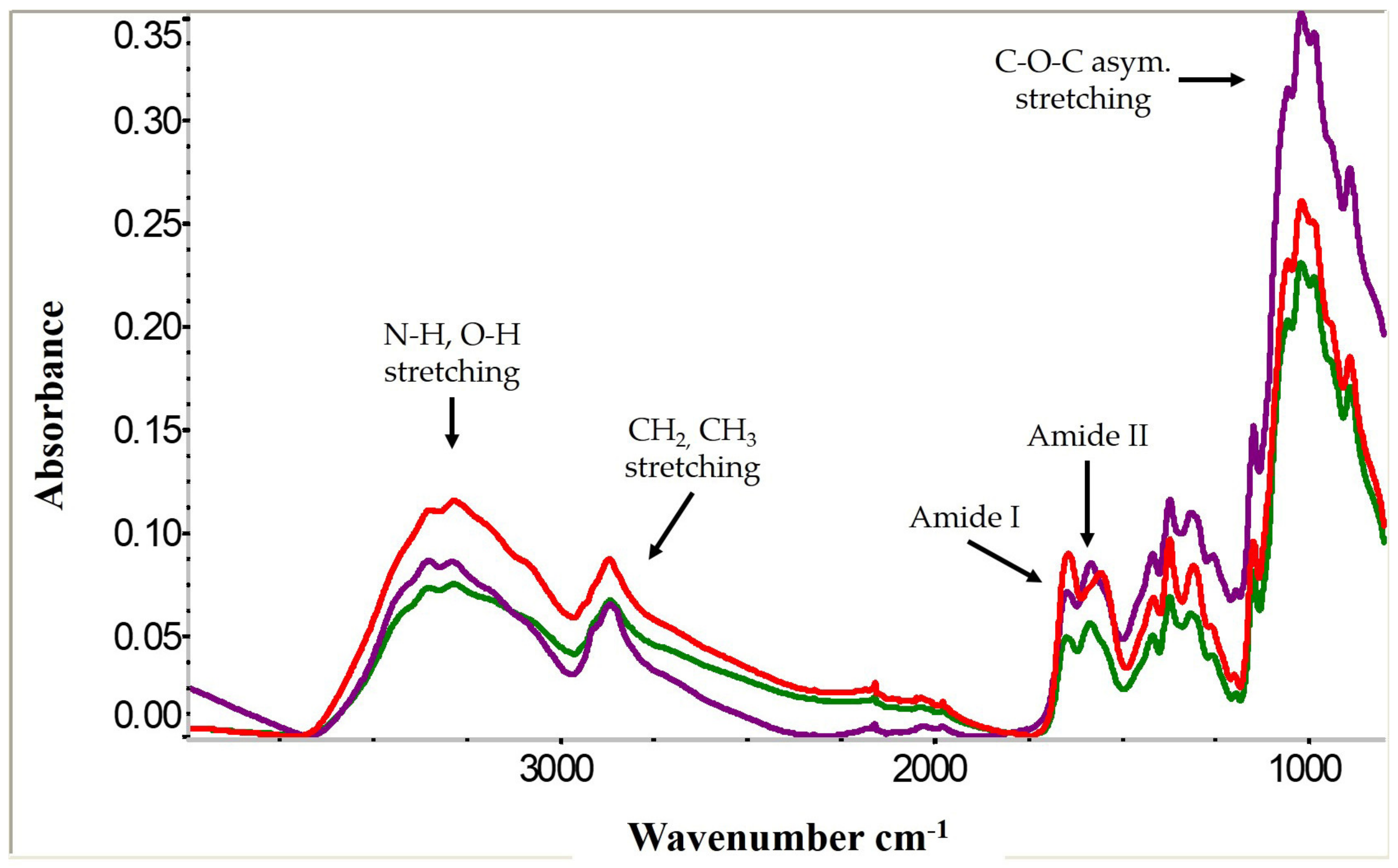
2.3. Chitosan Conversion and Characterization by Fourier Transform Infrared Spectroscopy (FTIR) and Degree of Deacetylation

Purified, lyophilized cricket chitin was converted to chitosan with 67% w/v NaOH (1:20 w/v) for 2, 4, 6 and 10 h to vary the degree of deacetylation [13]. Due to employing the traditional conversion method for chitosan with concentrated alkali solution, chitosan was first filtered (100 mesh) with water until effluent ran clear. Chitosan was then washed with a minimum of 5 L of water, until effluent reached neutrality (pH 6.5), to reduce the presence of residual NaOH. Finally, chitosan was collected, lyophilized and stored at −20 °C until needed. Chitosan conversion yield (dwb) for each duration treatment was determined using Equation (3):
C h i t o s a n   y i e l d   % =   m a s s   c h i t o s a n   g   m a s s   c h i t i n   g   ×   100
The eight cricket chitosan samples were evaluated by FT-IR (Thermo Scientific, Waltham, MA, USA) from 3500 to 800 cm−1 with a resolution of 8 cm−1 (n = 8) to determine the chemical structure and degree of deacetylation (DDA) [1,13,14]. Intensity of FTIR peaks with appropriate baselines at 1650 cm−1 (A1652) and 3350 cm−1 (A3450) were used to determine DDA Equation (4) [13,30,31].
D D A   % = 100 A 1650 A 3350   ×   100 1.33
Additionally, commercial shrimp chitosan spectra were used as a reference. Cricket chitosan samples from each species, which were determined to have similar DDA values (~72%, 76% and 80%), were used in the lipid-binding capacity and antibacterial activity experiments.

2.4. Molecular Weight Determination

The average molecular weight of cricket chitosan was determined viscometrically following the method of Czechowska-Biskup, Wach [32] and Roberts and Domszy [33]. In brief, solutions (1.5–4.5 mg/mL) were prepared from ~80% DDA cricket chitosan (10 h samples) in a solvent system consisting of equal parts of 0.1 mol L−1 CH3OOH and 0.2 mol L−1 NaCl and stirred overnight. The flow time of each chitosan solution and solvent system (5 mL) was measured in triplicate using a capillary viscometer (Cannon-Fenske, Size 75) in a 25 °C water bath. Relative viscosity, specific viscosity and reduced viscosity were calculated using measured flow times [34]. The linear relationship between reduced viscosity and concentration for each cricket chitosan was extrapolated to determine its intrinsic viscosity ( η ). Finally, using the averaged intrinsic viscosity (n = 3), the viscosity average molecular weight (Mν) for each chitosan was calculated using the Mark–Houwink Equation:
η = K M v α
with previously determined solvent system constants: K =1.81 × 10⁻³ α   =0.93 [12,33].

2.5. Anti-Obesity Effects (Lipid-Binding Capacity) of Cricket Chitosan

The in vitro lipid-binding capacity of shrimp and cricket (72, 76 and 80% DDA) chitosan samples was determined in triplicate following established procedures [35,36]. Each chitosan sample (20 mg) was dissolved in 1.25 mL 0.6 mol L−1 HCl and incubated for 30 min in a 37 °C water bath with constant shaking. Then, 25 g of olive oil was added to each tube, vortexed and incubated under the same conditions for 2 h. After incubation, 8 mL of phosphate buffer (pH 7.4) was added and the solution’s pH was adjusted to 6.8 with 1 mol L−1 NaOH and incubated again for 30 min to mimic duodenal conditions. Finally, tubes were centrifuged at 697× g for 10 min and the supernatant representing unbound lipids was gravimetrically measured. The lipid-binding capacity of chitosan was calculated using Equation (6):
l i p i d   b i n d i n g   c a p a c i t y   g   o i l g   c h i t o s a n = 25   g   o i l g   u n b o u n d   o i l 0.02   g   c h i t o s a n

2.6. Determination of Antimicrobial Activity

The antimicrobial activity of cricket and shrimp chitosan samples against two bacteria strains was determined using methods described by Aguilar-Toalá and Deering [37] with slight modifications [38]. Specifically, antibacterial activity against Escherichia coli ATCC 25922 and Listeria innocua ATCC 33090, as surrogates for foodborne pathogens Escherichia coli O157:H7 and Listeria monocytogenes, were studied [39].

2.6.1. Bacterial Stock Cultures and Sample Preparation

The lyophilized bacteria were inoculated in Brain Heart Infusion (BHI) and Mueller Hinton Broth (MHB) for L. innocua and E. coli, respectively, following ATCC guidelines. Inoculums were used to prepare freezer stocks with a final glycerol concentration of 20% (w/v), cryovials were stored at −80 °C until needed. Prior to use, 50 μL of each stock was transferred into 5 mL MHB and BHI, for E. coli and L. innocua, grown to stationary phase. As determined by plate counts, E. coli inoculums were 1.1 × 10⁸ cfu/mL and L. innocua inoculums were 7.2 × 10⁸ cfu/mL. Fresh cultures were prepared from freezer stocks following the same incubation procedure and used immediately for each assay.
Shrimp and cricket chitosan (72, 76 and 80% DDA) solutions (8 mg/mL) were prepared in 1% (v/v) acetic acid and stirred overnight to ensure homogenous dissolution. Solutions were then sterilized by autoclaving for 20 min at 121 °C. Additionally, 1% acetic acid was sterilized under the same conditions.

2.6.2. Antibacterial Determination

The antimicrobial activity of cricket chitosan samples was determined using 96-well sterile microplates. Chitosan samples, positive and negative control wells were prepared aseptically with a final volume of 220 µL (n = 6). For chitosan wells, 13.75 µL of each chitosan sample was added with 204.25 µL BHI, resulting in a final chitosan sample concentration of 0.5 mg/mL in each well. Positive control and negative control wells were prepared with 220 µL and 218 µL BHI, respectively. Chitosan sample and negative control wells were inoculated with 2 µL of bacterial culture (~106 cfu/mL) (n = 6). Finally, an additional solvent control was prepared to deconvolute its effects from chitosan on bacterial growth. Similar to the chitosan wells, 13.75 µL of 1% CH3OOH was combined with 204.25 µL BHI and inoculated (2 µL) (n = 6).
Following preparation, microplates were incubated for 24 h at 37 °C with optical density measurements (OD = 620 nm) conducted at 0, 6, 12 and 24 h. Growth curves of samples were created by plotting optical density over the duration of the experiment. Additionally, the percent of bacterial growth inhibition at 24 h was calculated (n = 6) using Equation (7):
%   i n h i b i t i o n = 1   Δ O D s a m p l e ,   24   h r O D c o n t r o l ,   24   h r × 100

2.7. Statistical Analysis

All experiments and analyses were conducted in triplicate, unless otherwise indicated. Statistical analysis of chitosan samples’ degree of deacetylation, chitosan yield, lipid-binding capacities and percent of bacterial inhibition were performed using a one way ANOVA with 95% confidence level (Minitab 18® State College, PA, USA).

3. Results and Discussion

3.1. Cricket Chitin and Chitosan Processing

Cricket chitin and chitosan were successfully extracted, purified and converted with similarities to the commercial products. Overall, the extraction and purification processing steps produced cricket chitin with low protein and ash compositions, in comparison to their original compositions (Table 1).
The demineralization process is an important first step to remove the minerals present in the exoskeleton matrix. Traditionally, crustacean shells are demineralized with hydrochloric acid solutions, to react with the calcium carbonate intrinsically present at high concentrations (30–50%) [4,27,40]. In the case of insects, due to their environmental differences, they do not contain a large amount of minerals in their exoskeletons when compared to crustaceans [41]. Yet, the majority of insect chitin studies commonly employ the demineralization procedures optimized for crustaceans [41]. For example, two studies on grasshoppers (Decticus verrucivorus) and cockroach wings (Blaberus giganteus), employed HCl demineralization parameters that were highly concentrated (2 M), hot (75 °C) and prolonged (>2 h) [7,11]. Demineralization of cicada sloughs, silkworms, beetles and black soldier flies were performed at 100 °C for 20–30 min with 1 mol L−1 HCl and resulted in ash values of 3–5% [29,42,43]. Whereas silkworms, mealworms and grasshoppers were treated with the same acid concentration at lower temperatures (25–30 °C) for more than 2 h and ash contents were reported to be less than 1% [44]. In this study, A. domesticus and G. sigillatus chitin was demineralized with 0.25 mol L−1 HCl for 15 min at 85 °C with final chitin ash contents less than 0.09% (Table 1). The results of this study show that cricket chitin and likely other insect-derived chitin, do not need extensive demineralization treatment as those used for crustacean (shellfish) sources.
A. domesticus chitin had comparable yields (5.68%) to that extracted in other studies for other insects including other cricket species (5.1%), grasshoppers (5.7%), beetles (5.0%) and wasp pupa (6.2%) [8,13,44,46]. In contrast, the G. sigillatus had lower chitin yields (3.38%); however, it was still comparable to chitin extracted from silkworm chrysalis (3.1%), mealworms (2.5%), honeybees (2.5%) and wasp larvae (2.2%) [8,44,46]. This chitin yield difference between the two cricket species is likely a result of the larger body size and exoskeleton of the A. domesticus in comparison to G. sigillatus. Other insect chitin studies reported higher chitin yields (18–37%); however, these studies used molted exoskeletons or wings, which naturally have higher chitin contents, when compared to the intact insect [29,44,47].

3.2. Cricket Chitin and Chitosan Characterization

FTIR analysis of all cricket chitin and chitosan samples showed strong chemical and structural similarities to those from shrimp. Chitin from both cricket species (Figure 1) contained characteristic peaks known to chitin from both crustaceans and other insects [12,48]. The two cricket chitins did not differ from each other. Chitin has three crystalline forms, either alpha, beta or gamma, although chitin is most commonly present in its alpha form [48]. The FTIR spectra peak between 1700 and 1500 cm−1, known as the Amide I band, shows that cricket chitin from both species is in the alpha crystalline form, due to the two peaks observed at ~1660 and ~1630 cm−1. These two peaks represent alpha chitin’s intra- and inter-molecular hydrogen bonds forming its antiparallel chain arrangement [48,49]. The hydrogen bonds between –C=O (Amide I) and –NH– (Amide II) are responsible for the peak at ~1660 cm−1, while the second peak occurring at ~1630 cm−1 is due to hydrogen bonding between the –CH2O side chain and –C=O [50].
Spectral peaks related to chitosan’s chemistry have been extensively reported. Following deacetylation of chitin, chitosan samples produced from both cricket species were in good alignment with commercially purchased shrimp chitosan (Figure 2). Two spectral peaks, the OH peak (~3450 cm−1) and the –CO–NH (1650 cm−1) of the Amide I band, were used to calculate the DDA of the cricket and shrimp chitosans [13,30,31] (Table 2). Deacetylating both the A. domesticus and G. sigillatus chitin for 2 h resulted in chitosan with a DDA of ~73%, while the commercial shrimp chitosan was ~70%. Colorado potato beetle (Leptinotarsa decemlineata) and larvae chitin processed under similar conditions for 3 h produced chitosan with 71% and 64% DDA, respectively [12]. A. domesticus chitin reached 76% DDA after 4 h, whereas G. sigillatus chitin required 6 h to reach a similar DDA. After 10 h of deacetylation, cricket chitosan reached a DDA of ~ 80%, similar to that reported by Chae, Shin [13], which produced 84% DDA chitosan from the African field cricket (Gryllus bimaculatus) chitin after 9 h of deacetylation under similar conditions. In Figure 2, as the deacetylation time of cricket chitin increased, acetyl groups were continuously replaced with amine groups leading to a higher DDA [30]. Shrimp chitosan had a lower DDA (70%) and therefore a greater peak intensity at ~1650 cm−1 compared to the two cricket chitosans deacetylated for 10 h (~80% DDA).
The chemical deacetylation of cricket chitin to chitosan had similar yields (Table 2) comparable to other insects and crustaceans [45]. Approximately 70–80% of the A. domesticus chitin mass was recovered after deacetylation, whereas of G. sigillatus cricket chitin yielded about 60–65% chitosan. Additionally, deacetylation duration had little effect on the overall conversion yields. G. sigillatus chitosan conversion yields were not affected by duration time and A. domesticus chitosan yield was the highest after 6 h and the lowest after 10 h. However, the 6 and 10 h deacetylation had chitosan yields that were not significantly different after 2 and 4 h of deacetylation.

3.3. Molecular Weight Determination

The average molecular weight was determined to be 344 kDa for A. domesticus chitosan, while G. sigillatus chitosan had a larger molecular weight of 524 kDa (Table 2). While the A. domesticus cricket is physically larger than G. sigillatus, its molecular weight was determined to be smaller. This suggests that A. domesticus may have more chitin fibers in its exoskeleton of a smaller weight, while G. sigillatus cricket’s exoskeleton may have larger, but fewer, chitin fibers in its exoskeleton. The size of chitosan from these two cricket species is in good agreement with another cricket chitosan study. Kim, Song [22] studied the molecular weight of G. bimaculatus crickets using dynamic light scattering and found its molecular weight to be ~308 kDa. As a result, A. domesticus and G. bimaculatus crickets have similar molecular weights, but are smaller than G. sigillatus crickets. Additionally, cricket chitosan is relatively similar in size to other insect chitosan previously studied, such as that sourced from blowfly (501 kDa) and housefly larvae (426 kDa) [51,52]. However, compared to other species of insect chitosan, A. domesticus and G. sigillatus cricket chitosan are much larger in size. Cicada sloughs, grasshoppers, mealworms, silkworm chrysalis, black soldier flies and beetles ranged in molecular weights between 2.7 and 15 kDa [12,44,53]. As a result, insect chitosan has repeatedly been stated to be of low molecular weight compared to crustacean chitin and chitosan, which can range from 50 to 2000 kDa [54,55]. The results of this study, as well as others, confirm that insect chitosan may also be characterized as high molecular weight.

3.4. Lipid-Binding Capacity

Commercial shrimp chitosan and its water-soluble derivatives, has been reported as an anti-obesity or hyperlipidemic treatment in a variety of in vitro and in vivo studies [24]. Chitosan exhibits both hypocholesteremic and hypolipidemic properties through its ability to regulate lipid metabolism, achieved by electrostatic interaction with or physical entrapment of, targeted molecules [24]. The focus of this study was to evaluate the lipid-binding capacity of chitosan through interaction and entrapment of dietary lipids in vitro, one of the reported mechanisms of chitosan to reduce lipid metabolism in a high lipid diet. The consumption of lipids in the presence of chitosan, is thought to lead to the glucosamine in chitosan to be positively charged in the stomach due to its low pH, followed by its binding to negatively charged lipid molecules such as triglycerides and fatty acids [56,57]; this emulsion formation has been demonstrated previously with fluorescent microscopy [35]. When the lipid–chitosan emulsion is transferred from the acidic stomach conditions to the alkaline conditions of the duodenum, the chitosan precipitates and physically entraps the emulsified lipid droplets through gel formation [35,36,56]. Ultimately, this entrapment prevents lipids to be accessed and digested by the body and results in their excretion through feces.
Chitosan from both cricket species showed high lipid-binding capacity, between ~160 and 220 g of oil per gram of chitosan (Table 3). To the best of our knowledge, this is the first study evaluating the lipid-binding capacity of chitosan from edible insects. There were no significant differences on lipid-binding capacity between the different samples; therefore, no correlation between physicochemical properties such as molecular weight or DDA can be made from this study. However, these results suggest that edible cricket chitosan could be as effective as commercial shrimp chitosan in binding lipids under gastric conditions. Panith, Wichaphon [35] showed that chitosan from shrimp shells, produced with a low molecular weight (~30 kDa), had inferior lipid-binding capacity compared to medium and high molecular weight chitosan samples (890 kDa and 8900 kDa). In contrast, low molecular weight chitosan (~25 kDa), from an unspecified marine species, had superior-binding capacity compared to larger molecular weight chitosan (408 kDa) [56]. In this study, the molecular weights of cricket and shrimp chitosans were determined to be high molecular weight. Therefore, it is possible the molecular weights of the samples of this study were too similar to result in significant changes between their lipid-binding capacities. Further research on this topic is required since many insect chitosan studies report low molecular weights [12,44,58] and it is unknown how cricket chitosans’ lipid-binding capacities would compare to other insect-based chitosans due to opposing literature conclusions.
Overall, the lipid-binding capacities of edible cricket and shrimp chitosan in this study were found to be different than those reported for crustacean chitosan. Panith, Wichaphon [35] showed the highest-binding capacity to be at ~28 g oil/g chitosan, which is much lower than the results of this study. These differences are likely due to the different in vitro methods applied. For example, our study gravimetrically measured unbound oil, whereas Panith et al. (2016) solvent-extracted entrapped lipids and then gravimetrically measured bound oil. These differences in methods could lead to some over-estimation in our study. Additionally, Panith et al. (2016) centrifuged at 4000× g, whereas in this study, our method called for 696× g to separate bound lipids from unbound lipids. It is possible the larger centrifugal force used in other studies destabilized the chitosan-lipid gel and physically removed chitosan entrapped lipids; this could result in an under-estimation of lipid-binding capacity reported by Panith et al. (2016). In contrast, Zhou and Xia [36] reported a much larger lipid-binding capacity value of ~1200 g of oil bound per gram of chitosan. However, the methods for determining the-binding capacity did not differ, making it difficult to understand the differences in values reported.

3.5. Antimicrobial Activity

The effect of edible cricket chitosan on inhibiting growth of Listeria innocua ATCC 33090 and Escherichia coli ATCC 25922 over 24 h is shown in Figure 3. For each bacteria strain, the effect of shrimp chitosan on growth was also evaluated as a reference for current commercial (shrimp) chitosan. Additionally, the solvent used, 1% acetic acid, is an antimicrobial agent itself, and thus its effect on measured antibacterial properties was evaluated (Table 4) [59,60]. At a concentration of 0.50 mg/mL, all chitosan samples were able to effectively inhibit growth of L. innocua over 24 h, with ~100% inhibition at hour 24 (Table 4), as shown by no increase in the optical density of the samples during the duration of the experiment (Figure 3). Acetic acid, at a final concentration of 0.06%, resulted in L. innocua growth to be inhibited by 38% (Table 4). Therefore, L. innocua inhibition by chitosan samples was due to both the presence of chitosan, as well as acetic acid.
One generally accepted mechanism of chitosan’s antibacterial activity is a result of the positive charged +NH3 glucosamines interacting with negatively charged surface components of bacterial cells, which ultimately interrupts or prevents vital cell functions [54]. Therefore, it is thought that an increase of DDA increases the number of positively charged glucosamines that are able to bind with cell components, leading to an increase in antimicrobial activity. This was seen when the DDA of G. sigillatus cricket chitosan increased from 72 to 76 and 80%, which resulted in a sequential increase of growth inhibition of E. coli; showing approximately 90% growth inhibition at 80% DDA (Table 4). However, DDA did not have an apparent effect on the inhibition of L. innocua growth at the chitosan concentration chosen (Figure 3). Decreasing the chitosan sample concentrations sequentially may allow for differentiation between chitosan samples with different degrees of deacetylation on the efficacy of inhibiting L. innocua growth, while minimizing the contribution due to acetic acid. For both bacteria, growth inhibition achieved by 72% DDA cricket chitosan was similar to that of commercial shrimp chitosan, suggesting that cricket chitosan can be as effective as current market options.
The results of this study are similar to other insect chitosan antimicrobial assays; however, direct comparisons are difficult to make due to differing microorganisms, chitosan concentrations employed and assaying methods (i.e., MIC, MBC, zone of inhibition, etc.). For example, chitosan derived from larvae and adult Colorado potato beetles were found to have minimal bactericidal concentrations (MBC) of 0.32 and 1.25 mg/mL against L. monocytogenes, where MBC values represent the lowest concentration of an antimicrobial agent required to kill bacteria in an inoculated sample. Chitosan derived from two different grasshopper species were found to have a MBC of 0.32 and 0.63 mg/mL against L. monocytogenes. Another study quantified the effect of mealworm (Tenbrio molitor) and crustacean-derived chitosan on cell counts of foodborne pathogens E. coli O157:H7, L. monocytogenes and Salmonella enterica serovar Typhimurium, over 48 h [61]. The study found in general that crustacean and insect chitosan antibacterial activity at 1.5 mg/mL led to unchanged or reduced following 24 h after inoculation for all three bacteria, with recovery of bacterial counts detected between 24 and 48 h (when inoculated with 106 cfu/mL, pH 6.2). At 24 h, the antibacterial activity of crustacean chitosan was slightly greater for E. coli and S. Typhimurium, whereas mealworm chitosan was more effective against L. monocytogenes. However, the study did not characterize or report the specifications of the chitosan samples, such as molecular weight or % deacetylation, so it is difficult to understand the basis of these differences. Another study evaluated the antimicrobial efficacy of two different grasshopper species against a variety of organisms, including that of L. monocytogenes. The MBC of one grasshopper chitosan (Calliptamus barbarous) was 0.32 mg/mL, whereas the MBC of chitosan from the other grasshopper species (Oedaleus decorus) was 0.63 mg/mL [38]. The chitosan from the two species were reported to have a DDA between 70 and 75%, although the specific DDA of each chitosan was not reported. It is possible the differences in MBC are due to slight differences in DDA. As shown in this current study (Figure 3), a 4% change in the cricket chitosan DDA (i.e., from 72 to 76 and 80%) significantly improved E. coli inhibition (Table 4). No difference in chitosan antimicrobial efficacy was seen between the two cricket species, likely a result of standardizing DDA prior to antimicrobial testing.
The results of this study, in conjunction with others, elucidate the importance to evaluate each newly derived chitosan against specific bacterial strains to determine its anticipated antimicrobial activity. Overall, cricket chitosan derived from A. domesticus and G. sigillatus were as effective at inhibiting microbial growth of surrogate foodborne pathogens, as the traditional and commercial crustacean (shrimp) chitosan. A concentration of 0.50 mg/mL was an effective concentration for all chitosan samples against L. innocua; however, higher concentrations of the lower DDA chitosan samples may be required to enhance the antimicrobial effect against E. coli. Further studies on the effect of edible cricket chitosan on bacterial cell wall material are required to understand the mechanisms of inhibition by these chitosan samples.

4. Conclusions

Chitin from two edible cricket species commonly reared in the United States were successfully collected as a by-product of cricket protein hydrolysis and adequately isolated through demineralization and deproteinization processing steps. A. domesticus crickets were found to yield slightly higher amounts of purified chitin, compared to G. sigillatus crickets. FTIR results confirmed that cricket chitin was in its alpha-crystalline form, similar to that isolated from most crustacean and insect species. Cricket chitin was successfully converted to chitosan with approximately 72%, 76% and 80% degree of deacetylation, achieved by varying deacetylation times using concentrated alkaline treatments. Structural analysis of cricket chitosan samples was chemically similar to that of commercial shrimp chitosan. Finally, the average molecular weight of chitosan derived from A. domesticus crickets was determined to be 344 kDa, while that from G. sigillatus had a larger molecular weight of 524 kDa. In contrast to the findings of other insect-derived chitosan studies, the results of this study suggest that cricket chitosan can be recognized as a high molecular weight polymer, similar to commercially available chitosan with potential bioactive properties.
Lipid-binding capacity of all chitosan samples were determined to be between 160 g and 220 g oil per 1 g chitosan, although physicochemical properties did not have any significant effect on lipid binding. Additionally, the edible cricket chitosan samples effectively inhibited E. coli and L. innocua. The degree of deacetylation did not have an effect on the antibacterial properties of cricket chitosan against L. innocua, whereas the antimicrobial activity of A. domesticus and G. sigillatus chitosan was more effective against E. coli at higher DDA values, compared to shrimp chitosan. Thus, edible cricket chitosan has the potential to inhibit the growth of foodborne pathogens, as a naturally derived antimicrobial agent.
As the consumption of insects and insect-containing food products is rapidly growing, an increase of insect protein production and thereof chitin by-products will become more available. This study concludes that chitosan derived from U.S. reared edible crickets have physicochemical and bioactive properties similar to commercial crustacean (e.g., shrimp) chitosan. As a result, there is potential for the mass production of cricket-based chitosan as the consumer acceptability for arthropods widens outside the traditional source (crustaceans).

Author Contributions

Conceptualization, M.M. and A.M.L.; methodology, M.M. and A.M.L.; investigation, M.M.; data curation, M.M. and A.M.L.; writing—original draft preparation, M.M.; writing—review and editing, M.M. and A.M.L.; supervision, A.M.L.; funding acquisition, A.M.L. All authors have read and agreed to the published version of the manuscript.

Funding

This work is supported by the USDA National Institute of Food and Agriculture, Hatch Act formula funds project 1019794.

Institutional Review Board Statement

Not Applicable.

Informed Consent Statement

Not Applicable.

Data Availability Statement

All findings discussed are based on the data contained within this current study.

Acknowledgments

The authors acknowledge Aguilar-Toala, Deering and Hansel for their support with the antimicrobial assays performed.

Conflicts of Interest

The authors declare no conflict of interest.

References

  1. Sivashankari, P.R.; Prabaharan, M. 5-Deacetylation modification techniques of chitin and chitosan. In Chitosan Based Biomaterials; Jennings, J.A., Bumgardner, J.D., Eds.; Woodhead Publishing: Cambridge, UK, 2017; Volume 1, pp. 117–133. [Google Scholar]
  2. Kumar, M.N.V.R. A review of chitin and chitosan applications. React. Funct. Polym. 2000, 46, 1–27. [Google Scholar] [CrossRef]
  3. Hamed, I.; Özogul, F.; Regenstein, J.M. Industrial applications of crustacean by-products (chitin, chitosan, and chitooligosaccharides): A review. Trends Food Sci. Technol. 2016, 48, 40–50. [Google Scholar] [CrossRef]
  4. Zargar, V.; Asghari, M.; Dashti, A. A Review on Chitin and Chitosan Polymers: Structure, Chemistry, Solubility, Derivatives, and Applications. ChemBioEng Rev. 2015, 2, 204–226. [Google Scholar] [CrossRef]
  5. No, H.K.; Meyers, S.P. Preparation and Characterization of Chitin and Chitosan—A Review. J. Aquat. Food Prod. Technol. 1995, 4, 27–52. [Google Scholar] [CrossRef]
  6. Majtán, J.; Bíliková, K.; Markovič, O.; Gróf, J.; Kogan, G.; Šimúth, J. Isolation and characterization of chitin from bumblebee (Bombus terrestris). Int. J. Biol. Macromol. 2007, 40, 237–241. [Google Scholar] [CrossRef]
  7. Kaya, M.; Lelešius, E.; Nagrockaitė, R.; Sargin, I.; Arslan, G.; Mol, A.; Baran, T.; Can, E.; Bitim, B. Differentiations of Chitin Content and Surface Morphologies of Chitins Extracted from Male and Female Grasshopper Species. PLoS ONE 2015, 10, e0115531. [Google Scholar] [CrossRef]
  8. Kaya, M.; Sofi, K.; Sargin, I.; Mujtaba, M. Changes in physicochemical properties of chitin at developmental stages (larvae, pupa and adult) of Vespa crabro (wasp). Carbohydr. Polym. 2016, 145, 64–70. [Google Scholar] [CrossRef] [PubMed]
  9. Zelencova, L.; Erdoǧan, S.; Baran, T.; Kaya, M.; Erdogan, S. Chitin extraction and chitosan production from Chilopoda (Scolopendra cingulata) with identification of physicochemical properties. Polym. Sci. Ser. A 2015, 57, 437–444. [Google Scholar] [CrossRef]
  10. Greven, H.; Kaya, M.; Sargin, I.; Baran, T.; Kristensen, R.M.; Sørensen, M.V. Characterisation of chitin in the cuticle of a velvet worm (Onychophora). Turk. J. Zooöl. 2019, 43, 416–424. [Google Scholar] [CrossRef]
  11. Kaya, M.; Sargin, I.; Sabeckis, I.; Noreikaite, D.; Erdonmez, D.; Salaberria, A.M.; Labidi, J.; Baublys, V.; Tubelytė, V. Biological, mechanical, optical and physicochemical properties of natural chitin films obtained from the dorsal pronotum and the wing of cockroach. Carbohydr. Polym. 2017, 163, 162–169. [Google Scholar] [CrossRef]
  12. Kaya, M.; Baran, T.; Erdoğan, S.; Menteş, A.; Özüsağlam, M.A.; Çakmak, Y.S. Physicochemical comparison of chitin and chitosan obtained from larvae and adult Colorado potato beetle (Leptinotarsa decemlineata). Mater. Sci. Eng. C 2014, 45, 72–81. [Google Scholar] [CrossRef] [PubMed]
  13. Chae, K.-S.; Shin, C.-S.; Shin, W.-S. Characteristics of cricket (Gryllus bimaculatus) chitosan and chitosan-based nanoparticles. Food Sci. Biotechnol. 2018, 27, 631–639. [Google Scholar] [CrossRef] [PubMed]
  14. Ibitoye, B.E.; Lokman, I.H.; Hezmee, M.N.M.; Goh, Y.M.; Zuki, A.B.Z.; Jimoh, A.A. Extraction and physicochemical characterization of chitin and chitosan isolated from house cricket. Biomed. Mater. 2017, 13, 025009. [Google Scholar] [CrossRef] [Green Version]
  15. Liceaga, A.M. Approaches for Utilizing Insect Protein for Human Consumption: Effect of Enzymatic Hydrolysis on Protein Quality and Functionality. Ann. Entomol. Soc. Am. 2019, 112, 529–532. [Google Scholar] [CrossRef]
  16. Hall, F.; Johnson, P.E.; Liceaga, A. Effect of enzymatic hydrolysis on bioactive properties and allergenicity of cricket (Gryllodes sigillatus) protein. Food Chem. 2018, 262, 39–47. [Google Scholar] [CrossRef] [PubMed]
  17. Hall, F.; Liceaga, A. Effect of microwave-assisted enzymatic hydrolysis of cricket (Gryllodes sigillatus) protein on ACE and DPP-IV inhibition and tropomyosin-IgG binding. J. Funct. Foods 2020, 64, 103634. [Google Scholar] [CrossRef]
  18. Hall, F.G.; Jones, O.G.; O’Haire, M.E.; Liceaga, A.M. Functional properties of tropical banded cricket (Gryllodes sigillatus) protein hydrolysates. Food Chem. 2017, 224, 414–422. [Google Scholar] [CrossRef]
  19. Berezina, N.; Hubert, A.; Berro, F.; Levon, J.-G.; Le Roux, K.; Socolsky, C. Chitin, Hydrolysate and Production of at Least One Desired Product from Insects by Means of Enzymatic Hydrolysis, Comprising a Combination of Steps Performed Prior to the Enzymatic Hydrolysis. U.S. Patent Application No. 15/541,162, 2019. [Google Scholar]
  20. Berezina, N.; Hubert, A.; Berro, F.; Levon, J.-G.; Le Roux, K.; Socolsky, C. Chitin, Hydrolysate and Method for the Production of One or More Desired Products by Means of Enzymatic Hydrolysis, Including Pre-Treatment with an Oxidising Agent. U.S. Patent Application No. 15/541,174, 2018. [Google Scholar]
  21. Berezina, N.; Hubert, A.; Berro, F.; Levon, J.-G.; Le Roux, K.; Socolsky, C. Chitin, Hydrolysate and Method for the Production of One or More Desired Products from Insects by Means of Enzymatic Hydrolysis. U.S. Patent Application No. 15/541,186, 2018. [Google Scholar]
  22. Kim, M.-W.; Song, Y.-S.; Han, Y.S.; Jo, Y.H.; Choi, M.H.; Park, Y.-K.; Kang, S.H.; Kim, S.-A.; Choi, C.; Jung, W.-J. Production of chitin and chitosan from the exoskeleton of adult two-spotted field crickets (Gryllus bimaculatus). Entomol. Res. 2017, 47, 279–285. [Google Scholar] [CrossRef]
  23. Jin, Q.; Yu, H.; Wang, X.; Li, K.; Li, P. Effect of the molecular weight of water-soluble chitosan on its fat-/cholesterol-binding capacities and inhibitory activities to pancreatic lipase. PeerJ 2017, 5, e3279. [Google Scholar] [CrossRef] [PubMed] [Green Version]
  24. Xia, W.; Liu, P.; Zhang, J.; Chen, J. Biological activities of chitosan and chitooligosaccharides. Food Hydrocoll. 2011, 25, 170–179. [Google Scholar] [CrossRef]
  25. Rabea, E.I.; Badawy, M.E.-T.; Stevens, C.V.; Smagghe, G.; Steurbaut, W. Chitosan as Antimicrobial Agent: Applications and Mode of Action. Biomacromolecules 2003, 4, 1457–1465. [Google Scholar] [CrossRef]
  26. Luna, G.C.; Martin-Gonzalez, F.S.; Mauer, L.; Liceaga, A. Cricket (Acheta domesticus) protein hydrolysates’ impact on the physicochemical, structural and sensory properties of tortillas and tortilla chips. J. Insects Food Feed. 2021, 7, 109–120. [Google Scholar] [CrossRef]
  27. Percot, A.; Viton, A.C.; Domard, A. Optimization of Chitin Extraction from Shrimp Shells. Biomacromolecules 2003, 4, 12–18. [Google Scholar] [CrossRef] [PubMed]
  28. Elshaarawy, R.F.M.; Mustafa, F.H.A.; Herbst, A.; Farag, A.E.M.; Janiak, C. Surface functionalization of chitosan isolated from shrimp shells, using salicylaldehyde ionic liquids in exploration for novel economic and ecofriendly antibiofoulants. RSC Adv. 2016, 6, 20901–20915. [Google Scholar] [CrossRef]
  29. Sajomsang, W.; Gonil, P. Preparation and characterization of α-chitin from cicada sloughs. Mater. Sci. Eng. C 2010, 30, 357–363. [Google Scholar] [CrossRef]
  30. Czechowska-Biskup, R.; Jarosińska, D.; Rokita, B.; Ulański, P.; Rosiak, J.M. Determination of degree of deacetylation of chitosan-comparision of methods. Prog. Chem. Appl. Chitin Deriv. 2012, 17, 5–20. [Google Scholar]
  31. Domard, A.; Rinaudo, M. Preparation and characterization of fully deacetylated chitosan. Int. J. Biol. Macromol. 1983, 5, 49–52. [Google Scholar] [CrossRef]
  32. Czechowska-Biskup, R.; Wach, R.A.; Rosiak, J.M.; Ulański, P. Procedure for determination of the molecular weight of chitosan by viscometry. Prog. Chem. Appl. Chitin its Deriv. 2018, XXIII, 45–54. [Google Scholar] [CrossRef]
  33. Roberts, G.A.; Domszy, J.G. Determination of the viscometric constants for chitosan. Int. J. Biol. Macromol. 1982, 4, 374–377. [Google Scholar] [CrossRef]
  34. Chen, R.H.; Tsaih, M.L. Effect of temperature on the intrinsic viscosity and conformation of chitosans in dilute HCl solution. Int. J. Biol. Macromol. 1998, 23, 135–141. [Google Scholar] [CrossRef]
  35. Panith, N.; Wichaphon, J.; Lertsiri, S.; Niamsiri, N. Effect of physical and physicochemical characteristics of chitosan on fat-binding capacities under in vitro gastrointestinal conditions. LWT 2016, 71, 25–32. [Google Scholar] [CrossRef]
  36. Zhou, K.; Xia, W.; Zhang, C.; Yu, L. (Lucy) In vitro binding of bile acids and triglycerides by selected chitosan preparations and their physico-chemical properties. LWT 2006, 39, 1087–1092. [Google Scholar] [CrossRef]
  37. Aguilar-Toalá, J.E.; Deering, A.J.; Liceaga, A.M. New Insights into the Antimicrobial Properties of Hydrolysates and Peptide Fractions Derived from Chia Seed (Salvia hispanica L.). Probiotics Antimicrob. Proteins 2020, 12, 1571–1581. [Google Scholar] [CrossRef]
  38. Kaya, M.; Baran, T.; Asan-Ozusaglam, M.; Cakmak, Y.S.; Tozak, K.O.; Mol, A.; Mentes, A.; Sezen, G. Extraction and characterization of chitin and chitosan with antimicrobial and antioxidant activities from cosmopolitan Orthoptera species (Insecta). Biotechnol. Bioprocess Eng. 2015, 20, 168–179. [Google Scholar] [CrossRef]
  39. Hu, M.; Gurtler, J.B. Selection of Surrogate Bacteria for Use in Food Safety Challenge Studies: A Review. J. Food Prot. 2017, 80, 1506–1536. [Google Scholar] [CrossRef]
  40. Kurita, K. Chitin and Chitosan: Functional Biopolymers from Marine Crustaceans. Mar. Biotechnol. 2006, 8, 203–226. [Google Scholar] [CrossRef] [PubMed]
  41. Berezina, N.; Hubert, A. Marketing and Regulations of Chitin and Chitosan from Insects. Chitin Chitosan 2019, 2019, 477–489. [Google Scholar] [CrossRef]
  42. Zhang, M.; Haga, A.; Sekiguchi, H.; Hirano, S. Structure of insect chitin isolated from beetle larva cuticle and silkworm (Bombyx mori) pupa exuvia. Int. J. Biol. Macromol. 2000, 27, 99–105. [Google Scholar] [CrossRef]
  43. Purkayastha, D.; Sarkar, S. Physicochemical Structure Analysis of Chitin Extracted from Pupa Exuviae and Dead Imago of Wild Black Soldier Fly (Hermetia illucens). J. Polym. Environ. 2019, 28, 445–457. [Google Scholar] [CrossRef]
  44. Luo, Q.; Wang, Y.; Han, Q.; Ji, L.; Zhang, H.; Fei, Z.; Wang, Y. Comparison of the physicochemical, rheological, and morphologic properties of chitosan from four insects. Carbohydr. Polym. 2019, 209, 266–275. [Google Scholar] [CrossRef]
  45. Mohan, K.; Ganesan, A.R.; Muralisankar, T.; Jayakumar, R.; Sathishkumar, P.; Uthayakumar, V.; Chandirasekar, R.; Revathi, N. Recent insights into the extraction, characterization, and bioactivities of chitin and chitosan from insects. Trends Food Sci. Technol. 2020, 105, 17–42. [Google Scholar] [CrossRef] [PubMed]
  46. Marei, N.H.; El-Samie, E.A.; Salah, T.; Saad, G.R.; Elwahy, A.H. Isolation and characterization of chitosan from different local insects in Egypt. Int. J. Biol. Macromol. 2016, 82, 871–877. [Google Scholar] [CrossRef] [PubMed]
  47. Kaya, M.; Baran, T. Description of a new surface morphology for chitin extracted from wings of cockroach (Periplaneta americana). Int. J. Biol. Macromol. 2015, 75, 7–12. [Google Scholar] [CrossRef] [PubMed]
  48. Kumirska, J.; Czerwicka, M.; Kaczyński, Z.; Bychowska, A.; Brzozowski, K.; Thöming, J.; Stepnowski, P. Application of Spectroscopic Methods for Structural Analysis of Chitin and Chitosan. Mar. Drugs 2010, 8, 1567–1636. [Google Scholar] [CrossRef] [PubMed] [Green Version]
  49. Liu, S.; Sun, J.; Yu, L.; Zhang, C.; Bi, J.; Zhu, F.; Qu, M.; Jiang, C.; Yang, Q. Extraction and Characterization of Chitin from the Beetle Holotrichia parallela Motschulsky. Molecules 2012, 17, 4604–4611. [Google Scholar] [CrossRef] [PubMed]
  50. Jang, M.-K.; Kong, B.-G.; Jeong, Y.-I.; Lee, C.H.; Nah, J.-W. Physicochemical characterization of α-chitin, β-chitin, and γ-chitin separated from natural resources. J. Polym. Sci. Part A Polym. Chem. 2004, 42, 3423–3432. [Google Scholar] [CrossRef]
  51. Ai, H.; Wang, F.; Yang, Q.; Zhu, F.; Lei, C. Preparation and biological activities of chitosan from the larvae of housefly, Musca domestica. Carbohydr. Polym. 2008, 72, 419–423. [Google Scholar] [CrossRef]
  52. Song, C.; Yu, H.; Zhang, M.; Yang, Y.; Zhang, G. Physicochemical properties and antioxidant activity of chitosan from the blowfly Chrysomya megacephala larvae. Int. J. Biol. Macromol. 2013, 60, 347–354. [Google Scholar] [CrossRef]
  53. Khayrova, A.; Lopatin, S.; Varlamov, V. Black Soldier Fly Hermetia illucens as a Novel Source of Chitin and Chitosan. Int. J. Sci. 2019, 8, 81–86. [Google Scholar] [CrossRef] [Green Version]
  54. Raafat, D.; Sahl, H.G. Chitosan and its antimicrobial potential—A critical literature survey. Microb. Biotechnol. 2009, 2, 186–201. [Google Scholar] [CrossRef] [Green Version]
  55. Varun, T.K.; Senani, S.; Jayapal, N.; Chikkerur, J.; Roy, S.; Tekulapally, V.B.; Gautam, M.; Kumar, N. Extraction of chitosan and its oligomers from shrimp shell waste, their characterization and antimicrobial effect. Veter. World 2017, 10, 170–175. [Google Scholar] [CrossRef] [Green Version]
  56. Czechowska-Biskup, R.; Rokita, B.; Ulanski, P.; Rosiak, J. Radiation-induced and sonochemical degradation of chitosan as a way to increase its fat-binding capacity. Nucl. Instrum. Methods Phys. Res. Sect. B Beam Interact. Mater. Atoms 2005, 236, 383–390. [Google Scholar] [CrossRef]
  57. Dimzon, I.K.D.; Ebert, J.; Knepper, T.P. The interaction of chitosan and olive oil: Effects of degree of deacetylation and degree of polymerization. Carbohydr. Polym. 2013, 92, 564–570. [Google Scholar] [CrossRef]
  58. Kaya, M.; Sargin, I.; Mulerčikas, P.; Labidi, J.; Salaberria, A.M.; Cakmak, Y.S.; Kazlauskaitė, S.; Erdonmez, D.; Baublys, V. Conversion of Waste Parasitic Insect (Hylobius abietis L.) into Antioxidative, Antimicrobial and Biodegradable Films. J. Renew. Mater. 2019, 7, 215–226. [Google Scholar] [CrossRef] [Green Version]
  59. Fraise, A.; Wilkinson, M.; Bradley, C.; Oppenheim, B.; Moiemen, N. The antibacterial activity and stability of acetic acid. J. Hosp. Infect. 2013, 84, 329–331. [Google Scholar] [CrossRef] [PubMed]
  60. Halstead, F.D.; Rauf, M.; Moiemen, N.S.; Bamford, A.; Wearn, C.M.; Fraise, A.P.; Lund, P.A.; Oppenheim, B.A.; Webber, M.A. The Antibacterial Activity of Acetic Acid against Biofilm-Producing Pathogens of Relevance to Burns Patients. PLoS ONE 2015, 10, e0136190. [Google Scholar] [CrossRef] [PubMed] [Green Version]
  61. Ibañez-Peinado, D.; Ubeda-Manzanaro, M.; Martínez, A.; Rodrigo, D. Antimicrobial effect of insect chitosan on Salmonella Typhimurium, Escherichia coli O157:H7 and Listeria monocytogenes survival. PLoS ONE 2020, 15, e0244153. [Google Scholar] [CrossRef]
Figure 1. FTIR spectra of A. domesticus (blue) and G. sigillatus (red) purified chitin.
Polysaccharides 02 00022 g001
Figure 2. FTIR Characterization of A. domesticus (green) and G. sigillatus (purple) chitosan samples after 10 h of deacetylation, compared to commercial shrimp chitosan (red).
Polysaccharides 02 00022 g002
Figure 3. Antimicrobial effect of chitosan from edible A. domesticus (empty shapes) and G. sigillatus (filled shapes) crickets, with varying degrees of deacetylation including 72% (triangles), 76% (circles) and 80% (squares), on the growth of Listeria innocua (left column) and Escherichia coli (right column) over 24 h of incubation (37 °C).
Polysaccharides 02 00022 g003
Table 1. Chitin composition and yield, after proteolysis and purification, with reference to whole crickets.
Composition
Analysis (%)
A. domesticusG. sigillatus
Whole crickets, ground
Protein67.4 ± 1.5 156.8 ± 0.01 2
Ash4.0 ± 0.90 118.1 ± 0.60 2
Chitin 35.7 ± 0.103.4 ± 0.10
Chitin, extracted
and purified
Nitrogen<6.97.1
Protein (calculated) 4ND1.4
Ash0.09<0.01
1 [26]; 2 [18]; 3 yield of chitin, extracted and purified, following Equation (2); 4 corrected protein concentration following Equation (1), where protein content in chitin with N% < 6.9% (due to residual inorganic materials), is determined to be “none detected” (ND) [45].
Table 2. Yield and degree of deacetylation (DDA) of cricket chitin converted to chitosan.
Conversion Time (Hours)A. domesticus3G. sigillatus3
DDA (%) 1
272.5 ± 1.0 a73.5 ± 1.4 a
476.3 ± 1.3 b74.9 ± 1.3 a
679.1 ± 1.9 c77.2 ± 1.8 b
1079.4 ± 1.3 c81.3 ± 1.1 c
Chitosan yield (%) 2
276.0 ± 6.7 ab65.0 ±1.6 a
477.3 ± 1.9 ab63.7 ± 1.2 a
680.5 ± 2.1 b60.3 ± 3.3 a
1069.0 ± 2.2 a62.3 ± 0.9 a
Chitosan molecular weight 4-344 kDa524 kDa
1 DDA (%) for each cricket species, average of triplicate determinations ± standard deviation, was calculated using Equation (4); 2 chitosan yield (%), average of triplicate determinations ± standard deviation, was calculated using Equation (3); 3 values that do not share the same letter (a–c) within a column, for DDA% or chitosan yield, are statistically different (p < 0.05); 4 intrinsic viscosity, measured in triplicate, was used to calculate molecular weight average using Equation (5).
Table 3. Cricket and shrimp chitosan lipid-binding capacity.
Lipid-Binding Capacity (g Oil per g Chitosan) 1
DDA (%)A. domesticusG. sigillatusShrimp
~72210.8 ± 21.1163.5 ± 17.8168.5 ± 36.8
~76221.8 ± 25.4159.0 ± 15.9-
~80168.7 ± 10.2180.5 ± 21.6-
1 Average of triplicate determinations ± standard deviation, calculated with Equation (3). DDA= degree of deacetylation. Values were not statistical different (p > 0.05).
Table 4. Effect of edible cricket chitosan (0.50 mg/mL) with varying degrees of deacetylation on bacteria growth inhibition (%) after 24 h incubation.
Bacteria Growth Inhibition (%) 1
Inhibitors Degree of Deacetylation (%)
727680
Escherichia coli ATCC 25922
A. domesticus chitosan -26.6 ± 0.7 a65.6 ± 2.5 b89 ± 1.2 c
G. sigillatus chitosan-33.8 ± 1 a64.2 ± 2.7 b94.2 ± 0.9 c
Commercial shrimp chitosan 21.4 ± 2.4---
Acetic acid 35.4 ± 0.2---
Listeria innocua ATCC 33090
A. domesticus chitosan -100 2100100
G. sigillatus chitosan-100100100
Commercial shrimp chitosan 100---
Acetic acid 337.7 ± 0.7---
1 Inhibition (%) values represent mean of six replicates ± standard error, determined using Equation (7), where different letters (a, b, c) indicate significant difference (p < 0.05) between degree of deacetylation for each cricket chitosan (rows); 2 100% represents complete inhibition in chitosan samples inoculated with L. innocua, where no change in optical density at 620 nm was observed (Figure 3, left); 3 acetic acid (0.06% v/v final concentration) quantifies the contribution of solvent to chitosan’s antimicrobial activity.
Publisher’s Note: MDPI stays neutral with regard to jurisdictional claims in published maps and institutional affiliations.

