Review

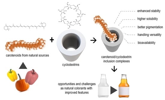
Encapsulation of Carotenoids as Food Colorants via Formation of Cyclodextrin Inclusion Complexes: A Review

by
Carlos A. Fuenmayor
1,*,
Omar G. Baron-Cangrejo
2 and
Paula A. Salgado-Rivera
3
1
Institute of Food Science and Technology (ICTA), Universidad Nacional de Colombia, Bogotá 111321, Colombia
2
Food Chemistry Research Group, Department of Chemistry, Universidad Nacional de Colombia, Bogotá 111321, Colombia
3
Department of Chemical and Environmental Engineering, Universidad Nacional de Colombia, Bogotá 111321, Colombia
*
Author to whom correspondence should be addressed.
Submission received: 2 May 2021 / Revised: 25 May 2021 / Accepted: 2 June 2021 / Published: 5 June 2021
(This article belongs to the Collection Current Opinion in Polysaccharides)

Abstract

:
The use of natural carotenoids as food colorants is an important trend of innovation in the industry due to their low toxicity, their potential as bio-functional ingredients, and the increasing demand for natural and organic foods. Despite these benefits, their inclusion in food matrices presents multiple challenges related to their low stability and low water solubility. The present review covers the main concepts and background of carotenoid inclusion complex formation in cyclodextrins as a strategy for their stabilization, and subsequent inclusion in food products as color additives. The review includes the key aspects of the molecular and physicochemical properties of cyclodextrins as complexing agents, and a detailed review of the published evidence on complex formation with natural carotenoids from different sources in cyclodextrins, comparing complex formation methodologies, recovery, inclusion efficiency, and instrumental characterization techniques. Moreover, process flow diagrams (PFD), based on the most promising carotenoid-cyclodextrin complex formation methodologies reported in literature, are proposed, and discussed as a potential tool for their future scale-up. This review shows that the inclusion of carotenoids in complexes with cyclodextrins constitutes a promising technology for the stabilization of these pigments, with possible advantages in terms of their stability in food matrices.

Graphical Abstract

1. Introduction

Pigments and dyes play an important role in the food industry as enhancers of homogeneity and stability of color and appearance of the products. The most widely used colorants in the industry are synthetic azo-compounds, classified as artificial colorants, due to their high solubility in water and chemical stability towards multiple stimuli. These compounds are generally recognized as safe, however, there are rising concerns due to evidence of their long-term intake toxicity [1,2,3,4], which ultimately has increased the distrust in their use by consumers. Pigment compounds naturally present in foodstuffs are an interesting alternative to artificial colorants, in virtue of their low toxicity in recommended doses, and the growing ‘clean-label’ consumption trend, as well as their potential nutritional value [5,6,7]. However, some of the most used natural colorants, such as carotenoids, have low water solubility and stability [8]. Different approaches have been proposed to solve these disadvantages, such as micro- and nano-encapsulation with host molecules, of which nano-encapsulation by formation of inclusion complexes has been highlighted as a straightforward and effective alternative, with possible benefits in terms of increased stability, solubility, and bioavailability of the guest compounds.
Most of the published evidence on the use of inclusion complexes as encapsulation systems relies on a model or proof-of-concept guest compounds, and the complex formation methodologies have been tested at laboratory or bench scale. Several authors have employed encapsulation with β-cyclodextrin since it is a molecule of great commercial interest, cheaper compared to its α and γ counterparts, and allows the encapsulation of hydrophobic molecules as it has a non-polar center due to the arrangement of the internal hydrogens of the truncated cone, while increasing the hydrophilicity of these molecules due to the conformation of the external hydrogens [9,10]. Another benefit of using this molecule in the food industry is its toxicity as it is considered a GRAS (generally recognized as safe) molecule by the FDA [11]. The use of these encapsulations in natural extracts has been of great interest in the food industry, since they generate great thermal, chemical and color stability in the product and permit exploring the use of natural ingredients as colorants to replace the controversial use of artificial colorants [9,12,13]. In order to enhance the commercial application of this encapsulation strategy for natural carotenoids in the food industry, attempts must be made to propose possible scaleups based on the published evidence on encapsulation of carotenoids, or carotenoid-rich extracts, via the formation of cyclodextrin inclusion complexes.
The present review covers the basic concepts and literature related to the use of cyclodextrin/carotenoid inclusion complexes formation as an encapsulation method for this type of natural pigments and sets a basis for future studies focused on the use of this strategy for the enhancement of the solubility and the stability of carotenoids from plant sources, under an industrial perspective.

2. Food Colorants: Artificial vs. Natural

In food, an additive is purposely added to an edible matrix to impart a desired characteristic. In the specific case of color, the addition of a coloring compound or substance is aimed at guaranteeing homogeneity and stability over time, or at intensifying or providing non-previously existing color to the matrix [14]. A definition of a colorant as an additive, proposed by the FDA is as follows: “a color additive is a dye, pigment, or other substance, which is capable of imparting color when added or applied to a food, medicine, cosmetic or human body” [15]. All over the world, there has been management in the food additives regulation, risks, and safety assessments, evaluating the exposition, analytical methods and the development general principles had been handled through the Joint FAO/WHI Committee on Food Additives (JECFA), and the regulations are implemented by the respective entities in each country, such as, FDA in the United States or EFSA in the European Union.
Most of the regulatory frameworks recognize two classification of food colorants as additives: natural and synthetic (also known as artificial) [16,17,18]. Natural colorants are extracted from nature by physical methods. When they are synthesized in the laboratory with a structure similar to their natural analogues and are chemically modified from natural colorants or fully synthetized in the laboratory, they are called synthetic.
The principal advantage of artificial colorants is their high solubility in aqueous medium. Being in their majority sodium salts or sulphonic acids of azo-derivatives, they have polar groups that allow water solubility, an important fact in the industry, due to the wide variety of existing food high in water. They can also be used as lacquers, making them insoluble in water or grease, working as a coverage. These colorants are divided into two main groups: allowed and not allowed [1]. The main problem of these colorants is the increasing concern about their toxicity. For example, the tartrazine is officially declared by the FDA to be allergenic [2]. Allura red has been studied by the EFSA as teratogenic in mice, which has led to a prohibition on its use in animal feed, as well as to reports of allergy and hives in humans [2,3]. Another problem of artificial colorants lies in their purification, because it is necessary to eliminate subsidiary colorants and residual non-coloring reagents from their chemical synthesis [19].
In contrast, several natural colorants have been part for a long period of the human diet, so their toxicity is low in the doses recommended by national and international organizations. This asset becomes advantageous for the market trend towards organic and healthy food, in which the use of solely natural ingredients generates security in the consumers of all ages [5,6,7].
These advantages offered by natural colorants, combined with the increase of bans that artificial colorants have faced in recent years, mainly in the United States, have led to the change of formulations in different countries [20], a trend of “clean label” began, which, although not regulated and does not have a standardized definition by organizations or legislation, refers to the use of additives of natural origin in foods, transparency in their origin, greener and cleaner processes, or simply less chemical substances [21,22]. Thus, the food industry is focusing on the development, design and improvement of formulations with natural additives that preserve and conserve properties in the same way as synthetic additives do, generating an innovative challenge for the industry [22].
Certain natural colorants can exert toxic or allergenic effects, although to a minor extent compared to their artificial counterparts. Some examples include cochineal extract (carminic acid), which has shown severe cases of allergy possibly attributed to protein hypersensitivity due to the glycoproteins remaining in the extraction [23]. Charcoal has polycyclic aromatic compounds and tar, which have shown a carcinogenic effect, being prohibited in the United States, and strongly regulated in the European Union [24]. Another study has shown that annatto extract generates hypersensitivity and hives in mice [25]. The main technological drawbacks for the use of natural colorants are their lower stability, higher cost compared to their artificial analogues, the need for extraction processes, and difficulties to reproduce the color due to variability in the extraction source [6].
Different studies have been carried out on the functionalization of different natural pigments to take advantage of the benefits that natural colorants offer, as well as their use as biomolecules to give additional food properties. For example, Enache et al. [26] formed co-microencapsulates using chitosan, inulin and soy protein isolates, encapsulating anthocyanins and Lactobacillus casei ssp. generating antioxidant activity and inhibition of α-amylase and α-glucosidase, with the aim of being released into the body for use in food. Milea et al. [27] performed an extraction of anthocyanins in the essential oil of black rice and lavender and generated a microencapsulation of this with a mixture of soy protein isolate, soy protein hydrolysates and casein, which they lyophilized and obtained α-glucosidase inhibitory activity, radical scavenging and release in the intestine by in vitro assays. Other studies show the functionalization of carotenoids in different matrices. Nogueira et al. [28], using soy protein encapsulation and lyophilization in order to protect carotenoids extracted from Phaffia rhodozyma, obtained an inclusion efficiency of 65% and a protection against temperature determined by DSC. Deng et al. [29] encapsulated β-carotenoids in soy protein isolate and octenylsuccinic anhydride-modified starch, obtaining a significant improvement in terms of stability at low temperatures and protection against sunlight, avoiding carotenoid degradation.
In this context, there is an ongoing search for new sources of natural pigments, as well as for technological strategies for their enhancement in terms of stability, solubility, pigmentation quality, and ease for inclusion in food matrices.

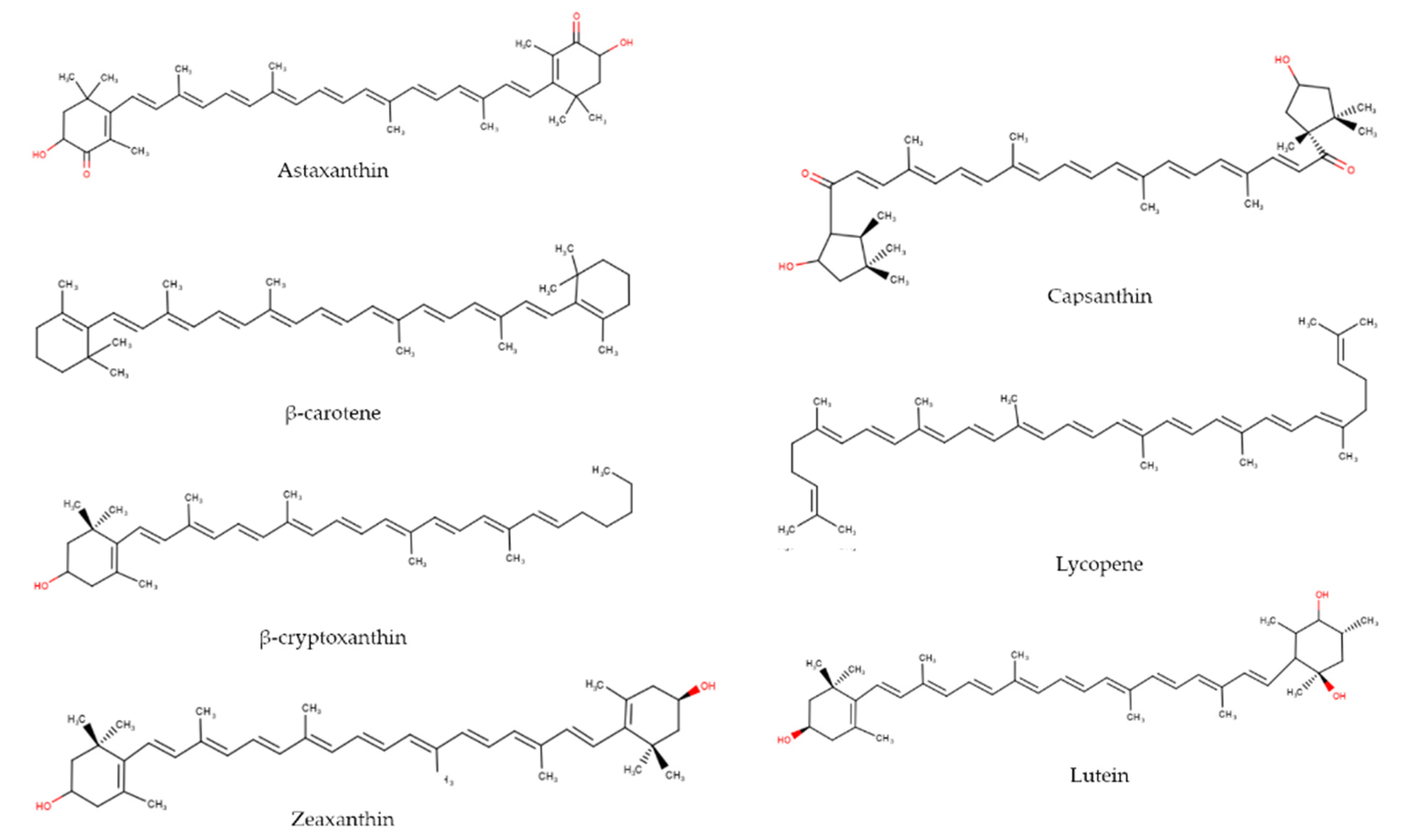
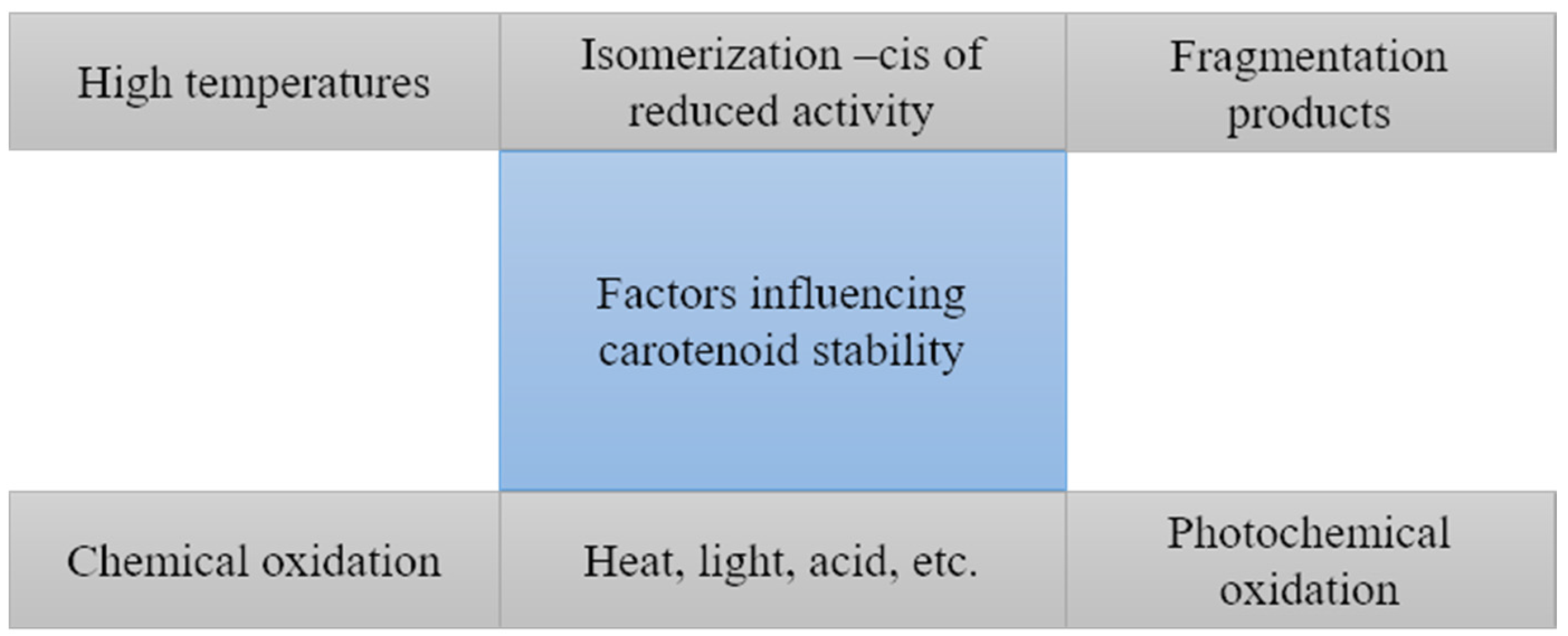
3. Carotenoids: Sources, Physicochemical Characteristics, and Stability

Carotenoids are natural pigments, generally composed of eight isoprenoid units; they are lipid compounds, and for that reason they are insoluble in water and soluble in nonpolar organic solvents. These compounds are synthesized by plants and some fungi and bacteria [8,30] and they have the capacity to absorb light between 400 and 500 nm [8]. They are generally classified as carotenes or xanthophylls according to the absence or presence of oxygen functional groups in their molecular structure, respectively.
Carotenoids are the main responsible of the red, yellow, or orange pigmentation of fruits, vegetables, flowers, fungi, birds, insects, crustaceans, among others [31], some of which acquire them via dietary intake of carotenoid-rich plants and microorganisms. Indeed, they are considered essential for their role in the photosynthesis process by helping to capture light, photo-protecting chlorophyll in the visible spectrum near to the UV and dissipating excess energy [32]. Their coloration is due to the oscillation of electrons in the unsaturated hydrocarbon chain [32]. Table 1 presents the general classification of carotenoids and their primary sources and is directly associate with Figure 1.
Carotenoids have been of interest in food science and nutrition, initially because of the pro-vitamin A activity that some of them exhibit, in particular β-carotene, and over time they have become more important due to their association with a variety of beneficial health properties, such as antioxidant activity [33]. About 700 varieties of carotenoids were characterized and but only around 20 have been detected in human tissues and bloodstream [34].
Table 1. Classification and sources of common carotenoids and their associated colors.
Classification Name SourcesAssociated Color
Caroteneβ-caroteneRed and yellow pepper, ñame, bee pollen, carrot, tomato, squash, ahuyama, spinach, pumpkin, guayaba, apricotRed, orange, yellow [35]
LycopeneCarrot, tomato, purple cabbageRed, orange [36]
XanthophyllsAstaxanthineFish, crustaceans, salmon, pomegranatePink-red [36]
β-criptoxanthinLemons, paprika, oranges, green pepper, papaya, squash, peach, pumpkinYellow-orange [35]
CanthaxantinMushrooms, crustaceans, troutOrange [36]
Capsanthin-capsorubinRed paprika, rice endospermYellow-orange [35]
LuteinPotato, carrot, corn, tomatoes, egg yolk, papaya, pumpkin, red and yellow pepperYellow [36]
ZeaxanthinPapaya, egg, orange, honey, pumpkinYellow [36]
Besides their insolubility in water, the use of carotenoids as food ingredients or additives can be limited by their promptness to isomerization and oxidation reactions, which is, on the other hand, part of their mechanism as antioxidants [37].
Carotenoids are relatively stable compounds while they are within the food matrix where they were synthesized; once extracted, they become highly labile, creating limitations for their use as ingredients, including the loss or decrease of their nutritional value, as well as undesired changes in their coloring features. Namely, carotenoids are sensitive to high temperatures and exposure to oxygen [8], which can induce either isomerization or oxidation reactions, as the main types of chemical instability.

3.1. Isomerization

An increase in temperature can provide the necessary energy that allows for geometric isomerization of both carotenes and xanthophylls, thus increasing the proportion of z-isomers. The 5,6-epoxides are not stable and undergo rapid isomerization to the furan form of 5,8-epoxide [38]. This type of isomerization is closely linked to the carotenoid structure, and it is induced by protonation. This process follows nine different mechanisms, including formal hemolytic or heterolytic addition of nucleophiles, radicals, or protons [39].

3.2. Oxidation

The oxidation is one of the main reactions that affect carotenoids, and it is favored by temperature, UV or visible light exposure, enzymes, and the presence of metals. It is counteracted by the presence of antioxidants, such as tocopherols or vitamin C. The mechanism of oxidation of carotenoids is not yet fully known, however, it is assumed that the process involves epoxidation reactions, apocarotenoids formation and hydroxylation, leading to lower molecular weight compound [32]. The oxidative reactions lead to the production of volatiles with a high stoichiometric influence, and different relations between cis or trans isomers and the volatile molecules produced. The oxidation can be also enzymatically catalyzed; when enzymes are present, they can be inactivated by rapid heat exposure, therefore, bleaching before cutting or grinding helps to avoid carotenoid losses [39]. Self-oxidation by oxygen results in a large and complex mixture of epoxides, aldehydes, ketones, peroxides, and other secondary reactions that produce short-chain carbonyl compounds, carbon dioxides and carboxylic acids [39,40].
Figure 2 summarizes the factors influencing carotenoid stability. Due to the chemical reactivity of carotenoids, alternatives have been sought to improve their stability, via encapsulation, from light, temperature, and oxygen exposure.

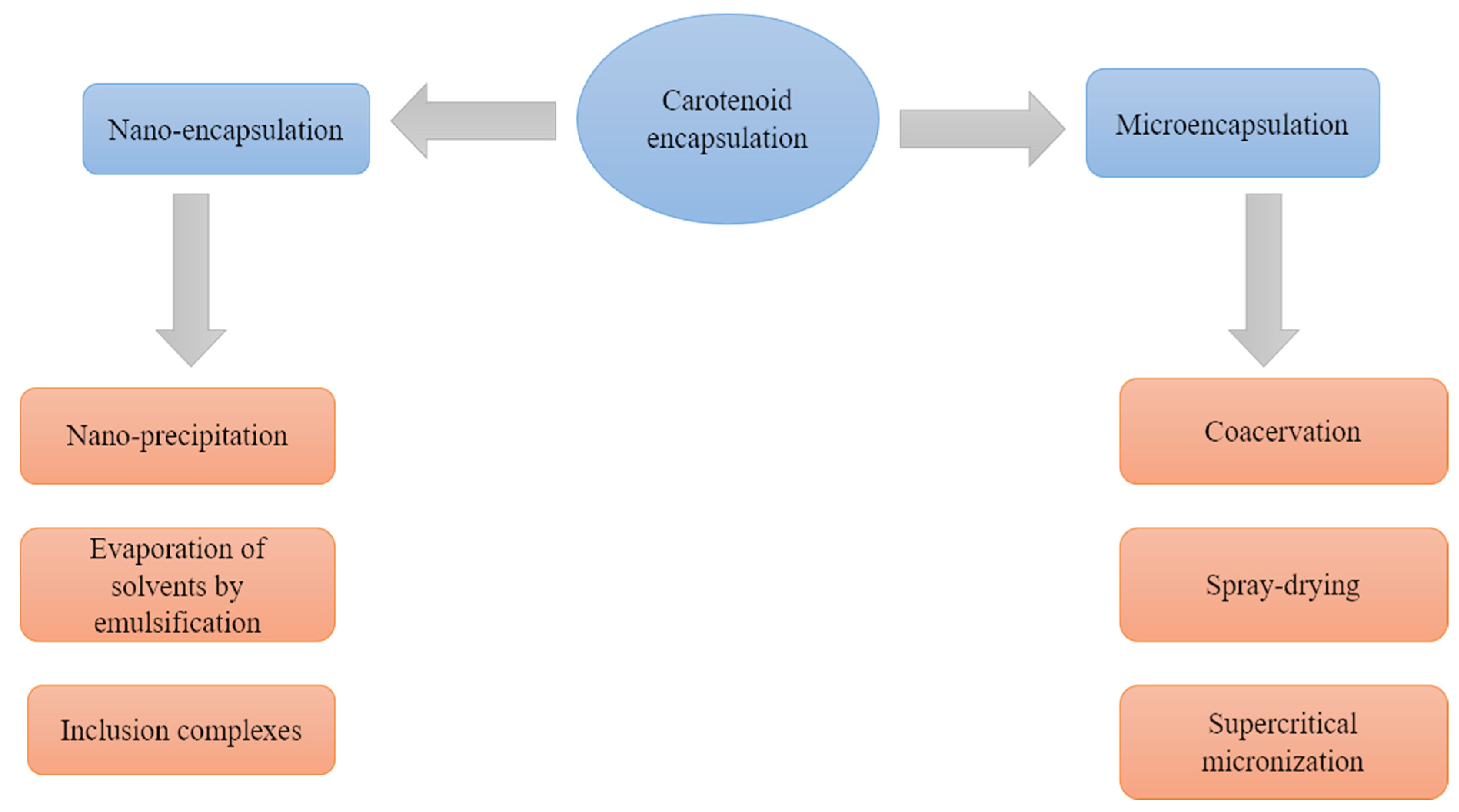
4. An Overview of Carotenoid Encapsulation Technologies

Encapsulating is an expanding technological strategy, which refers to the incorporation or dispersion of active components (cargo compounds) in the form of small vesicles, of micrometer or nanometer diameters in structures generally formed by polymers or oligomers that function as a vehicle. The encapsulation of bioactive compounds commonly seeks to improve their stability against degrading factors, e.g., electromagnetic radiation, high temperatures, presence of reactive chemical species, their solubility (especially in water) and their bioavailability when ingested [8,41].
For the encapsulation of carotenoids there are techniques of microencapsulation, which involve the formation of particles between 1 and 5000 μm, and nanoencapsulation, referring to particles between 10–1000 nm [42] (Figure 3). Such technologies have been the subject of multiple studies, and some key aspects, related specifically to carotenoids, are summarized below (Table 2); however, their in-deep discussion is outside the scope of the present review.

4.1. Microencapsulation

The process involves the coating of a solid, liquid, or gaseous substance with a coating material. The aim is to make the material non-reactive and to achieve long-term stability in structures larger than 1 μm [45] through various techniques, of which the principles and background of their use in carotenoid encapsulation are discussed below.

4.1.1. Coacervation

The technique involves liquid–liquid separation into a solution containing charged macro ions, producing a polymer-rich phase; the separation of the polymer from the supernatant is followed by deposition of the resulting coacervate containing the polymer around the active ingredient [8].
The cargo is dispersed in the solution, and the particle size is defined by the stirring speed, the shape of the stirrer, the surface tension and the viscosity. Coacervation starts with the change of the pH value by adding a strong acid, such as sulfuric or hydrochloric or organic acids. As a result, there is a reduction in the solubility of the dispersed phases. Depending on the number of polymers used, coacervation can be classified as simple (only one type pf polymers) and complex coacervation (two or more polymers) [50]. The use of this technique has been reported in palm oil with high carotenoid content [43], and in the encapsulation of β-carotene [44].

4.1.2. Spray-Drying

The objective of this technique is to increase the area of heat and mass transfer between a liquid that contains both the cargo and vehicle materials (usually a polymer or polymer combination) dispersed or dissolved in a solvent (usually water or an aqueous solution), and a drying gas. To carry out this technique, a series of steps must be considered. Initially, the main material to be encapsulated, filling or internal phase [57] is solubilized, dispersed or emulsified with the encapsulating agent in a solution, suspension or emulsion. It is homogenized and fed to the spray-dryer where it is atomized by a nozzle and a hot gas, generally air, so that the contact between drops and the drying gas occurs. After the evaporation of the water, the powder produced is separated by a cyclone and subsequently recovered [45].
The homogenization of the mixtures before spray formation and the drop size are key factors, and they can be controlled by speed, flow and air temperature, as well as by the nozzle design [45].
Despite carotenoid sensitivity towards air and high temperatures the technique has been used for carotenoid encapsulation such as β-carotene and lycopene [45], with great variation in recovery efficiencies, but there are no generalizable parameters for its application, which must be found experimentally, depending on the specific carotenoids [8].

4.1.3. Supercritical Micronization

This technique is used to avoid high temperatures that can affect the quality of carotenoids; carbon dioxide in supercritical conditions is commonly used as a solvent due to its low cost and ease of downstream removal, given by its low critical pressure and temperature (31.2 °C and 7.38 MPa), high volatility and diffusivity. The supercritical anti-solvent procedure seems to have the more suitable results for carotenoid encapsulation and also the higher level of carotenoid protection [45]. In this process, the solvent (CO2) is used as an anti-solvent because it does not dissolve the solute but is miscible with the solvent [58]. First, the cargo material is dissolved in an organic solvent to reach supercritical conditions required by the CO2 in a tank. Due to the reduction of solubility when de CO2 is added a local supersaturation conditions are created [58], as a result of this phenomena that take place between the supercritical phase of CO2 and the other solvents, precipitation of the active and the vehicle substances occurs in the form of crystals [45].
This method of supercritical anti-solvent has shown good yields or efficiency in the encapsulation of astaxanthin with an efficiency of 20%–74% using polymers as carriers in a small scale [46], also lutein with an efficiency of 90% using as carrier phosphatidylcholine [47] and in zeaxanthin with yields between 48%–74% [48].

4.1.4. Emulsification

Emulsion formation has generally been used for encapsulation of bioactives in aqueous solutions, which can be used directly in a liquid form, or can be dried to form powders after emulsification, for instance by freeze-drying or spray-drying techniques. The emulsion consists of two immiscible phases, where one of the liquids is dispersed in the form of small spheres in the other. In general, emulsions can be water in oil W/O or oil in water O/W [50].
The emulsions oil in water are considered to be a low-cost, efficient method to increase the stability and bioavailability of β-carotene [49]. Different types of carotenoids have improved their stability because of the use of oil water emulsions—some examples are lycopene [51] and β-carotene [52].

4.2. Nano-Encapsulation

Nano-encapsulation seeks to form structures or particles that function as nano-carriers, which implies that at least one of the characteristic dimensions of the capsules is smaller than 100 nm (or 1 µm in some approaches). One of the main advantages of these techniques over microencapsulation is that they provide larger surface areas, which in turn can improve dispersibility in aqueous media, as well as penetrability throughout epithelial tissues of the encapsulated compounds, thus potentially enhancing their inclusion in liquid matrices, as well as their bioavailability [8]. The most widely used nanoencapsulation methodologies that have been applied to carotenoids are briefly described below.

4.2.1. Nano-Precipitation

This technique refers to the precipitation of a polymer from an organic solvent and the subsequent diffusion into aqueous medium [53]. It is based on the spontaneous emulsification between an internal organic phase containing the dissolved polymer, the cargo compound and the organic solvent, and an external aqueous phase [42]. In the procedure, the polymer is in the organic phase, which is poured into the aqueous phase under slight magnetic agitation. When both phases are in contact, the solvent diffuses into the water carrying some polymer chains that are in solution, from the organic phase. Then, when the solvent diffuses into the water, the polymer chains are aggregated forming the nano-particles [53]. It should be noted that to improve the efficiency of the technique, it is usually complemented with freeze drying as the final step. The technique has been used to encapsulated β-carotene in polymers as ethylcellulose with results of a size particle of 60 ± 9 nm and encapsulation efficiency of 74 ± 2% and in zein with a size of 83 ± 8 nm and encapsulation efficiency of 93 ± 4% [54].

4.2.2. Solvent Evaporation after Emulsification

This technique involves two main stages: the emulsification of the polymer solution containing the cargo compound in an aqueous phase, and the evaporation of the solvent where the polymer is dissolved, inducing the precipitation of the polymer in the form of nano-spheres. In detail, an organic solution of the polymer containing the dissolved active component is dispersed and homogenized. Finally, the polymer precipitates in the form of nano-spheres in which the bioactive component is finely dispersed in the polymer matrix network [59]. Usually, polymers such as PLA or PLGA are used [42]. The size of the sphere is controlled by the stirring mode, the amount of dispersing agent, viscosity of the organic and aqueous phases, and temperature. This technique has been used to produce nanoemulsions where β-carotene has been encapsulated, the authors report a diameter from 9 to 280 nm [55].

4.2.3. Inclusion Complexes

Inclusion complexes are defined as the supramolecular association of a ligand (encapsulated bioactive) in the cavity of a substrate (cover material) using a hydrogen bond, Van der Waals forces or hydrophobic effect directed by entropy [56]. This technique is used to encapsulate volatile or small-molecule compounds, such as vitamins and essential oils, greatly improving their stability. Only a few components can be used as a substrate for this purpose, for example β-cyclodextrin and β-lactoglobulin. It gives the encapsulated substance high stability and provides high encapsulation yields.
This approach has been explored for the encapsulation of carotenoids; this review focuses on inclusion complexes formed with cyclodextrins, especially β-cyclodextrin, due to their larger commercial availability, and its proven ability to protect and increase solubility of carotenoids in aqueous matrices [9,12].

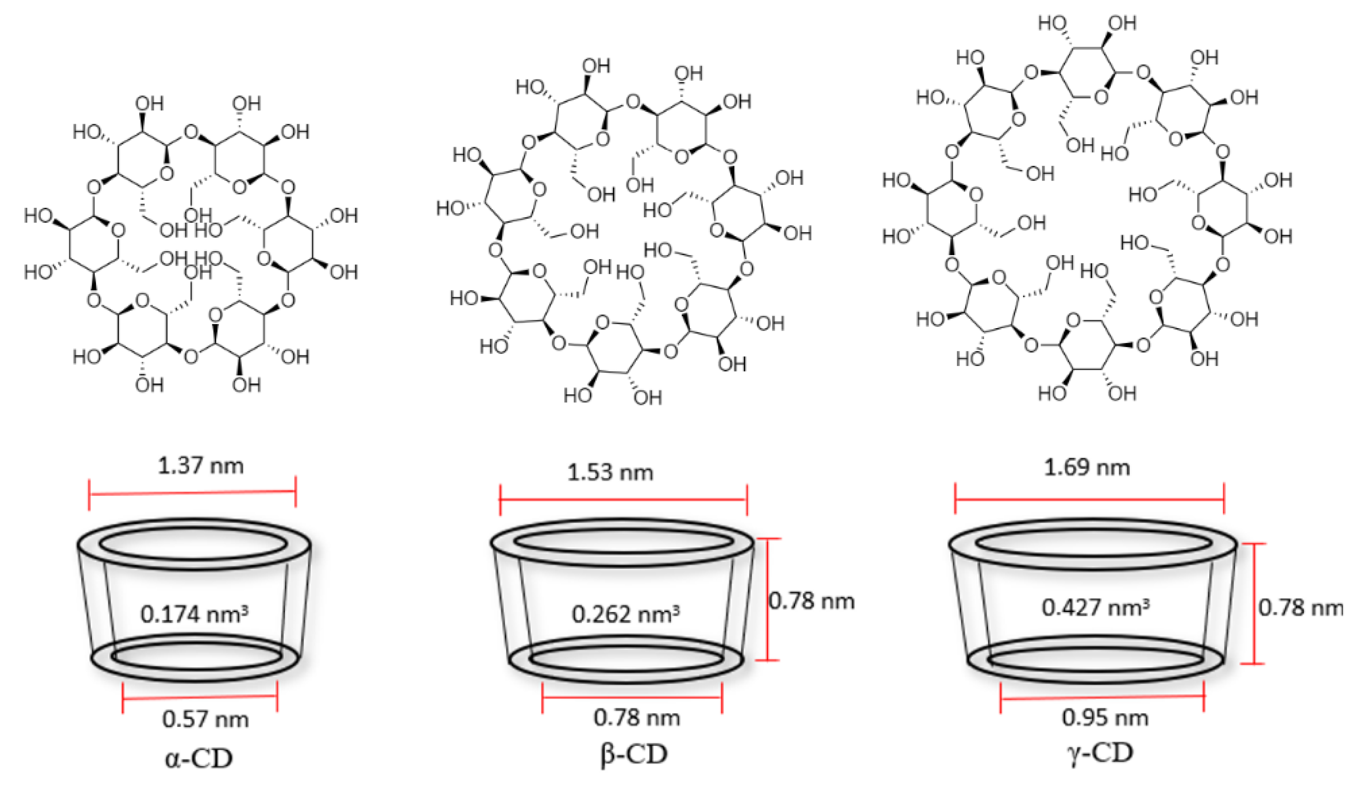
5. Cyclodextrins: Physicochemical Properties and Complex Formation Ability

Cyclodextrins are rings linked by α (1→4) of d-glucopyranosyl units, composed by 6, 7 or 8 carbons, being α-, β-, and γ-cyclodextrin those of greater commercial interest (Figure 4). Table 3 presents relevant physicochemical properties of the most common cyclodextrins. These compounds are produced on an industrial scale from starch by means of the enzyme cyclodextrin glycosyltransferase, obtained by different bacteria [60]. Its chemical synthesis has been reported, but it is quite complex for its industrial scaling. A selective synthesis of some of the three varieties can be made by precipitation of the bacterial growth media in specific organic solvents, e.g., toluene, decanol or butanone [61].
Table 3. Physical properties of cyclodextrins.
PropertiesCyclodextrinRef.
α-CDβ-CDγ-CD
Glucose units678-
Molecular formulaC36H60O30C42H70O35C48H80O45-
Molecular weight (g/mol)972.811351297.1-
Melting point (°C)278290–300No data[62]
Boiling point (°C)DiscomposesDiscomposesNo data[62]
Water solubility (25 °C) (g/100 mL)12.81.825.6[63,64]
Water solubility (45 °C) (g/100 mL)29.04.558.5[63,64]
Water solubility (60 °C) (g/100 mL)66.29.1129.2[63,64]
Internal volume (nm3)0.1740.2620.427[10]
Internal diameter (nm)0.570.780.95[65]
External diameter (nm)1.371.531.69[65]
Ring height (nm)No data0.780.78[65]
Figure 4. Chemical structure and cavity dimensions of cyclodextrins. α-, β-, and γ-cyclodextrin (α-CD, β-CD, and γ-CD) from left to right adapted from [10,65].
Polysaccharides 02 00028 g004
As observed in Figure 4, the cyclodextrin forms a truncated cone with a hydrophobic center and a hydrophilic exterior. This allows its hydrophobic center to generate weak interactions (Van der Waals forces) with nonpolar molecules that remain in its interior, while its exterior makes it more soluble or dispersible in an aqueous media, a feature that is of great interest for the protection of poorly water-soluble molecules with use in medicine, food or even cosmetics [9,10].
Inclusion complex formation takes advantage of the truncated cone shape of cyclodextrins to encapsulate molecules capable of occupying the same or a smaller volume of the cavity. This interaction turns the cyclodextrin into a host and the internal molecule into a guest, which interact by non-covalent molecular forces such as Van der Waals forces, or by hydrogen bridges. It is used extensively in the encapsulation of nonpolar molecules in order to increase their solubility and protect them from the environment, since cyclodextrins are not toxic to humans [9,10].
This interaction occurs through a dynamic balance between a recombination constant and a complex dissociation constant. As the guest molecule increases in size, ionizes or increases in polarity, the complex formation constant [10]:
C y c l o d e x t r i n   ( C D ) + M o l e c u l e   ( M ) K D K R C D M ; K = K R K D
where K is the formation constant in equilibrium, K R is the formation constant of the complex and K D corresponds to the dissociation constant of the complex.
Among cyclodextrins, β-cyclodextrin is the most widely reported molecule as vehicle for encapsulation in inclusion complexes of carotenoids and carotenoid-rich extracts, under different methodologies for its encapsulation from different, as discussed below (see Section 6 and Section 7).

6. Characterization Methods for Inclusion Complexes

The physicochemical assessment of cyclodextrin inclusion complexes is a key aspect of the use of this technique as an encapsulation strategy for carotenoids. The main instrumental techniques that can be used to study the physical and chemical features of cyclodextrin inclusion complexes are briefly described below.

6.1. Fourier Transform Infrared Spectroscopy (FTIR)

This spectroscopic technique employs wavelengths in the medium infrared to obtain the characteristic molecular vibration spectra. It is not a quantitative technique, but it offers an approach to the formation of the inclusion complex by comparing the IR spectra of the guest molecule, the cyclodextrin, and the complex formed. In the latter, both the characteristic bands of cyclodextrin should be observed, being the O–H stretching wide band by the inter- and intramolecular hydrogen bridges formed in the region of 3500 cm−1, the aliphatic C–H stretching bands in 2900 cm−1, and the stretching of the C–O and C–C bonding bands between 1200 to 1000 cm−1.
The infrared spectrum of the guest carotenoid will depend on its nature, but molecules that distinguish with functional groups in clean zones of the cyclodextrin spectrum will be preferred, for example, the C=O stretch is in the range of 1650 to 1800 cm−1, which is a clean zone in the cyclodextrin spectrum. Changes have also been reported in the intensity and width of the O–H stretch bands of cyclodextrin compared to the inclusion complex due to the formation of hydrogen bonds with the host molecule [9,12].

6.2. Differential Scanning Calorimetry (DSC)

Using this technique, changes in matter can be analyzed as a function of temperature, such as fusion, boiling, decomposition, oxidation, glass transition, and crystallization, among others. These will be of endothermic or exothermic character according to the reference material used, which requires a well-defined calorific capacity. As in FTIR, this technique seeks to compare the characteristic peaks of degradation in cyclodextrin (which is a fairly high peak close to 300 °C) and the peaks of degradation, fusion, boiling or oxidation in the guest molecule (in this case the carotenoids), which will be at a lower temperature. If this peak appears at a higher temperature in the inclusion complex formed, it indicates that the host has increased its thermal stability [9]. For instance, an increase in the exothermic degradation peak of a red pepper extract was observed from a range of 115 °C to 160 °C, to a range of 300 °C to 320 °C, where degradation of both β-cyclodextrin and carotenoid-rich extract occurred in the inclusion complex formed, exhibiting thermal protection [12]. In addition, a DSC allows the observation of melting enthalpies or evaporation of remaining compounds in the molecules such as water.

6.3. Raman Spectroscopy

Raman spectroscopy gives a good indication of the formation of an inclusion complex by the presence of the host molecule in the cyclodextrin cavity as long as the host molecule has Raman dispersion [66]. In Raman spectroscopy, characteristic vibration bands are observed, similarly as in infrared spectroscopy. In the case of carotenoids, the main vibration modes are observed between 900 and 1600 cm−1, corresponding to a C=C stretch (ν1 near 1500 cm−1), an asymmetric angular deformation in the C=C–H plane (ν2 near 1150 cm−1) and a C–CH3 deformation (ρ3 near 1000 cm−1). These Raman displacements will change according to the carotenoid molecule as guest [67]. Raman spectra between the host molecules and the cyclodextrin inclusion complex are compared, observing a change both the intensities of ν1, ν2 and ρ3 bands, and their chemical displacement due to the matrix effect attributed to the positioning of the guest in the cyclodextrin cavity. De Oliveira et al. (2011) report a change in the pronunciation of the more intense band ν1, being greater as the inclusion is more effective. Performing the experiment with different carotenoids, being astaxanthin (AST), β-carotene (BCT), lycopene (LYC) and norbixin (NOR), the authors found that those with a less voluminous group at their extremes (LYC and NOR) generated a deeper inclusion with the β-cyclodextrin cavity through a theoretical and experimental Raman study.

6.4. Proton Nuclear Magnetic Resonance (1H-NMR)

This technique allows to observe the formation of the inclusion complex through the analysis of the change in the chemical displacement that is generated due to the interaction between the internal hydrogens of cyclodextrin and those of the guest molecule. Ficarra et al. (2000) conducted a 1H-NMR study between two types of drugs β-blockers and β-cyclodextrin, using a 300 MHz Gemini Variation Spectrophotometer in D2O, finding that the delta chemical displacements between the complex and β-cyclodextrin (defined as Δδ = δcomplexδfree) were negative, which suggests a protection of the protonic nucleus by the electrons of the guest molecule, displacing these internal protons of the β-cyclodextrin in the inclusion complex at low frequencies.
This NMR behavior also occurs in the inclusion complex between β-caroten-8’-oic acid and β-cyclodextrin performed by Polyakov et al. on Bruker AM-360 and AM-500 NMR spectrometers (360 and 500 MHz operating frequency) [68]. Gomes et al [12] prepared an inclusion complex of carotenoids from an extract of red pepper and β-cyclodextrin, while Petito et al [13] carried out another inclusion among carotenoids from a yellow pepper extract with 2-hydroxypropyl-β-cyclodextrin; both investigations, using a Varian VNMRS equipment at 500 MHz in D2O, observed a displacement at low frequencies in the inclusion complex compared with the free β-cyclodextrin, but with a very low Δδ, arguably due to a low coupling constant between the guest and the internal protons in the cavity of the β-cyclodextrin, as it was described by Wimmer and collaborators previously [69], which suggests that the 1H-NMR technique commonly used in the laboratory allows us to know both the formation of the complex and stoichiometry as long as the coupling constants between the host and the cyclodextrin are high, otherwise it does not allow a conclusive result and the use of other techniques is necessary.

6.5. Scanning Electron Microscopy (SEM)

Through this qualitative technique, it is possible to know the morphology of the inclusion complexes obtained by different strategies. Although it does not allow us to know precisely the formation of the inclusion complex, it enables us to generate a differentiation between its crystalline structure compared to its precursors [70,71,72]. Ribeiro et al. (2008) carried out the inclusion of miconazole (MCZ) in methyl-β-cyclodextrin (MβCD) in a JEOL JSM-5310 electronic microscope, Tokyo, Japan, and found that the crystalline structure of the MCZ corresponded to a regular parallelogram, while the structure of the MβCD was composed of spherical particles; these authors showed that in some experiments with different inclusion strategies (co-evaporation, spray-drying and freeze-drying) no differentiation occurred, while by kneading a new solid phase and decrease in particle size was obtained. Wang et al. (2019) observed the inclusion of a complex between lycopene and β-cyclodextrin using an SEM electronic microscope, SU8020, Hitachi, Japan, and found that while lycopene aggregates corresponded to irregular block-shaped particles, and β-cyclodextrin aggregates corresponded to ellipsoidal crystalline shapes of different sizes, those of the corresponding inclusion complex showed a compact structure, different from its precursors both in shape and size.

6.6. X-ray Diffraction (XRD)

Through this technique it is possible to know the crystallinity of the precursors and compare them with the changes of crystallinity in the inclusion complex [69,71]. When both precursors are crystalline, an overlap of the peaks in the inclusion complex could be observed, as reported by Ficarra et al. (2000) in a study of β-blockers at β-cyclodextrin using a Philips PW 1050/25 diffractometer with Cu radiation Kα (λ = 1.5418 Å). Furthermore, these authors also reported that, although the precursors presented crystallinity, the inclusion product changed to an amorphous structure. This behavior was also observed by Ribeiro et al. (2008) in the study of the inclusion of miconazole (MCZ) with methyl-β-cyclodextrin (MβCD) using a Philips X’Pert diffractometer system, with Bragg-Bentano geometry and a cobalt X-ray source, finding that the MCZ has a crystalline structure, the MβCD has an amorphous structure and the inclusion complex obtained has a crystalline structure with a different X-ray pattern than the MCZ, which was attributed to the promotion of a crystalline structure through the inclusion strategy.

7. Previous Studies of Encapsulation of Carotenoids by Formation of Inclusion Complexes in Cyclodextrins

In order to review the background of the use of inclusion in complexes of β-cyclodextrin as a technique for the encapsulation of carotenoids, a synthesis of the inclusion methodologies, recovery, inclusion efficiency and instrumental characterization techniques, as reported by different authors, is presented in Table 4. The methodologies for the preparation of carotenoid/cyclodextrin complexes can be classified in three main types: ultrasound-assisted, mixture with mechanical stirring, and physical mixture. In general, the three of them, although variable, present high inclusion efficiencies and yields.
As shown in Table 4, the extract:cyclodextrin ratios vary between 1:2 and 1:10, except for one study [66] that uses excess cyclodextrin to ensure complete encapsulation. The ratio most commonly reported as optimal is 1:4, which offers the highest recoveries (mass of complex recovered/mass of initial material*100) and inclusion efficiencies (measured extract content/theoretical extract content *100, assessed by UV-vis spectrophotometry); likewise, Lobo et al. [9] reported that with lower ratios, it is possible to obtain the same pigmentation power as with higher ratios, suggesting to avoid excesses of cyclodextrin.
Recovery rates have a wide range of variation, from 40.79% ± 2.76% to 96.9%. When the reaction is performed with 2-hydroxypropyl-β-cyclodextrin using the ultrasound-assisted preparation methodology, recoveries from 90.75% ± 1.24 to 93.03% ± 1.99 were obtained. On the other hand, the inclusion efficiency reports have a range of variation between 37% and 81.87% ± 2.44%. For the case of the 1:4 ratio, according to Gomes et al. [12] and Lobo et al. [9], a recovery percentage of 40.79% ± 2.76% and 63% ± 4%, respectively, was obtained, and for the inclusion efficiencies of 52.95% ± 9.39% and 73% ± 8% were reported.
In addition to the characterization techniques previously described [9,12], the stability of the complexes has been evaluated by tristimulus colorimetry, using CIELAB space (L*, a* and b* coordinates), through the observation of the loss of color over time of the synthesized complexes: in the cases in which carotenoids were used, the authors reported an improvement in color stability over time. In addition, Lobo et al. [9], and De Oliveira et al. [66], confirmed an improvement in the solubility of the carotenoids used through the formation of inclusion complexes.
According to Durante et al. [73], the highest oil-dispersion for tomato oil supercritical extract (TO) was achieved with β-cyclodextrin, significantly improving lycopene stability and antioxidant activity.
This information provides a solid ground for the use of cyclodextrins, in particular β-cyclodextrin, to nanoencapsulate carotenoids to be used as coloring additives and bioactive ingredients in food matrices.

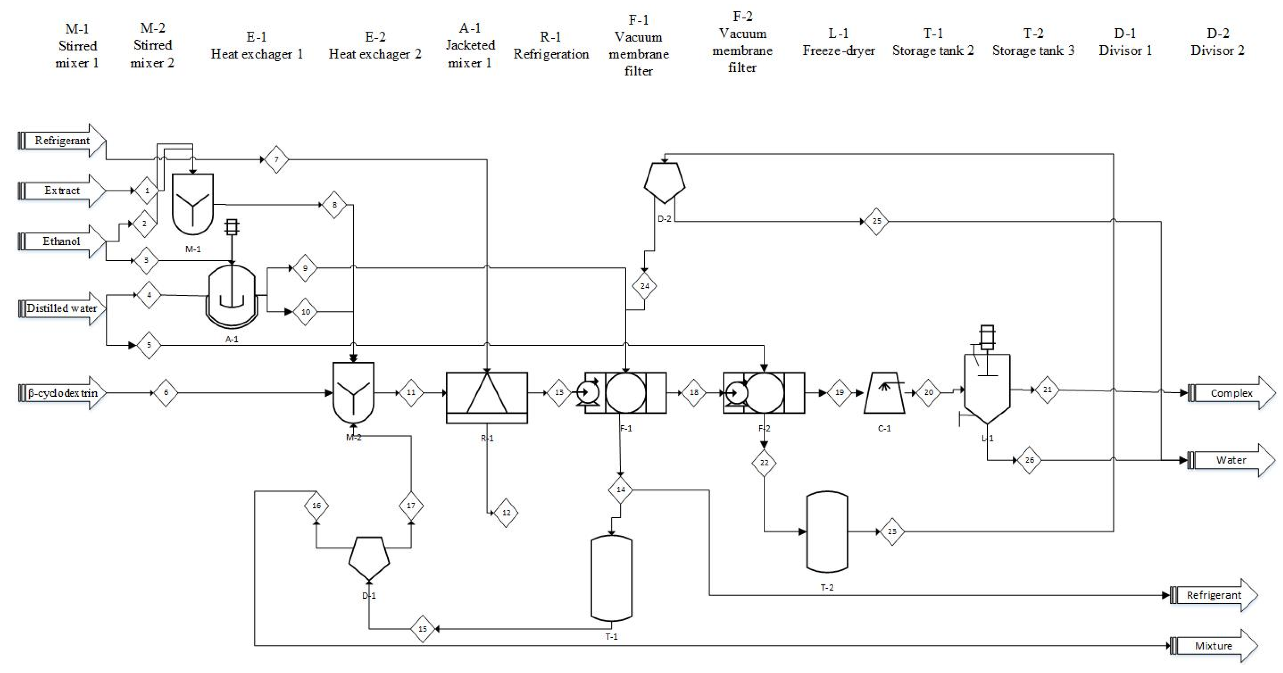
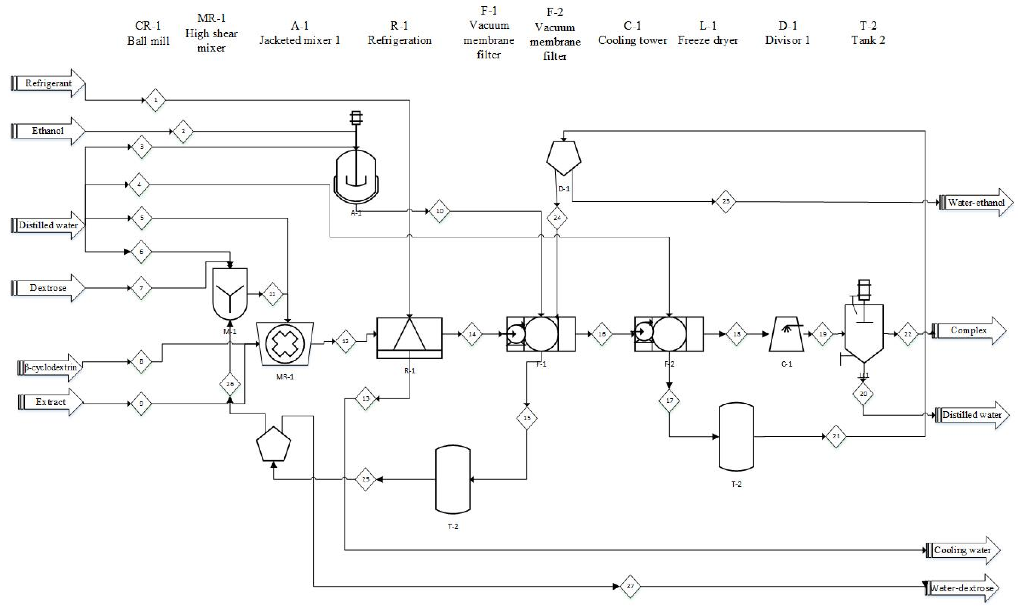
8. Carotenoid/Cyclodextrin Inclusion Complex Manufacturing: A Preliminary Proposal for Industrial Scale-Up

There is an information gap regarding the scale-up of the preparation of inclusion complexes of carotenoids with cyclodextrins at pilot or industrial levels, with further studies needed to validate the feasibility and performance of the different processing stages regarding cost-efficiency with larger processing volumes. Indeed, there is a lack of certainty about whether there is currently commercial manufacturing of carotenoid-based food colorants by means of cyclodextrin complexation. Fully based on the literature review presented above, two process flow diagrams have been proposed, with preliminary mass balances, as a contribution to future studies, according to the most promising preparation methodologies reported.

8.1. Method 1: Co-Precipitation

The process schematized in Figure 5 is mainly based on the preparation methodology and results of Gomes et al. [12] at 1:4 extract:cyclodextrin ratio. Initially, the carotenoid, or carotenoid-rich extract (e.g., paprika), enters through Stream 1 to be dissolved or disperse in azeotropic ethanol, in the mixer, M-1. Parallelly, azeotropic ethanol and water are mixed at a 1:2 v/v ratio in the mixer A-1, which must be jacketed for cooling. The output current A-1 is split into Streams 9 and 10. Stream 10 takes fresh ethanol-water mixture fed to stirred tank M-2, where it is mixed with β-cyclodextrin (Stream 6) and the ethanol-dissolved/dispersed extract (Stream 8) from M-1. The mass ratios of the M-2 feed (Streams 8, 10, and 6) are presented in the mass balance (Table 5). These ratios can vary if Stream 17 is not considered, which is the recirculated proportion of Stream 14, a downstream current of the first permeate of filter F-1 that contains carotenoids and cyclodextrin.
The resulting dispersion (Stream 11) is kept at 10 °C for 12–18 h in the refrigeration chamber R-1. Under these conditions, the formation and precipitation of carotenoid/cyclodextrin inclusion complexes should be thermodynamically favored, and the refrigeration step is expected to maximize the encapsulation efficiency. However, this unit operation leads to an intrinsically batch process, and refrigeration time and temperature must be optimized to enhance the energetic sustainability of the whole manufacturing process.
The recovery of the precipitated complexes from the output of R-1 (Stream 13) is carried out in a three-step filtration process at filter F-1. First, Stream 13 is membrane-filtered (e.g., using cellulose acetate membranes with 0.2 µm nominal pore size), which can be carried out using either dead-end or crossflow filtration. A first permeate (Stream 14) corresponds to the ethanol-water mixture containing the carotenoids and β-cyclodextrin that were not retained in the precipitate. As mentioned above, this permeate should be partially recycled as a feed to M-2 (Stream 17) in a proportion that has be determined at the operating conditions on the basis of its cyclodextrin and carotenoid content; according to the results of Gomes et al. [12] by studying the complexation of carotenoids from red bell pepper extracts, considering a powder recovery of approximately 50% (recovered powder (complex)/initial dry matter (red bell pepper extract + β-cyclodextrin), the estimated proportion of recirculation of permeate could be up to approximately 65–70%, allowing to reduce the quantity of fresh raw materials in the process. In a second filtration step, the precipitate (or retentate) is re-suspended with an ethanol-water solution (a mixture of the fresh Stream 8 and the recirculated Stream 23), and filtered, to ensure that non-complexed carotenoids and excess cyclodextrin are washed out from the precipitate. The second permeate (Stream 21), is stored to recover ethanol by conventional distillation, or, depending on its purity, to be partially recirculated as the Stream 23. Finally, in a third step, the precipitate (or retentate) is re-suspended with a water, and filtered, to remove remaining ethanol. A crossflow membrane filtration process design, with subsequent ethanol-water washing solution feeds, can be implemented to reduce this to a single-step unit operation.
The precipitate or retentate obtained (Stream 18), is frozen in the cooling tower C-1, and subsequently subjected to a freeze-drying process (L-1), in which the moisture content of the mixture is reduced, and the complex is obtained in the form of a powder. This powder can be subsequently ground to obtain a suitable granulometry according to its use.
In this process flow diagram design, the use of a stirred tank is proposed (M-2). A higher performance should be obtained, in terms of encapsulation efficiency and recovery, if this step is substituted by an ultrasound-assisted homogenization process [12]; however, the feasibility of implementing such unit operation can be limited, since the design of ultrasonic equipment for industrial purposes remains challenging.
The recirculation is carried out to reduce the consumption of the ethanol–water mixture, which leads to require only the total fresh amount of ethanol-water for mixing and washing at the start of the process. The material balances show the importance of recovering the solvents as well as the β-cyclodextrin, highlighting that the profitability of the process depends on the implementation of the recycling.
The calculations made for the mass balance by stream presented in Table 5 were made using a calculation base of 1 kg of extract: also, the recovery efficiency of the extract given by [12] of 53%, was used. To implement sustainable co-precipitation process at industrial scales, further studies are required to characterize the recovery of carotenoids, as well as the sources and mechanisms of losses.

8.2. Method 2: Kneading

Figure 6 is mainly based on the results of Lobo et al. [9], in which the wet milling/kneading is proposed as an alternative for obtaining the complex of the β-cyclodextrin extract using a similar ratio, 1:4 extract:cyclodextrin, than the method present above. Initially, a wet milling is done in a high shear mixer (MR1). Currents 5, 8, 9, and 11 enter to this equipment, which allows for obtaining a semi-solid paste formed by the β-cyclodextrin, distilled water, ethanol, and dextrose. In parallel, in A-1, a mixture between ethanol and distilled water at 1:2 v/v proportion is prepared, giving as a result stream 10 that will be used to wash the precipitate in F-1.
After the wet milling, the paste mixture enters Stream 11 to a 24 h refrigeration process (around 4 °C), and, afterwards, a vacuum filtration process, using a 2 µm membrane in F-1. The filtrate (Stream 13) will contain some β-cyclodextrin, ethanol, water and dextrose that might be recirculated. Stream 14 will contain most of the β-cyclodextrin inclusion complex with the extract, as well as remaining ethanol and dextrose, which can be washed off with water in F-2. In addition, water in Stream 17 can be recirculated, to make the process more efficient. However, the convenience of these recirculations need to be validated experimentally. Finally, Stream 18 enters a cooler where the complex is frozen and finally lyophilized.
As in the co-precipitation method, in this case, a calculation base of 1 kg was used for the mass balances, and a 67% recovery efficiency of the extract as reported by [9]; the same recovery percentage is assumed for the β-cyclodextrin. The mass balance of the process is shown in Table 6.
Both processes could be carried out at an industrial level, but from the preliminary mass balances, kneading seems to be a more sustainable option in terms of water consumption; however, these process flow diagrams fully are based on the reported works, and therefore, there is a need of pilot-scale studies to carry out detail designs and accurate mass balances.

9. Conclusions

Inclusion complex formation with cyclodextrins is a suitable strategy for enhancing the use of carotenoids as natural food colorants. The incorporation of carotenoids in these supramolecular systems, either isolated or in carotenoid-rich extracts, allows for tackling some of the drawbacks related to their use as secondary ingredients, i.e., poor chemical and sensory stability, low water-solubility, and high viscosity. Co-precipitation and kneading, also known as physical mixture or wet milling, are promising preparation strategies, however, the latter implies much less water consumption, and therefore could be a more sustainable way of processing. There is a great variability regarding the preparation time of inclusion complexes, according to the reviewed literature, with faster methods of 5–15 min for ultrasound-assisted homogenization, and 20–60 min for physical mixing and kneading, and slower methods, such as five days or 24 h in physical mixing. However, mild reaction conditions are always used, such as temperatures between 35 °C and 40 °C for physical mixing, without the need for heat for ultrasound and kneading, as well as solvents with low or no toxicity (natural extracts, water, and ethanol). Limitations of these methods include the control of light and temperature that is necessary throughout the encapsulation to avoid degradation of carotenoids, reported by all authors, especially in ultrasound, arguably related to the effects of cavitation and temperature uprisals. The inclusion efficiencies reported range from less than 50% to around 80%. Carotenoids from natural sources, especially those obtained by supercritical fluid extraction, might be especially interesting as cargo compounds, due to the higher concentration in the extracts, and the possibility of using green solvents. Although there is a great variety of instrumental techniques for the characterization of carotenoid–cyclodextrin inclusion complexes, some physicochemical properties, in particular, their water solubility and dispersibility have been poorly described in the scientific literature.
As evidenced above, the encapsulation of carotenoids using cyclodextrin offer positive and promising results that are potentially applicable for obtaining novel coloring additives. Further research focused on the insights of the molecular and physicochemical characteristics of the complexes carotenoid, as well as on the efficiency, recovery, and mechanisms of losses during their formation, are of great scientific interest for their application in the industry of natural food additives, and functional ingredients.

Author Contributions

All the authors have contributed equally to the conceptualization, investigation, data curation, writing—original draft preparation, review, and editing. Supervision, and funding acquisition, C.A.F. All authors have read and agreed to the published version of the manuscript.

Funding

This review was developed as a part of the project “Spectroscopic Study and Profiling of Carotenoid Compounds of Food Matrices with High Agro-Industrial Potential for the Extraction and Microencapsulation of Yellow-Orange Pigments”, funded by the Colombian Ministry of Science through the call “Patrimonio Autónomo Fondo Nacional de Financiamiento para la Ciencia, la Tecnología y la Innovación Francisco José de Caldas“(Project Code FP44842-271-2018).

Institutional Review Board Statement

Not applicable.

Informed Consent Statement

Not applicable.

Data Availability Statement

Not applicable.

Acknowledgments

We are grateful to Universidad Nacional de Colombia, in particular to the Institute of Food Science and Technology (ICTA) for providing the space, equipment and computer resources.

Conflicts of Interest

The authors declare no conflict of interest.

References

  1. Zahra, N.; Fatima, Z.; Kalim, I.; Saeed, K. Identification of synthetic food dyes in beverages by thin layer chromatography. Pak. J. Food Sci. 2015, 25, 178–181. [Google Scholar]
  2. FDA. Electronic Code of Federal Regulations. Title 21, Chapter 1, Subchapter A, 2020. Available online: https://www.ecfr.gov/cgi-bin/text-idx?gp=&SID=9a60ccddf74a018d5771bc29f4c9134f&mc=true&tpl=/ecfrbrowse/Title21/21CIsubchapA.tpl (accessed on 5 April 2020).
  3. EFSA Panel on Additives and Products or Substances used in Animal Feed (FEEDAP). Scientific Opinion on the safety and efficacy of Allura Red AC (E 129) in feed for cats and dogs. EFSA J. 2012, 10, 2675. [Google Scholar] [CrossRef]
  4. European Food Safety Authority (EFSA). EFSA statement on the review of the risks related to the exposure to the food additive titanium dioxide (E 171) performed by the French Agency for Food, Environmental and Occupational Health and Safety (ANSES). EFSA J. 2019, 17, e05714. [Google Scholar] [CrossRef] [Green Version]
  5. Schoefs, B. Plant Pigments: Properties, Analysis, Degradation. Adv. Food Nutr. Res. 2005, 49, 41–91. [Google Scholar] [CrossRef]
  6. Hendry, G.A.F.; Houghton, J.D. (Eds.) Natural Food Colours. In Natural Food Colorants, 2nd ed.; Blackie Academic & Professional: Glasgow, UK, 1996; pp. 40–80. [Google Scholar]
  7. Solymosi, K.; Latruffe, N.; Morant-Manceau, A.; Schoefs, B. Food colour additives of natural origin. In Colour Additives for Foods and Beverages; Scotter, M.J., Ed.; Elsevier BV: Amsterdam, The Netherlands, 2015; pp. 3–34. [Google Scholar]
  8. Rehman, A.; Tong, Q.; Jafari, S.M.; Assadpour, E.; Shehzad, Q.; Aadil, R.M.; Iqbal, M.W.; Rashed, M.M.A.; Mushtaq, B.S.; Ashraf, W. Carotenoid-loaded nanocarriers: A comprehensive review. Adv. Colloid Interface Sci. 2020, 275, 102048. [Google Scholar] [CrossRef]
  9. Lobo, F.A.T.; Silvana, R.; Domingues, J.; Rodrigues, S.; Costa, V.; Falcão, D.; Araújo, K.G.D.L. Inclusion complexes of yellow bell pepper pigments with β-cyclodextrin: Preparation, characterisation and application as food natural colorant. J. Sci. Food Agric. 2018, 98, 2665–2671. [Google Scholar] [CrossRef]
  10. Astray, G.; Gonzalez-Barreiro, C.; Mejuto, J.C.; Rial-Otero, R.; Simal-Gándara, J. A review on the use of cyclodextrins in foods. Food Hydrocoll. 2009, 23, 1631–1640. [Google Scholar] [CrossRef]
  11. FDA. GRAS Notices. 2001. Available online: https://www.cfsanappsexternal.fda.gov/scripts/fdcc/?set=GRASNotices&id=74&sort=GRN_No&order=DESC&startrow=1&type=basic&search=cyclodextrin (accessed on 22 May 2021).
  12. Gomes, L.M.M.; Petito, N.; Costa, V.G.; Falcão, D.Q.; Araújo, K.G.D.L. Inclusion complexes of red bell pepper pigments with β-cyclodextrin: Preparation, characterisation and application as natural colorant in yogurt. Food Chem. 2014, 148, 428–436. [Google Scholar] [CrossRef] [PubMed]
  13. Petito, N.; Dias, D.D.S.; Costa, V.G.; Falcão, D.Q.; Araujo, K.G.D.L. Increasing solubility of red bell pepper carotenoids by complexation with 2-hydroxypropyl-β-cyclodextrin. Food Chem. 2016, 208, 124–131. [Google Scholar] [CrossRef] [PubMed]
  14. Sasaki, Y.F.; Kawaguchi, S.; Kamaya, A.; Ohshita, M.; Kabasawa, K.; Iwama, K.; Taniguchi, K.; Tsuda, S. The comet assay with 8 mouse organs: Results with 39 currently used food additives. Mutat. Res./Genet. Toxicol. Environ. Mutagen. 2002, 519, 103–119. [Google Scholar] [CrossRef]
  15. FDA. Food Ingredient & Packaging Terms. 2018. Available online: https://www.fda.gov/food/food-ingredients-packaging/food-ingredient-packaging-terms (accessed on 5 April 2020).
  16. Barrow, J.; Lipman, A.; Bailey, C. Color Additives History. Reprinted from Food Safety Magazine October/November 2003 Issue, 2003. Available online: https://www.fda.gov/industry/color-additives/color-additives-history#authors (accessed on 5 March 2021).
  17. European Food Safety Authority (EFSA). Food Colours. 2016. Available online: https://www.efsa.europa.eu/en/topics/topic/food-colours (accessed on 5 March 2021).
  18. Secretaria de Salud de los Estados Unidos Mexicanos. ACUERDO por el que se determinan los aditivos y coadyuvantes en alimentos, bebidas y suplementos alimenticios, su uso y disposiciones sanitarias (Continúa en la Cuarta Sección). DOF, 2006. Available online: https://dof.gob.mx/nota_detalle_popup.php?codigo=5259470 (accessed on 5 March 2021).
  19. FAO/WHO. All specifications monographs from the 1st to the 65th meeting (1956–2005). In Combined Compendium of Food Additive Specifications; FAO JECFA Monogr: Rome, Italy, 2006. [Google Scholar]
  20. Lehto, S.; Buchweitz, M.; Klimm, A.; Straßburger, R.; Bechtold, C.; Ulberth, F. Comparison of food colour regulations in the EU and the US: A review of current provisions. Food Addit. Contam. Part A 2017, 34, 335–355. [Google Scholar] [CrossRef] [Green Version]
  21. Asioli, D.; Aschemann-Witzel, J.; Caputo, V.; Vecchio, R.; Annunziata, A.; Næs, T.; Varela, P. Making sense of the “clean label” trends: A review of consumer food choice behavior and discussion of industry implications. Food Res. Int. 2017, 99, 58–71. [Google Scholar] [CrossRef]
  22. Nascimento, K.D.O.D.; Paes, S.D.N.D.; Augusta, I.M. A Review ’Clean Labeling’: Applications of Natural Ingredients in Bakery Products. J. Food Nutr. Res. 2018, 6, 285–294. [Google Scholar] [CrossRef]
  23. Gallen, C.; Pla, J. Allergie et intolérance aux additifs alimentaires. Rev. Fr. d’Allergol. 2013, 53, 9–18. [Google Scholar] [CrossRef]
  24. European Food Safety Authority (EFSA). Scientific Opinion on the re-evaluation of vegetable carbon (E 153) as a food additive. EFSA J. 2012, 10, 2592. [Google Scholar] [CrossRef]
  25. Auttachoat, W.; Germolec, D.R.; Smith, M.J.; White, K.L.; Guo, T.L. Contact sensitizing potential of annatto extract and its two primary color components, cis-bixin and norbixin, in female BALB/c mice. Food Chem. Toxicol. 2011, 49, 2638–2644. [Google Scholar] [CrossRef] [PubMed]
  26. Enache, I.M.; Vasile, A.M.; Enachi, E.; Barbu, V.; Stănciuc, N.; Vizireanu, C. Co-Microencapsulation of Anthocyanins from Cornelian Cherry Fruits and Lactic Acid Bacteria in Biopolymeric Matrices by Freeze-Drying: Evidences on Functional Properties and Applications in Food. Polymers 2020, 12, 906. [Google Scholar] [CrossRef] [PubMed] [Green Version]
  27. Milea, Ş.A.; Dima, C.V.; Enachi, E.; Dumitraşcu, L.; Barbu, V.; Bahrim, G.E.; Alexe, P.; Stănciuc, N. Combination of freeze drying and molecular inclusion techniques improves the bioaccessibility of microencapsulated anthocyanins from black rice (Oryza sativa L.) and lavender (Lavandula angustifolia L.) essential oils in a model food system. Int. J. Food Sci. Technol. 2020, 55, 3585–3594. [Google Scholar] [CrossRef]
  28. Nogueira, M.B.; Prestes, C.F.; Burkert, J.F.D.M. Microencapsulation by lyophilization of carotenoids produced by Phaffia rhodozyma with soy protein as the encapsulating agent. Food Sci. Technol. 2017, 37, 1–4. [Google Scholar] [CrossRef] [Green Version]
  29. Deng, X.-X.; Chen, Z.; Huang, Q.; Fu, X.; Tang, C.-H. Spray-drying microencapsulation of β-carotene by soy protein isolate and/or OSA-modified starch. J. Appl. Polym. Sci. 2014, 131, 40399. [Google Scholar] [CrossRef]
  30. Saini, R.K.; Keum, Y.-S. Carotenoid extraction methods: A review of recent developments. Food Chem. 2018, 240, 90–103. [Google Scholar] [CrossRef]
  31. Delgado-Vargas, F.; Jimenez, A.R.; Paredes-Lopez, O. Natural Pigments: Carotenoids, Anthocyanins, and Betalains—Characteristics, Biosynthesis, Processing, and Stability. Crit. Rev. Food Sci. Nutr. 2000, 40, 173–289. [Google Scholar] [CrossRef]
  32. Meléndez-Martínez, A.J.; Vicario, I.M.; Heredia, F.J. Pigmentos carotenoides: Consideraciones estructurales y fisicoquímicas. Arch. Latinoam. Nutr. 2007, 57, 109–117. [Google Scholar]
  33. Britton, G. Carotenoid research: History and new perspectives for chemistry in biological systems. Biochim. Biophys. Acta (BBA)-Mol. Cell Biol. Lipids 2020, 1865, 158699. [Google Scholar] [CrossRef]
  34. Wu, L.; Lin, D. Molecular Aspects of Carotenoid Metabolizing Enzymes and Implications for Ophthalmology. In Handbook of Nutrition, Diet, and the Eye, 2nd ed.; Preedy, V.R., Watson, R.R., Eds.; Elsevier BV: Amsterdam, The Netherlands, 2019; pp. 415–424. [Google Scholar]
  35. Khoo, H.-E.; Prasad, N.; Kong, K.-W.; Jiang, Y.; Ismail, A. Carotenoids and Their Isomers: Color Pigments in Fruits and Vegetables. Molecules 2011, 16, 1710–1738. [Google Scholar] [CrossRef] [PubMed]
  36. De Carvalho, C.C.C.R.; Caramujo, M.J. Carotenoids in Aquatic Ecosystems and Aquaculture: A Colorful Business with Implications for Human Health. Front. Mar. Sci. 2017, 4. [Google Scholar] [CrossRef] [Green Version]
  37. Eun, J.-B.; Maruf, A.; Das, P.R.; Nam, S.-H. A review of encapsulation of carotenoids using spray drying and freeze drying. Crit. Rev. Food Sci. Nutr. 2019, 60, 1–26. [Google Scholar] [CrossRef] [PubMed]
  38. Britton, G.L.; Khachik, F. Carotenoids in Food. Carotenoids 2009, 5, 45–66. [Google Scholar]
  39. Schieber, A.; Weber, F. Handbook on Natural Pigments in Food and Beverages, 1st ed.; Carle, R., Schweiggert, F., Eds.; Woodhead Publishing: Duxford, UK, 2016; pp. 101–123. [Google Scholar]
  40. Martin, H.D.; Ruck, C.; Schmidt, M.; Sell, S.; Beutner, S.; Mayer, B.; Walsh, R. Chemistry of carotenoid oxidation and free radical reactions. Pure Appl. Chem. 1999, 71, 2253–2262. [Google Scholar] [CrossRef]
  41. De Boer, F.Y.; Imhof, A.; Velikov, K.P. Encapsulation of colorants by natural polymers for food applications. Color. Technol. 2019, 135, 183–194. [Google Scholar] [CrossRef] [Green Version]
  42. Anandharamakrishnan, C. Techniques for Nanoencapsulation of Food Ingredients; Springer: New York, NY, USA, 2013. [Google Scholar]
  43. Jain, A.; Thakur, D.; Ghoshal, G.; Katare, O.; Shivhare, U. Characterization of microcapsulated β-carotene formed by complex coacervation using casein and gum tragacanth. Int. J. Biol. Macromol. 2016, 87, 101–113. [Google Scholar] [CrossRef] [PubMed]
  44. Rutz, J.K.; Borges, C.D.; Zambiazi, R.C.; Crizel-Cardozo, M.M.; Kuck, L.S.; Noreña, C.P. Microencapsulation of palm oil by complex coacervation for application in food systems. Food Chem. 2017, 220, 59–66. [Google Scholar] [CrossRef]
  45. Janiszewska-Turak, E. Carotenoids microencapsulation by spray drying method and supercritical micronization. Food Res. Int. 2017, 99, 891–901. [Google Scholar] [CrossRef]
  46. Mezzomo, N.; De Paz, E.; Maraschin, M.; Martín, Á.; Cocero, M.J.; Ferreira, S.R. Supercritical anti-solvent precipitation of carotenoid fraction from pink shrimp residue: Effect of operational conditions on encapsulation efficiency. J. Supercrit. Fluids 2012, 66, 342–349. [Google Scholar] [CrossRef]
  47. Xia, F.; Hu, D.; Jin, H.; Zhao, Y.; Liang, J. Preparation of lutein proliposomes by supercritical anti-solvent technique. Food Hydrocoll. 2012, 26, 456–463. [Google Scholar] [CrossRef]
  48. Lin, K.-L.; Chng, L.-M.; Lin, J.C.-T.; Hsu, S.-L.; Young, C.-C.; Shieh, C.-J.; Chang, C.-M.J. Effect of Anti-solvent Conditions on Low Density Supercritical Fluids Precipitation of Zeaxanthin Palmitates from Lycium barbarum Fruits. J. Supercrit. Fluids 2014, 87, 104–110. [Google Scholar] [CrossRef]
  49. Gul, K.; Tak, A.; Singh, A.K.; Singh, P.; Yousuf, B.; Wani, A.A. Chemistry, encapsulation, and health benefits of β-carotene-A review. Cogent Food Agric. 2015, 1, 1–12. [Google Scholar] [CrossRef]
  50. Özkan, G.; Bilek, S.E. Microencapsulation of Natural Food Colourants. Int. J. Nutr. Food Sci. 2014, 3, 145–156. [Google Scholar] [CrossRef]
  51. Bou, R.; Boon, C.; Kweku, A.; Hidalgo, D.; Decker, E.A. Effect of different antioxidants on lycopene degradation in oil-in-water emulsions. Eur. J. Lipid Sci. Technol. 2011, 113, 724–729. [Google Scholar] [CrossRef]
  52. Qian, C.; Decker, E.; Xiao, H.; McClements, D.J. Inhibition of β-carotene degradation in oil-in-water nanoemulsions: Influence of oil-soluble and water-soluble antioxidants. Food Chem. 2012, 135, 1036–1043. [Google Scholar] [CrossRef]
  53. Galindo-Rodriguez, S.; Allemann, E.; Fessi, H.; Doelker, E. Physicochemical Parameters Associated with Nanoparticle Formation in the Salting-Out, Emulsification-Diffusion, and Nanoprecipitation Methods. Pharm. Res. 2004, 21, 1428–1439. [Google Scholar] [CrossRef] [PubMed]
  54. Afonso, B.S.; Azevedo, A.G.; Gonçalves, C.; Amado, I.R.; Ferreira, E.C.; Pastrana, L.M.; Cerqueira, M.A. Bio-Based Nanoparticles as a Carrier of β-Carotene: Production, Characterisation and In Vitro Gastrointestinal Digestion. Molecules 2020, 25, 4497. [Google Scholar] [CrossRef]
  55. Silva, H.D.; Cerqueira, M.A.; Souza, B.W.; Ribeiro, C.; Avides, M.C.; Quintas, M.A.; Coimbra, J.S.; Carneiro-Da-Cunha, M.G.; Vicente, A.A. Nanoemulsions of β-carotene using a high-energy emulsification–evaporation technique. J. Food Eng. 2011, 102, 130–135. [Google Scholar] [CrossRef] [Green Version]
  56. Ezhilarasi, P.N.; Karthik, P.; Chhanwal, N.; Anandharamakrishnan, C. Nanoencapsulation Techniques for Food Bioactive Components: A Review. Food Bioprocess Technol. 2013, 6, 628–647. [Google Scholar] [CrossRef]
  57. Gonçalves, A.; Estevinho, B.N.; Rocha, F. Microencapsulation of vitamin A: A review. Trends Food Sci. Technol. 2016, 51, 76–87. [Google Scholar] [CrossRef] [Green Version]
  58. Ghosh, S.; Mishra, S.; Singh, T. Antisolvents in Perovskite Solar Cells: Importance, Issues, and Alternatives. Adv. Mater. Interfaces 2020, 7, 1–24. [Google Scholar] [CrossRef]
  59. Reis, C.P.; Neufeld, R.J.; Ribeiro, A.J.; Veiga, F. Nanoencapsulation I. Methods for preparation of drug-loaded polymeric nanoparticles. Nanomedicine Nanotechnol. Biol. Med. 2006, 2, 8–21. [Google Scholar] [CrossRef] [Green Version]
  60. Horikoshi, N.K.; Nakamura, N.; Matsuzawa, N.; Yamamoto, M. Industrial Production of Cyclodextrins. In Proceedings of the First International Symposium on Cyclodextrins, Budapest, Hungary, 30 September–2 October 1981; Szejtli, J., Ed.; Springer Science and Business Media: Berlin/Heidelberg, Germany, 1982; pp. 25–39. [Google Scholar]
  61. Hedges, A.R. Industrial Applications of Cyclodextrins. Chem. Rev. 1998, 98, 2035–2044. [Google Scholar] [CrossRef] [PubMed]
  62. Caligur, V. Cyclodextrins. BioFiles 2008, 3, 32. [Google Scholar]
  63. Wiedenhoff, N.; Lammers, J.N.J.J. Properties of cyclodextrins. Part II. Preparation of a stable β-cyclodextrin hydrate and determination of its water content and enthalpy of solution in water from 15–30°. Carbohydr. Res. 1968, 7, 1–6. [Google Scholar] [CrossRef]
  64. Jozwiakowski, M.J.; Connors, K.A. Aqueous solubility behavior of three cyclodextrins. Carbohydr. Res. 1985, 143, 51–59. [Google Scholar] [CrossRef]
  65. Gidwani, B.; Vyas, A. A Comprehensive Review on Cyclodextrin-Based Carriers for Delivery of Chemotherapeutic Cytotoxic Anticancer Drugs. BioMed Res. Int. 2015, 2015, 1–15. [Google Scholar] [CrossRef] [Green Version]
  66. De Oliveira, V.E.; Almeida, E.W.C.; Castro, H.V.; Edwards, H.G.M.; Dos Santos, H.F.; De Oliveira, L.F.C. Carotenoids and β-Cyclodextrin Inclusion Complexes: Raman Spectroscopy and Theoretical Investigation. J. Phys. Chem. A 2011, 115, 8511–8519. [Google Scholar] [CrossRef] [PubMed]
  67. Merlin, J.C. Resonance Raman spectroscopy of carotenoids and carotenoid-containing systems. Pure Appl. Chem. 1985, 57, 785–792. [Google Scholar] [CrossRef]
  68. Polyakov, N.E.; Leshina, T.V.; Konovalova, T.A.; Hand, E.O.; Kispert, L.D. Inclusion complexes of carotenoids with cyclodextrins: 1HNMR, EPR, and optical studies. Free. Radic. Biol. Med. 2004, 36, 872–880. [Google Scholar] [CrossRef] [PubMed]
  69. Wimmer, R.; Aachmann, F.L.; Larsen, K.L.; Petersen, S.B. NMR diffusion as a novel tool for measuring the association constant between cyclodextrin and guest molecules. Carbohydr. Res. 2002, 337, 841–849. [Google Scholar] [CrossRef]
  70. Ficarra, R.; Ficarra, P.; Di Bella, M.R.; Raneri, D.; Tommasini, S.; Calabrò, M.L.; Gamberini, M.C.; Rustichelli, C. Study of β-blockers/β-cyclodextrins inclusion complex by NMR, DSC, X-ray and SEM investigation. J. Pharm. Biomed. Anal. 2000, 23, 33–40. [Google Scholar] [CrossRef]
  71. Ribeiro, A.M.; Figueiras, A.; Santos, D.; Veiga, F. Preparation and Solid-State Characterization of Inclusion Complexes Formed Between Miconazole and Methyl-β-Cyclodextrin. AAPS PharmSciTech 2008, 9, 1102–1109. [Google Scholar] [CrossRef] [PubMed] [Green Version]
  72. Wang, H.; Wang, S.; Zhu, H.; Wang, S.; Xing, J. Inclusion Complexes of Lycopene and β-Cyclodextrin: Preparation, Characterization, Stability and Antioxidant Activity. Antioxidants 2019, 8, 314. [Google Scholar] [CrossRef] [Green Version]
  73. Durante, M.; Milano, F.; De Caroli, M.; Giotta, L.; Piro, G.; Mita, G.; Frigione, M.; Lenucci, M. Tomato Oil Encapsulation by α-, β-, and γ-Cyclodextrins: A Comparative Study on the Formation of Supramolecular Structures, Antioxidant Activity, and Carotenoid Stability. Foods 2020, 9, 1553. [Google Scholar] [CrossRef]
Figure 1. Chemical structures of common carotenoids used as natural pigments.
Polysaccharides 02 00028 g001
Figure 2. Factors influencing carotenoids stability.
Polysaccharides 02 00028 g002
Figure 3. Commonly used carotenoid encapsulation methods and techniques.
Polysaccharides 02 00028 g003
Figure 5. Process flow diagram of inclusion complex formation by co-precipitation with recycling.
Polysaccharides 02 00028 g005
Figure 6. Process flow diagram of inclusion complex formation by kneading with recycling.
Polysaccharides 02 00028 g006
Table 2. Summary of the main encapsulation technologies applied for encapsulation of carotenoids or carotenoid-rich extracts.
Encapsulation TechnologyAdvantagesDisadvantagesReported Applicability to Carotenoids
Micro-encapsulationCoacervationThe particle size can be engineered by stirring speed, shape of the stirrer, surface tension, and viscosity.The required change of pH involves the use of denaturing substances, such as sulfuric or hydrochloric or organic acids.This technique has been reported in palm oil with high carotenoid content [43], and in the encapsulation of β-carotene [44].
Spray-dryingCommon technology easily scalable. Particle size and key factors can be controlled by speed, flow and air temperature, as well as by the nozzle design [45]. The use of hot air which can be deleterious, considering carotenoid sensitivity towards air and high temperatures.The technique has been extensively used for carotenoid encapsulation, such as β-carotene and lycopene [45].
Supercritical micronizationThe technique does not require high temperatures that can affect the quality of the carotenoids. In addition, using supercritical CO2 as solvent guarantees low cost and ease of downstream removal [45].It needs the use of more than one solventThis method has been used to encapsulated astaxanthin [46], lutein [47] and zeaxanthin [48].
EmulsificationThe oil-in-water emulsions are consider to be a low-cost, efficient method to increase the stability and bioavailability of β-carotene [49]. Multiple strategies, either high-energy and low-energy, are available.Emulsions can be dried to form powders, but further processing is required, such as freeze-drying or spray-drying [50].Different types of carotenoids have improved their stability because of the use of oil water emulsions some examples are lycopene [51] and β-carotene [52].
Nano-encapsulationNano-precipitationRelatively low energy requirements. It is based on the spontaneous emulsification between an internal organic phase containing the dissolved polymer, the cargo compound and the organic solvent, and an external aqueous phase [42].To improve the efficiency of the technique, further processing is needed, usually freeze-drying [53].The technique has been used to encapsulated β-carotene in polymers as ethylcellulose [54]
Solvent evaporation after emulsificationThe size of the sphere is controlled by the stirring mode, the amount of dispersing agent, viscosity of the organic and aqueous phases, and temperature [42].The use of solvents can be an environmental problem.This technique has been used to produce nano-emulsions where β-carotene has been encapsulated [55].
Inclusion complexesThe method gives the encapsulated substance high stability and provides high encapsulation yields [12]. Only a few components can be used as a substrate for this purpose, for example cyclodextrins, and β-lactoglobulin [56].The technique has been used to encapsulated β-carotene, Lycopene, yellow and red pepper extract
Table 4. Encapsulation by inclusion complex formation with β-cyclodextrin and different guests, the inclusion methodologies, recovery, inclusion efficiency and instrumental characterization techniques reported by different authors.
Inclusion ComplexPreparation MethodologiesMass RatioRecovery
(%) *
Inclusion Efficiency
(%) **
Characterization TechniquesSummaryRef.
Red pepper extract and β-cyclodextrinCo-precipitation with ultrasound homogenization (100 W, 15 min).(1:4)54.5 ± 3.662.4 ± 12.9FT-IR
DSC
Particle size
Size distribution
Zeta potential
1H-NMR
Color stability
Greater performance and inclusion efficiency were obtained by using ultrasound over conventional (magnetic) stirring. The formation of the inclusion complex is demonstrated, with FT-IR and DSC better. High color stability in yogurt matrix was demonstrated.[12]
Co-precipitation with conventional (magnetic) stirring (up to 22 ± 1 °C, ~30 min).(1:4)40.8 ± 2.852.9 ± 9.4
Kneading(1:4)No dataNo data
Yellow pepper extract and β-cyclodextrinCo-precipitation with ultrasound homogenization (50 W, 15 min)(1:2)78 ± 157 ± 5FT-IR
DSC
Staining power
FT-IR and CRP techniques confirmed the formation of the inclusion complexes. Also, through the staining power it was decided that the best inclusion corresponded to 1:2. Finally, a high color stability was demonstrated in isotonic beverages.[9]
(1:4)63 ± 473 ± 8
(1:6)51 ± 375 ± 6
Kneading (50 min)(1:2)9637
Red pepper extract and 2-hydroxypropyl-β-cyclodextrinCo-precipitation with ultrasound homogenization (100 W, 5 min)(1:4)91.2 ± 1.781.9 ± 2.4FT-IR
DSC
DLS
1H-NMR
Solubility essay
It was observed that the amount of cyclodextrin did not affect the inclusion efficiency and high yields were obtained. The techniques of CSD, FT-IR, DLS and solubility test were the techniques that identified the formation of the inclusion complex. Finally, the use of 2-HPβCD increased water solubility by 660 times.[13]
(1:6)92.7 ± 1.275.2 ± 7.5
(1:8)90.7 ± 1.269.0 ± 3.3
(1:10)93.0 ± 2.069.7 ± 3.7
Kneading (20 min)(1:4) (1:6) (1:8) (1:10)No dataNo data
β-carotene (BCT) and β-cyclodextrinCo-precipitation with conventional (magnetic) stirring (5 days, N2 purge, covered with light)(1:360)No dataNo dataFT-Raman
Computational Quantum Model
The most intense band ν1 assigned to the stretching -C=C- was the most sensitive to change at the time of inclusion in Raman, and it was higher in non-bulky groups of the studied molecules, generating greater inclusion. BCT, LYC and AST presented favorable inclusion energies. The computational calculations agreed mostly with the experiments carried out.[66]
Lycopene (LYC) and β-cyclodextrin
Astaxanthin (AST) and β-cyclodextrin
Tomato oil and α-cyclodextrinCo-precipitation with stirring and nitrogen sparging (24 h)(10:19)No data61.5FTIR-ATR
DSC
SEM
LCSM
Antioxidant activity
The β-cyclodextrin complex showed the best dispersion in oil. α and β showed the best antioxidant activity as well as oil-dispersing agents and antioxidant carrier systems in aqueous media. A significant improvement in the stability of the lycopenes analyzed was shown by the three types of cyclodextrins.[73]
Tomato oil and β-cyclodextrin(10:23)No data62.4
Tomato oil and γ-cyclodextrin(10:26)No data44.0
FT-IR: Fourier transform infrared spectroscopy/ATR: attenuated total reflectance; DSC: differential scanning calorimetry; 1H-NMR: proton nuclear magnetic resonance; DLS: dynamic light scattering; FR-Raman: Raman spectroscopy with Fourier transform; XRD: X-ray diffraction; HPLC: high performance liquid chromatography; LCSM: laser confocal scanning microscopy; * recovery (%) = 100 [powder of the recovered complex (g)/(initial material (extract–β-cyclodextrin)) (g)]; ** inclusion efficiency (%) = 100 (measured extract content/theoretical extract content) usually with UV-Vis spectrophotometry at 451 nm. The mass ratio is indicated as the mass:mass proportion of carotenoid or carotenoid-rich extract to cyclodextrin.
Table 5. Mass balance of co-precipitation process with recycling.
Streams (kg)1234567891011121314151617181920212223242526
Distilled water---120.060.0---19.821.860.0-60.057.057.018.838.23.03.03.0-60.060.040.219.83.0
Extract1.0------0.7--1.0-1.00.50.50.20.30.50.50.50.5-----
Ethanol-1.060.0----1.024.914.331.0-31.023.423.47.715.77.6---7.67.65.12.5-
β-cyclodextrin-----2.7----4.0-4.01.91.90.61.30.82.12.12.1-----
Refrigerant------49.0----49.0--------------
Table 6. Mass balance of kneading process with recycling.
Streams (kg)123456789101112131415161718192021222324252627
Distilled water--60.060.04.03.3---24.019.023.0-23.078.44.660.04.64.64.660.0-24.036.078.415.762.7
Extract--------1.0--1.0-1.00.20.9-0.90.9--0.9--0.20.00.1
Carotenoids---------------------------
Ethanol-30.0-------26.4----24.06.06.0---6.0-2.43.624.04.819.2
β-cyclodextrin-------4.0---4.0-4.00.63.4-3.43.4--3.4--0.60.10.5
Dextrose55.3-----1.0---1.01.055.31.00.20.80.20.60.60.60.20.1-0.10.20.00.2
Publisher’s Note: MDPI stays neutral with regard to jurisdictional claims in published maps and institutional affiliations.

