
Oxi-HA/ADH Hydrogels: A Novel Approach in Tissue Engineering and Regenerative Medicine


Department of Engineering of Materials and Bioprocesses, School of Chemical Engineering, University of Campinas (UNICAMP), Campinas, São Paulo 13083-852, Brazil
*
Author to whom correspondence should be addressed.
Polysaccharides 2021, 2(2), 477-496; https://0-doi-org.brum.beds.ac.uk/10.3390/polysaccharides2020029
Submission received: 26 April 2021
/
Revised: 25 May 2021
/
Accepted: 2 June 2021
/
Published: 7 June 2021
(This article belongs to the Collection Current Opinion in Polysaccharides)
Abstract
:Hyaluronic acid (HA) is a natural polyelectrolyte abundant in mammalian connective tissues, such as cartilage and skin. Both endogenous and exogenous HA produced by fermentation have similar physicochemical, rheological, and biological properties, leading to medical and dermo-cosmetic products. Chemical modifications such as cross-linking or conjugation in target groups of the HA molecule improve its properties and in vivo stability, expanding its applications. Currently, HA-based scaffolds and matrices are of great interest in tissue engineering and regenerative medicine. However, the partial oxidation of the proximal hydroxyl groups in HA to electrophilic aldehydes mediated by periodate is still rarely investigated. The introduced aldehyde groups in the HA backbone allow spontaneous cross-linking with adipic dihydrazide (ADH), thermosensitivity, and noncytotoxicity to the hydrogels, which are advantageous for medical applications. This review provides an overview of the physicochemical properties of HA and its usual chemical modifications to better understand oxi-HA/ADH hydrogels, their functional properties modulated by the oxidation degree and ADH concentration, and the current clinical research. Finally, it discusses the development of biomaterials based on oxi-HA/ADH as a novel approach in tissue engineering and regenerative medicine.
1. Introduction
Hyaluronic acid (HA) is a glycosaminoglycan comprising repeating disaccharide units of glucuronic acid and N-acetylglucosamine that is widely distributed in the extracellular matrix (ECM) and plays a vital role in vertebrate tissue morphogenesis [1]. Isolated first in 1934 by Meyer and Palmer [2], HA has an evolutionary history supported by profound structural, physicochemical, and biological studies that resulted in an extensive network of clinical applications in the late 20th century. Laurent [3] summarized HA history research in the form of a tree, in which the great stages of development mark the trunk. The initial structural studies and evolution of analysis techniques boosted research on the macromolecular and physiological properties, followed by molecular recognition, cell biology, and HA metabolism. These stages have extensive ramifications, and HA development from Balazs’ studies crowned HA for clinical applications [4].
In the last 40 years, with studies on cell receptors and technological development, tissue engineering has emerged as a science. Regenerative medicine is more recent, appearing during the 1990s. Until 2006, the focus of regenerative medicine was the research and production of knowledge about stem cells. Since 2006, the focus has shifted to the development of new products and therapies. Regenerative medicine is now much more than stem cell technology, covering basic biology to clinical applications [5]. Although the first exploration of exogenous avian HA, the fermentation product bio-HA is currently the most commonly used in clinical applications.
HA was first approved by regulatory authorities for use in human patients mainly as a viscoelastic fluid for pain in osteoarthritis and as sheet formulations for preventing surgical adhesions. Currently, extensive studies of HA hydrogels have led to applications in tissue engineering and regenerative medicine [6,7], diagnostics [8], cell immobilization [9], separation of biomolecules or cells [10], and the regulation of biological adhesions as barrier materials [11] because of their desirable properties, such as adaptive chemistry, biodegradability, biocompatibility, viscoelasticity, and chondrogenic potential [12].
Despite versatility as a biomaterial, HA requires chemical functionalization or cross-linking reactions to improve stability and ensure the fidelity of the shape of the hydrogel constructs [13]. Generally, the chemical or physical modifications of HA control maintain the biological functions of the polymer and overcome degradation. In vivo, the resilience of HA hydrogels depends on their rate of degradation by hyaluronidases and reactive oxygen and nitrogen species, which can limit their effectiveness [14,15,16,17,18]. However, most of the usual cross-linkers are medium or large molecules not metabolized by the human body after HA degradation. Moreover, the cross-linking reactions require initiators or catalyzers and further purification to remove excess reagents.
The partial oxidation of HA catalyzed by a periodate (NaIO4) introduces aldehyde groups in HA glucuronic acid, providing opportunities for cross-linking with small metabolizable molecules such as adipic dihydrazide (ADH) by click reaction [19,20,21,22,23]. First, studies have used HA/ADH hydrogels for nucleus pulposus regeneration [24,25,26]. Furthermore, the improved injectability and in situ gelation motivate physicochemical studies aiming at novel applications [21,27]. Recently, we characterized the structural changes modulated by the oxidation degree and ADH concentration and the thermosensitivity of HA/ADH hydrogels with different packings [28]. However, the lack of characterization studies still limits understanding and developments.
This review focuses on the preparation and characterization of oxi-HA-based biomaterials, compares the properties and benefits of oxi-HA/ADH regarding HA, presents clinical studies, and discusses the potential of oxi-HA/ADH-based biomaterials for tissue engineering and regenerative medicine.
2. Hyaluronic Acid Hydrogels
2.1. Molecular and Structural Domains
Hyaluronic acid (HA) is a nonsulfated glycosaminoglycan (GAG) comprising alternating units of d-glucuronic acid and N-acetyl-d-glucosamine connected by β-1,3- and β-1,4-glycosidic bonds (Figure 1a). HA is negatively charged in neutral aqueous solution because of its pKa value, approximately 3–4, referring to the carboxyl groups. Additionally, it forms a ß-sheet tertiary structure because of hydrophobic interactions and intermolecular hydrogen bonds, enabling the aggregation of polymeric chains and formation of an extended meshwork (Figure 1b) [29]. The porous structure formed among the chains allows the diffusion of small and large molecules such as proteins (Figure 1c).
2.2. HA Hydrogels
The HA networks form three-dimensional (3D) hydrogels due to physical cross-links among the chains, maintaining a large amount of water in their swollen state [30]. However, physically cross-linked hydrogels exhibit faster degradation behavior in vivo [12,27,31]. To overcome degradation and maintain the integrity of native HA, chemical or physical modification of the functional groups of native HA (carboxyl, hydroxyl, and amide) is exploited. Various chemical conjugations and covalent bonds improve mechanical strength and control stiffness, viscosity, solubility, degradation, and biological properties [14,15,16,17,18,27,32,33,34], maintaining biocompatibility and biodegradability [15,27]. HA, in the salt form of hyaluronate, is preferably functionalized at the carboxyl and hydroxyl groups of d-glucuronic and in the amino group of N-acetyl-glucosamine. Using carbodiimides or carbonyl diimidazole, the carboxyl group is covalently modified to form amide bonds, while the hydroxyl group can be modified to ether formation, ester formation, hemiacetal formation, and oxidation, and –NHCOCH3 is modified by deacetylation, amidation, hemiacetylation, and hemiacetal formation [16].
2.3. HA Hydrogels in Tissue Engineering and Regenerative Medicine
HA hydrogels are 3D carriers for growth factors and cells, resulting in the successful regeneration of different tissues, including bone and cartilage, and the stimulation of angiogenesis [31,35,36]. Additional biological functionalities come from coupling proteins, amino acids, or therapeutic drugs [24,37]. Presently, HA is an appealing starting material for biomedical applications due to its biocompatibility, native biofunctionality, biodegradability, high viscoelasticity, chondroprotectivity, nonimmunogenicity, and versatility [12,38,39]. A wide range of studies include tissue engineering and regenerative medicine [6,7], diagnostics [8], cell immobilization [9], the separation of biomolecules or cells [10], and the regulation of biological adhesions as a barrier material [11]. Table 1 illustrates some HA-cross-linked hydrogels and their current applications.
2.4. HA/ADH Hydrogels
The most common method described in the literature to cross-link high-molar-mass HA with ADH uses 1-ethyl-3-(3-dimethyl aminopropyl)carbodiimide (EDCI) as a catalyst [68,69].
Pouyani et al. (1994) developed a methodology for the chemical modification of high-molar-mass HA (1.5 × 10⁶ Da) with ADH in excess and EDCI. The reaction was carried out in an aqueous acid solution (pH 4.0–4.75) and occurred only in the presence of the catalyst. ADH is a bifunctional cross-linking agent used in excess for coupling to HA carboxylate. The reaction ended by increasing the pH to 7.0, followed by elimination of the catalyst due to toxicity. The obtained hydrogel showed potential as a three-dimensional matrix for incorporation and controlled release of active compounds [70].
From the methodology of Pouyani et al. (1994), Lou et al. (2000) introduced modifications for the derivatization of HA with ADH, followed by cross-linking with a macromolecular homobifunctional reagent, poly(ethylene glycol)–propionaldehyde. The polymer network obtained under neutral aqueous conditions was suitable for in situ administration. Furthermore, the authors obtained an adequate film for hydrophobic steroidal anti-inflammatory or drug delivery at wound sites. The film did not require purification and could be used directly after gelling [39].
Wang et al. (2006) cross-linked the carboxyl groups of HA with the amine groups of glycol chitosan to form amide bonds. The carboxyl group of HA was initially activated in an aqueous solution by EDC at neutral pH and added to a glycol chitosan solution. As a result, the hydrogels had a soft and viscoelastic structure with adjustable biostability [71].
Zhang et al. (2010) studied collagen (Col) cross-linking with HA using genipin as a cross-linker. Genipin spontaneously reacts with the amine groups of Col, and Col/HA or Col/HA-ADH can be obtained directly by immersing the HA and HA/ADH hydrogels in a genipin solution. Regarding HA-ADH, the amine groups of Col and pendant hydrazide amino groups from HA-ADH promoted nucleophilic attack at C-3 of the genipin, opening the dihydropyran ring and forming a nitrogen-iridoid. The results suggested that HA-ADH improved the cross-linking efficiency of genipin, producing hydrogels with enhanced mechanical resistance, and the water absorption and degradation rate were reduced [72]. Despite the satisfactory results, the product required steps for purification that increase the production time and cost.
ADH is a small, nontoxic, and nucleophilic dihydrazide quickly metabolized by humans [73]. ADH forms spontaneous covalent bonds with HA, resulting in a 3D matrix in a few minutes. In addition, the hydrogels are stable, biocompatible, and thermoresistant biomaterials. In vivo, the HA/ADH hydrogels are gradually degraded by hyaluronidases, maintaining the gel state for up to 5 weeks after injection [24,31]. Despite these advantages, in vivo applications still face challenges because nonremoved EDCI can induce foreign or inflammatory reactions [74].
3. Partial Oxidation of Hyaluronic Acid (Oxi-HA)
Compounds with hydroxyl groups on adjacent atoms, such as HA, undergo oxidative cleavage when treated with periodically aqueous periodate (-IO4). This oxidation is a simple and effective method to introduce reactive groups into polysaccharide structures [22,23,75]. In addition, periodate treatment oxidizes the proximal hydroxyl groups (at the C2 and C3-carbons of glucuronic acid) to electrophilic aldehydes, thereby opening the sugar ring during oxidation and leaving them available for cross-linking (Figure 2) [24,27].
Partial oxidation leads to the introduction of highly flexible links in otherwise rather stiff and extended structures. In addition, the aldehyde groups along the backbone serve as reactive chemical anchors for further reactions with nucleophilic molecules, allowing the immobilization and stabilization of compounds (drugs, biomolecules, cells) [76,77] and hydrogel formation [78,79].
HA partially oxidized with NaIO4 requires a prolonged reaction time due to extensive intermolecular and intramolecular hydrogen bonding between the hydroxyl and carboxylate HA groups. This long time causes HA degradation and molecular weight reduction by at least an order of magnitude [21,80]. Furthermore, after the reaction, dialysis must be performed to remove by-products due to cytotoxicity [48].
Although NaIO4 oxidation reduces the HA molar mass [80], the intrinsic viscosity of the solution, at a constant HA molar mass, facilitates sterilization, passing through a 0.22 μm filter, the simplest method of sterilization [24]. Additionally, as a liquid solution, oxi-HA can easily incorporate drugs and cells [24,37]. Figure 2 describes the partial reaction between HA and NaIO4.
In most chains, the extent of oxidization is governed by electrostatic repulsion between carboxyl groups, resulting in a high sensitivity of chain extension or compaction concerning ionic strength (Figure 3a). This approximation of the chains allows long-range intermolecular associations to occur, benefiting cross-linking with small molecules, such as ADH [21].
4. Oxi-HA Cross-linkings and Conjugations
Different strategies for cross-linking oxi-HA hydrogels are found in the literature, providing the possibility to generate multifunctional materials. Sheu et al. (2013) developed an injection therapy by applying an oxi-HA hydrogel cross-linked with resveratrol (Res) as a chondrocyte carrier. The oxi-HA/Res hydrogel performed suitable cartilage restoration [48]. However, the matrix involves several steps of chemical reactions that use tetrahydrofuran, a toxic and carcinogenic solvent.
An injectable hydrogel matrix of a chitosan-HA-based hydrogel was prepared by Deng et al. (2017) using a similar strategy to Tan’s group (2009) for abdominal tissue regeneration. Gelation occurred via a Schiff base reaction between the amine group of chitosan and the aldehyde of oxi-HA. When applied in a rat model with a simulated open abdomen and a significant abdominal wall defect, the hydrogel matrix produced a rapid cellular response, suitable ECM deposition, and expressive neovascularization compared to the control and fibrin hydrogel groups [31].
Cai et al. (2017) cross-linked oxi-HA with chitosan at different ratios of chitosan oxi-HA to form in situ hydrogels for CD44-mediated chondrocyte binding studies. They used CD44-expressing human primary chondrocytes, isogenic MCF-7 CD44-expressing cells, and CD44-negative cells [83]; Liu et al. (2018) prepared oxi-HA/glycol chitosan (GC) cross-linked hydrogels. In vitro and in vivo results showed that the hydrogels provided an optimal environment for the proliferation of bone marrow mesenchymal stem cells, thus being a promising candidate biomaterial for cartilage tissue engineering [84].
In recent studies, Li et al. (2020) cross-linked oxi-HA-based hydrogel with cystamine dihydrochloride, a compound derived from cystamine, to treat gastric and mammary tumors. As a result, the hydrogels exhibited excellent self-healing capacity, faster gel formation, and better mechanical properties by increasing the cross-linker. Thus, the hydrogels were promising for various biomedical applications [85]. In addition, Bao et al. (2021) prepared a hydrogel film of oxi-HA/GP for topical ocular stepwise release of dexamethasone and levofloxacin as an alternative to conventional ophthalmic dosage forms treating postoperative endophthalmitis [86].
Additionally, Lee et al. (2020) incorporated graphene oxide (GO) in an injectable blended hydrogel of glycol chitosan (GC)/oxi-HA) with unique osteogenic functionalities. The GO systems exhibited robust mechanical properties and stability, enhancing bone tissue regeneration compared to the pure blend of hydrogel [87]. Han et al. (2020) exploited oxi-HA-based hydrogels for stem cell protection. They cross-linked methacrylate chitosan and oxi-HA through a Schiff-based reaction and electrostatic binding between chitosan and HA. In addition, methacrylate chitosan enabled photopolymerization, achieving hydrogels with high elasticity modulus and stability. Furthermore, the viability of stem cells in these hydrogels was approximately (~92%) after extrusion. Therefore, the hydrogels were considered candidates for therapeutic cell delivery and 3D printing of encapsulated cell scaffolds, both suitable for tissue engineering [88].
Injectable systems encapsulating cells for skin and cartilage tissue engineering were also studied by Kim et al. (2017) [89], and 95% of the encapsulated cells were viable, consistent with the results from Nair et al. (2011) [90] and Sun’s group (2013) [91].
Tan et al. (2009) synthesized composite hydrogels of N-succinyl-chitosan (more water soluble than chitosan) and oxi-HA and characterized their physicochemical and mechanical properties [36]. Zhu et al. (2020) used these hydrogels for integration with insulin-loaded micelles (ILMs) and epidermal growth factor (EGF) for diabetic wound healing. These hydrogels were pH-responsive and promising for wound healing due to fibroblast proliferation, collagen deposition, and myofibrils [92].
Martínez-Sanz et al. (2011) prepared different biomaterials suitable for injection and in situ gelation involving multistep reaction and purification. Initially, they modified the carboxylic groups of HA, adding 3-amino-1,2-propanediol in a reaction involving hydroxyl benzotriazole. Sodium periodate quickly (5 min) transformed the diol-modified HA into aldehyde-modified HA. The addition of rhBMP-2 provided specificity for bone augmentation, healing, and filling bone defects as a substitute for autologous bone grafting commonly present in clinical interventions [93]. In another example, Nimmo et al. (2011) developed a cytocompatible material with potential for soft tissue engineering and regenerative medicine, with an elastic modulus similar to that of central nervous system tissue. They used a clean and one-step click reaction (Diels–Alder type) to cross-link the HA. Furan-modified HA derivatives and cross-linked them with dimaleimide-PEG. The furan/dimaleimide-PEG molar ratio controlled the mechanical and degradation properties of the cross-linked hydrogels [94]. In addition to the multisteps, the synthesis and purification are complex because furan is flammable, toxic, and carcinogenic.
Sun et al. (2012) included dexamethasone (Dex) grafted onto N-succinyl-chitosan before being cross-linked to the aldehyde groups of oxi-HA [91]. Then, cross-linked HA/Dex produced beneficial results in the attenuation of osteoarthritis [95].
Fu et al. (2017) studied the cross-linking of oxi-HA with azide-modified polyethylene glycol (PEG). First, they produced cyclooctyne-modified HA and quadruply azide-terminated PEG. The components gelated in a few minutes and formed a robust HA-PEG hydrogel with a slow degradation rate, high cross-linking density, stability, adequate mechanical properties, and biocompatibility [96]. However, despite the low toxicity of PEG, the synthesis is complex and involves multiple steps.
Oxi-HA was also integrated into type I collagen to obtain hydrogels containing β-tricalcium phosphate (β-TCP) and the antibacterial drug tetracycline cross-linked with natural oligomeric proanthocyanidins. The hydrogels were biocompatible, provided gradual drug delivery, and are considered candidates for the treatment of advanced chronic periodontitis [97].
5. Oxi-HA/ADH Hydrogels
5.1. Partial Oxidation and Cross-linking Reactions
The partial oxidation of HA is an alternative to avoid the use of EDC in HA-ADH hydrogels. Additionally, the cross-linking of oxi-HA with ADH presents benefits in terms of the process because it is a spontaneous reaction. The oxidation degree is controlled by the sodium periodate concentration and time of reaction, while the cross-linking degree can be modulated by the ADH concentration and temperature, purity, and initial molar mass of HA. The versatility of the oxi-HA/ADH hydrogels opens possibilities for applications in tissue engineering [98] and regenerative medicine.
Figure 4 describes the cross-linking reaction of oxi-HA with ADH to form oxi-HA/ADH hydrogels.
Figure 5 summarizes the main properties of oxi-HA/ADH hydrogels.
5.2. Structural, Physicochemical, and Mechanical Characterizations of oxi-HA/ADH
The structural and physicochemical changes provided by the partial oxidation of HA and the ADH concentration were well characterized in recent studies by our group [28,99]. The partial oxidation of HA with NaIO4 introduces dialdehyde groups into HA that form highly flexible domains, leading to the compaction of chains and an increase in the oxidation degree (Figure 3b). In contrast, at physiological pH, the polymeric chains of nonoxidized HA overlap, forming a random coil structure with rigid domains and physical cross-linking. The approximation of the chains by compaction allows the formation of long-range intermolecular interactions and cross-linking with small molecules such as ADH.
The molar mass of oxi-HAs does not change substantially because molecular modifications occur only in the d-glucuronate residues. Thus, the N-acetyl glucosamine groups remain intact and protect against total oxidation and the scission of the chains. Additionally, the flexibility of the aldehyde groups reflects the increasing zeta potential, and the compaction of the structures causes decreasing hydrodynamic volumes and a reduction in the viscosity.
For a high degree of oxidation, steric hindrances induce diffusion limitations and incomplete cross-linking despite the high number of aldehyde groups available in oxi-HA. Therefore, an optimum oxidation degree and an optimum ADH concentration are needed to achieve optimal cross-linking. In addition, the structural changes and ADH cross-linking modulate functional properties, such as the gelation time, swelling, and stability of the oxi-HA/ADH hydrogels: the gelation time increases with increasing oxidation degree and ADH concentration; the moderate 65% oxidation degree and 4% ADH concentration produce the highest stability and least amount of swelling.
Mechanical properties include G′ (elastic modulus) and G″ (loss modulus) are essential to hydrogels for clinical applications. In the liquid state, the formation of the intermolecular network increases G′ over G′ “at the crossover point. The viscoelastic properties of oxi-HA/ADH hydrogels depend on oxidation and cross-linking degrees. According to França et al. (2019), G′ values at 37 °C to low, intermediate, and high cross-linking degrees were approximately 1, 3000, 2000 Pa, and gelation times of 1153, 231, and 71 seg, respectively and the compression force of the highly cross-linked hydrogels was approximately 3.50 ± 0.03 N [28].
Oxi-HA/ADH hydrogels are thermosensitive, an advantageous property due to the time available to handle the hydrogel before injection, fluidity of the hydrogel during injection, and in situ gelation. Additionally, hydrogel morphology with interconnected pores is essential for cell nutrition, proliferation, and migration related to cell growth. The degradation of oxi-HA 65%/ADH 4% in PBS was slow, lasting 25 days. Oxi-HA/ADH hydrogels have a high water-swelling capacity, which is essential for cell growth. This set of properties makes oxi-HA/ADH hydrogels promising for applications in tissue engineering and regenerative medicine.
The hydrogels cross-linked with GC had a modulus of elasticity G′ (6–19 Pa) slightly greater than the viscous modulus G″ (2–11 Pa), meaning a very soft and elastic hydrogel [71]. Oxi-HA integrated with type I collagen improved the swelling ratio by 420% compared to collagen, with a mechanical strength of 25 kPa [97].
5.2.1. Click Reactions
The concept of click synthesis has been used in pharmacology and has emerged as an innovative and versatile strategy to construct novel functional hydrogels that are particularly suitable for biological applications. [12,100]. Click reactions are advantageous over traditional cross-linking because they are irreversible and occur spontaneously under mild conditions, producing a single product with high yield and chemoselectivity and minimum by-products not requiring further purification [12].
HA and other biopolymers can potentially have a wide range of click reactions due to their reactive functional groups. Referring to Schiff base reactions, they typically occur in HA by condensing aldehyde groups with primary amines, forming imine linkages [27,31,36,101]. The structural changes from partial oxidation introducing aldehydes in the HA backbone allow spontaneous cross-linking with ADH by click reaction.
The most innovative application of click reactions is in situ gelation due to minimally invasive in vivo intervention. A mixture of the two components, in a liquid state, could be injected to become a gel inside the body at the desired site in a few minutes. Hydrogels can entrap growth factors or cells as a functional provisional ECM in tissue engineering and regenerative medicine. In general, hydrogels reduce cell death and the loss of bioactivity of encapsulated drugs or growth factors [100] because of the absence of toxicity and side effects. In situ click hydrogels have demonstrated outstanding potential for injectable systems for long-term sustained drug and protein release [102]. Notably, imine linkages undergo hydrolysis under acidic conditions, limiting their use as injectable hydrogels at slightly acidic pH [27,31,36,101]. Click reactions are also used to cross-link micro- and nanogels, forming patterned hydrogels to guide cell growth and tissue regeneration [103,104,105].
5.2.2. Thermosensitivity
The variation in the temperature affects the interactions between the polymer segments (hydrophilic and hydrophobic) and water molecules, resulting in new packing. Furthermore, the changes in solubility of the reticulated network cause the sol–gel transition phenomenon [106].
These hydrogels can be classified into two categories: thermosensitive negative or positive hydrogels [107]. For thermosensitive negative hydrogels, decreasing the temperature—i.e., a lower critical solution temperature (LCST)—increases their water solubility. In this case, the hydrogen bond between the hydrophilic segments of the polymer and molecules is dominant. However, as the temperature increases—that is, above the LCST—the hydrophobic interactions between hydrophobic groups become stronger, and the hydrogen bond becomes weaker, making them increasingly hydrophobic and insoluble and causing gelation [108]. In contrast, thermosensitive positive hydrogels are formed after cooling a polymer solution with an upper critical solution temperature (UCST). Thus, the water solubility of the hydrogel increases as the temperature increases, and gelation occurs with the decrease in temperature [108].
The most significant factor in polymeric systems is the level of solubility reached above the LCST due to the entanglement and collapse of the polymeric chains in the network. Factors such as polymer concentration, chemical structure, and molecular weight also affect the gelation process. Examples of polymers implemented as thermosensitive hydrogels include poloxamers or pluronics [109,110], chitosan [111,112], gelatin [113], cellulose derivatives [114], and synthetic poly(N-isopropylacrylamide) (PNIPAAm) and its derivatives [109], which have been identified for broad applications in the fields of drug administration and engineering of bone tissues [115].
Injectable hydrogels using hyaluronic acid have been explored as cell and biomolecule delivery systems because they can be readily integrated into the gelling matrix [116,117,118,119]. This hydrogel allows formation in situ and an easy and homogeneous cell distribution within any size or shape of the defect before gelation, in addition to allowing good physical integration to the defect, avoiding an open surgery procedure, and facilitating the use of minimally invasive approaches for material delivery [120,121]. Hydrogel formation for many systems occurs almost instantly when the gelation temperature is reached [122]. Each system has advantages and disadvantages, and the choice of hydrogel depends on its intrinsic properties and application [123].
5.2.3. Thermogelling Mechanisms
Several mechanisms underlying thermogelling in aqueous solutions are debated in the literature. The phenomenon of sol–gel phase separation is governed by the balance of hydrophilic and hydrophobic portions in the polymer chain and by the free energy of the mixture. The free energy of association varies with enthalpy, entropy, and temperature (Equation (1)) [122,124].
When dissolved in water, a polymer undergoes three types of interactions: between polymer molecules, polymer and water, and water molecules. For polymers that exhibit an LCST, the increase in temperature results in a negative free energy of the system, making the water–polymer association unfavorable and facilitating the other two types of interactions. In this case, the negative free energy () is attributed to the higher entropy () than to enthalpy () in the thermodynamic relationship (Equation (1)). Thus, the entropy increases because of water–water associations, which are the interactions that govern the system. This phenomenon is called the hydrophobic effect. Alternatively, some amphiphilic polymers, which self-assemble in solution, show gel formation due to polymer–polymer interactions when the temperature is increased [125,126,127].
For oxi-HA/ADH hydrogels at temperatures above 37 °C, the polymer shrinks, becoming hydrophobic, while at temperatures below 37 °C, the hydrogels are hydrophilic and swell (Figure 7). This responsive behavior by segregating water molecules from chains at temperatures above the phase temperature occurs due to the entropic gain of the system, superior to the enthalpic increment of the connections between the water molecules and polymer chains in this phase transition [28,128,129].
5.2.4. Injectable Thermosensitive Hydrogels
The implantation of premolded hydrogels in the body requires invasive surgical procedures that can cause pain and discomfort to the patient, in addition to cost and time, causing limitations in clinical uses. In contrast, injectable hydrogels not only have the distinct advantages of conventional hydrogels but can also be injected with minimal invasiveness at injured sites, acquiring a local irregular shape, providing more comfort (less pain) and a faster recovery period for patients, as well as lower costs and fewer complications and side effects [130,131].
Three main mechanisms for material injectability are described in the literature [131]:
In situ gelling liquids for materials in a flowable solution or in liquid phase form gels after injection only. A chitosan hydrogel with glycerol phosphate disodium salt is an example that promotes growth factor delivery and chondrocyte encapsulation for cartilage tissue engineering [111];
Injectable gels, for materials initially in the gel form but able to flow during injection due to shear forces, revert to the gel state after application. The hydrogel of hyaluronic acid with methylcellulose is an example of this case, with applications in drug delivery for the repair of spinal cord injuries [132];
Injectable nano, micro, or macroscale particle gels immersed in a liquid phase before and during the injection. In situ, they self-assemble or aggregate forming gels. Nano and microparticles were developed from HA previously cross-linked hydrogels [60,61,62] or HA coverage, including fluorescein isothiocyanate-conjugated mesoporous silica for target cancer tumors and delivery drugs [133].
Injectable hydrogels have been widely developed as biomaterials for delivering drugs, cells, and bioactive molecules such as growth factors and small stimulus biomolecules for treatment of inflammatory and infectious diseases, tissue repair, and regeneration [115,134,135]. HA is the most commonly used biomaterial in tissue engineering and regenerative medicine [136,137].
6. Oxi-HA/ADH Hydrogels in Tissue Engineering and Regenerative Medicine
The aldehyde–hydrazide reaction in oxy-HA/ADH hydrogels has currently attracted interest because of its high efficiency, cytocompatibility, simplicity, reversibility, and mild reaction conditions. In particular, these hydrogels are promising approaches for tissue engineering and regeneration [39].
Although still scarce, the works described in the literature show the potential that these hydrogels offer for the treatment of various diseases:
Su et al. (2010) studied an oxi-HA/ADH injectable highly cross-linked hydrogel for cell culture. The hydrogel filled the defect and was stable and biocompatible with nucleus pulposus cells, allowing the development of aggrecan and type II collagen, the main components of the ECM. These results suggest that the injectable hydrogel can play a role in nucleus pulposus replacement and regeneration through minimally invasive therapy [24].
Jia et al. (2004) obtained oxi-HA/ADH for gradual bupivacaine release through injection or in situ gelling. These hydrogels were considered safe and effective for prolonging the blocking of local anesthetics in surgical and nonsurgical applications [80].
Hu et al. (2017) evaluated the in vitro and in vivo cytocompatibility and anti-adherent effect of oxi-HA/ADH hydrogels with different human vertebral column cell types, aiming to prevent postoperative epidural fibrosis. The injectable hydrogels filled irregular surgical defects and gelled only in situ; therefore, they could prevent epidural fibrosis by inhibiting the adherence of fibroblasts and reducing scar tissue formation. Furthermore, the hydrogels presented reasonable biocompatibility with neural cells, Schwann cells, fibroblasts, and myoblasts. In vivo studies showed that gelation was suitable to cover the exposed neural structure at the laminectomy site [37].
Liang et al. (2018) investigated the viability and effects on radiotherapy treatment using a combination of the oxi-HA/ADH hydrogel with carboplatin (a common platinum-based antineoplastic agent). The application aimed to improve the control of tumor growth and treatment of malignant glioma. The results showed that the gelation time of the biomaterial, 17 s, was suitable for intratumoral injection and hydrogel stabilization in a mouse subcutaneous tumor model. However, for human brain tumor applications, the gelation time had to be increased to approximately 3 min. These results demonstrate that hy oxi-HA/ADH hydrogels are effective, convenient, and safe treatments that simplify drug administration and avoid excess radiation for the treatment of glioma [138].
Kim et al. (2019) exploited oxi-HA/GC/ADH in 3D bioprinting of cell-laden constructs. The chondrogenic differentiation of ATDC5 cells demonstrated potential application to cartilage tissue engineering [139].
The versatility of oxi-HA/ADH hydrogels enables improvements or combinations with other compounds to obtain novel properties. Ma et al. (2017) cross-linked oxi-HA in the presence of ADH and polyethylene glycol (PEG) to improve the toughness of oxi-HA/ADH hydrogels. The in situ formation of the hydrogel containing PEG occurred in approximately 20 s, and the PEG concentration was used to delay ADH cross-linking. The PEGylated oxi-HA/ADH hydrogels were pseudoplastic fluids, although part of the network structure was lost following PEG addition. PEG filled the internal pores of the 3D structure, shrinking the hydrogel and decreasing the compression force with increasing concentration [140]. Furthermore, PEG promoted better adhesion to the CD44 receptor when gelled in situ [141]. This higher stability induced by PEG is useful for bottom-up methods of cell culture, inhibiting biopolymer chain breaks, or avoiding damage inside bioreactors used in tissue engineering [142].
Hsiao et al. (2019) applied oxi-HA/ADH hydrogel as a sustained release of epigallocatechin gallate [143,144] and piracetam [144], a potent antioxidative and cytoprotective agent, respectively. They obtained synergic effects in the early intervention of tendinopathy because the hydrogel has dual effects as a drug carrier and a mitigating agent of symptoms. The dual effects were able to suppress nontenocyte lineage gene expression in Achilles tendon-derived cells in vivo after excessive mechanical loading, opening novel possibilities [144]. Hydrogels could mitigate tendinopathy changes, establishing new possibilities for therapies [143].
The oxi-HA/ADH coverage on nanoporous silicon microparticles decreased drug delivery and increased the compression and stress forces measured in the texturometer, in addition to enhancing microparticle biocompatibility, as expected [99].
Liang et al. (2020) developed oxi-HA covalently conjugated to chitosan nanoparticles for gene and drug delivery to treat bladder cancer. The nanoparticles (~100 nm) could achieve the CD44 receptor and deliver therapeutic siRNA in vitro (T24 bladder cancer cells). In vivo, nanoparticles could accumulate and interfere with the targeted oncogene Bcl2 in bladder cancer [145].
An in situ cross-linkable oxi-HA- and HA-ADH-based hydrogel was developed by Emoto et al. (2013) for the prolonged retention of cisplatin (for more than 4 days) against peritoneal carcinomatosis from dissemination of gastric cancer. In a mouse model, the cisplatin hydrogel decreased peritoneal cancer nodules, which were not observed for free cisplatin [146].
Jia et al. (2006) developed microgels cross-linking HA-ADH and oxi-HA for reduced enzymatic degradation in vocal fold restoration [73].
The rationale for the oxy-HA/ADH hydrogel actions in the reported studies are the structural modifications introduced from partial oxidation, which provide flexible domains in the HA chains, the dialdehyde formed allows for spontaneous cross-linking with ADH by click reaction type, and the thermosensitivity of oxy-HA/ADH hydrogels, which facilitates injectability and allows in situ gelation.
Despite its importance in medical applications, studies involving oxi-HA and HA/ADH hydrogels are still scarce and isolated. In general, there is a lack of necessary connections between the structure and functional properties to support the understanding of the results and enable technological approaches of these materials for desired purposes.
Aiming to contribute to oxi-HA/ADH applications in tissue engineering and regenerative medicine, in a previous study we prepared and characterized physicochemical, mechanical, and gelation properties of oxi-HA/ADH hydrogels by varying the oxidation degree and ADH concentration and compared them with HA [28].
The oxidation of the proximal hydroxyl in aldehyde groups promoted macromolecular compaction. The structural changes also increased the zeta potential and decreased the hydrodynamic volume and viscosity. In addition, the extrusion force, injectability, and ADH cross-linking benefited from click chemistry, where the ADH concentration modulated the functional properties such as the swelling, stability, gelation time, porosity, surface rugosity, and thermosensitivity of the oxi-HA/ADH hydrogels. Figure 8 summarizes the structural changes and acquired properties of oxi-HA/ADH in comparison with HA. In addition, micrographs of the surface and internal microstructure of the hydrogels are shown.
The changes in HA properties explain the benefits in mixtures with other polymers, such as alginate [51], chitosan [145], or PEG [140], modifying mechanical properties, and the incorporation of a diversity of hydrophobic and/or large drugs, such as bupivacaine [80] and epigallocatechin gallate [143,144], which would be difficult to incorporate in other cross-linked HA hydrogels. The lower viscosity of the oxi-HA/ADH hydrogel benefited the filling of irregular surgical defects and reduced epidural fibrosis, producing better-quality tissue. Moreover, it was suitable to cover the exposed neural structure at the laminectomy site [37].
The hydrogel morphology with interconnected pores promotes nutrition, proliferation, and migration of cells, along with the higher swelling capability justifying its compatibility with various types of cells [73,99,143,144,145,146,147]. The increased stability with ADH cross-linking is promising for cell cultivation in vitro and repair and regeneration in vivo.
Like HA, oxi-HA belongs to the class of soft hydrogels, with stiffness and elastic properties susceptible to modulation but adequate to tissue engineering of soft tissues. The oxi-HA/ADH hydrogels covered noncompatible surfaces and promoted cell adhesion and proliferation [99].
The surface groups of the oxi-HA structure promoted cross-linking with chitosan nanoparticles, and conjugation achieved the CD44 receptor. This targeting of the CD44 receptor is promising for extension to oxi-HA/ADH and opens possibilities of functionalities previously attributed to HA high molar mass, such as anti-inflammatory activity and modification of osteoarthritis and pain relief [4], with the advantages of higher viscoelasticity, lower viscosity, injectability, and in situ gelation of oxi-HA hydrogels.
Oxi-HA/ADH is also promising for the production of nanoparticles and microparticles in microfluidic devices, for drug delivery or cell encapsulation [148], and to produce 3D printed structures for therapy with adult cells or with stem cells, allowing for differentiation for regenerative purposes [135].
The present review compares HA with oxi-HA and oxi-HA/ADH hydrogels, showing structural differences and the advantages, relevance, and potential of oxidized hydrogels in the applications described in the literature. Meanwhile, it highlights that, in addition to applications, oxy-HA/ADH hydrogels still demand much basic research to understand and interpret the results from tissue engineering and regenerative studies.
7. Conclusions
Oxi-HA/ADH hydrogels have unique properties that potentialize various approaches to developing novel biomaterials for tissue engineering and regenerative medicine that can improve existing therapies or motivate the development of novel therapies.
Further research must be conducted in the physicochemical, mechanical, and biological characterizations for a better understanding of oxi-HA/ADH hydrogel behavior and to enable the project and construction of scientifically based structures for tissue engineering and regenerative medicine. Furthermore, the developed hydrogels should be standardized, allowing comparison of results in vitro and in vivo and enabling translation for medical interventions.
Author Contributions
Writing—review and editing, C.G.F. and D.G.V.; supervision, M.H.A.S. All authors have read and agreed to the published version of the manuscript.
Funding
This research was funded by the National Council for Scientific and Technological Development (CNPq), grant number 140924/2017-5, and São Paulo Research Foundation (FAPESP), grant numbers 2019/12665-3 and 2016/10132-0.
Conflicts of Interest
The authors declare no conflict of interest.
References
- Spicer, A.P.; Tien, J.Y. Hyaluronan and morphogenesis. Birth defects research. Embryo Today 2004, 72, 89–108. [Google Scholar]
- Meyer, K.; Palmer, J.W. The polysaccharide of vitreous humor. J. Biol. Chem. 1934, 107, 629–634. [Google Scholar] [CrossRef]
- Laurent, T.G. Glycoforun The Tree: Hyaluronan Research in the 20th Century. Available online: https://www.glycoforum.gr.jp/article/06A1.html (accessed on 20 May 2020).
- Balazs, E.A.; Leschiner, A.; Leschiner, A.; Band, P. Chemically modified hyaluronic acid preparation and method of recovery thereof from animal tissues. U.S. Patent No. 4,713,448, 15 December 1987. [Google Scholar]
- Berthiaume, F.; Maguire, T.J.; Yarmush, M.L. Tissue engineering and regenerative medicine: History, progress, and challenges. Annu. Rev. Chem. Biomol. Eng. 2011, 2, 403–443. [Google Scholar] [CrossRef] [PubMed]
- Lee, K.Y.; Mooney, D.J. Hydrogels for tissue engineering. Chem. Rev. 2001, 101, 1869–1879. [Google Scholar] [CrossRef]
- Spiller, K.L.; Maher, S.A.; Lowman, A.M. Hydrogels for the repair of articular cartilage defects. Tissue Eng. Part B Rev. 2011, 17, 288–299. [Google Scholar] [CrossRef] [Green Version]
- Van Der Linden, H.J.; Herber, S.; Olthuis, W.; Bergveld, P. Stimulus-sensitive hydrogels and their applications in chemical (micro)analysis. Analyst 2003, 128, 325–331. [Google Scholar] [CrossRef]
- Jen, A.C.; Wake, M.C.; Mikos, A.G. Review: Hydrogels for cell immobilization. Biotechnol. Bioeng. 1996, 50, 357–364. [Google Scholar] [CrossRef]
- Wang, K.L.; Burban, J.H.; Cussler, E.L. Hydrogels as separation agents. Adv. Polym. Sci. 1993, 110, 67–79. [Google Scholar]
- Bennett, S.L.; Melanson, D.A.; Torchiana, D.F.; Wiseman, D.M.; Sawhney, A.S. Next-generation hydrogel films as tissue sealants and adhesion barriers. J. Card. Surg. 2003, 18, 494–499. [Google Scholar] [CrossRef] [PubMed]
- Radhakrishnan, J.; Subramanian, A.; Krishnan, U.M.; Sethuraman, S. Injectable and 3D bioprinted polysaccharide hydrogels: From cartilage to osteochondral tissue engineering. Biomacromolecules 2017, 18, 1–26. [Google Scholar] [CrossRef]
- Zhu, W.B.; Mow, V.C.; Rosenberg, L.C.; Tang, L.H. Determination of kinetic changes of aggrecan-hyaluronan interactions in solution from its rheological properties. J. Biomech. 1994, 27, 571–579. [Google Scholar] [CrossRef]
- Walimbe, T.; Panitch, A.; Sivasankar, P.M. A review of hyaluronic acid and hyaluronic acid-based hydrogels for vocal fold tissue engineering. J. Voice 2017, 31, 416–423. [Google Scholar] [CrossRef]
- Burdick, J.A.; Prestwich, G.D. Hyaluronic acid hydrogels for biomedical applications. Adv. Mater. 2011, 23, 41–56. [Google Scholar] [CrossRef]
- Ahadian, S.; Savoji, H.; Khademhosseini, A. Recent advances in hydrogels for tissue engineering. Chem. Eng. Prog. 2018, 114, 56–63. [Google Scholar]
- Hoare, T.R.; Kohane, D.S. Hydrogels in drug delivery: Progress and challenges. Polymer 2008, 49, 1993–2007. [Google Scholar] [CrossRef] [Green Version]
- Schanté, C.E.; Zuber, G.; Herlin, C.; Vandamme, T.F. Chemical modifications of hyaluronic acid for the synthesis of derivatives for a broad range of biomedical applications. Carbohydr. Polym. 2011, 85, 469–489. [Google Scholar] [CrossRef]
- Painter, T.; Larsen, B. Further illustration of nearest-neighbor autoinhibitory effects in oxidation of alginate by periodate ion. Acta Chem. Scand. 1973, 27, 1957–1962. [Google Scholar] [CrossRef]
- Jeanloz, R.W.; Forchielli, E. Studies on hyaluronic acid and related substances: 4-Periodate oxidation. J. Biol. Chem. 1951, 190, 537–546. [Google Scholar] [CrossRef]
- Kristiansen, K.A.; Dalheim, M.; Christensen, B.E. Periodate oxidation and macromolecular compaction of hyaluronan. Pure Appl. Chem. 2013, 85, 1893–1900. [Google Scholar] [CrossRef] [Green Version]
- Wong, S.S. Chemistry of Protein Conjugation and Cross-Linking; CRC Press: Boca Raton, FL, USA, 1991. [Google Scholar]
- Solomons, G.; Fryhle, C. Organic Chemistry, 7 ed.; John Wiley & Sons: Hoboken, NJ, USA, 1999; p. 1344. [Google Scholar]
- Su, W.Y.; Chen, Y.C.; Lin, F.H. Injectable oxidized hyaluronic acid/adipic acid dihydrazide hydrogel for nucleus pulposus regeneration. Acta Biomater. 2010, 6, 3044–3055. [Google Scholar] [CrossRef]
- Chen, Y.C.; Su, W.Y.; Yang, S.H.; Gefen, A.; Lin, F.H. In situ forming hydrogels composed of oxidized high molecular weight hyaluronic acid and gelatin for nucleus pulposus regeneration. Acta Biomater. 2013, 9, 5181–5193. [Google Scholar] [CrossRef] [PubMed]
- Collin, E.C.; Grad, S.; Zeugolis, D.I.; Vinatier, C.S.; Clouet, J.R.; Guicheux, J.J.; Weiss, P.; Alini, M.; Pandit, A.S. An injectable vehicle for nucleus pulposus cell-based therapy. Biomaterials 2011, 32, 2862–2870. [Google Scholar] [CrossRef]
- Khunmanee, S.; Jeong, Y.; Park, H. Crosslinking method of hyaluronic-based hydrogel for biomedical applications. J. Tissue Eng. 2017, 8. [Google Scholar] [CrossRef] [Green Version]
- França, C.G.; Sacomani, D.P.; Villalva, D.G.; Nascimento, V.F.; Dávila, J.L.; Santana, M.H.A. Structural changes and crosslinking modulated functional properties of oxi-HA/ADH hydrogels useful for regenerative purposes. Eur. Polym. J. 2019, 121, 109288. [Google Scholar] [CrossRef]
- Fallacara, A.; Baldini, E.; Manfredini, S.; Vertuani, S. Hyaluronic acid in the third millennium. Polymers 2018, 10, 701. [Google Scholar] [CrossRef] [Green Version]
- Buwalda, S.J.; Boere, K.W.M.; Dijkstra, P.J.; Feijen, J.; Vermonden, T.; Hennink, W.E. Hydrogels in a historical perspective: From simple networks to smart materials. J. Control. Release 2014, 190, 254–273. [Google Scholar] [CrossRef] [PubMed]
- Deng, Y.; Ren, J.; Chen, G.; Li, G.; Wu, X.; Wang, G.; Gu, G.; Li, J. Injectable in situ cross-linking chitosan-hyaluronic acid based hydrogels for abdominal tissue regeneration. Sci. Rep. 2017, 7, 1–13. [Google Scholar] [CrossRef] [Green Version]
- Bulpitt, P.; Aeschlimann, D. New strategy for chemical modification of hyaluronic acid: Preparation of functionalized derivatives and their use in the formation of novel biocompatible hydrogels. J. Biomed. Mater. Res. 1999, 47, 152–169. [Google Scholar] [CrossRef]
- Kolb, H.C.; Finn, M.G.; Sharpless, K.B. Click chemistry: Diverse chemical function from a few good reactions. Angew. Chem. Int. 2001, 40, 2004–2021. [Google Scholar] [CrossRef]
- Crescenzi, V.; Cornelio, L.; Di Meo, C.; Nardecchia, S.; Lamanna, R. Novel hydrogels via click chemistry: Synthesis and potential biomedical applications. Biomacromolecules 2007, 8, 1844–1850. [Google Scholar] [CrossRef]
- Kim, J.; Kim, I.S.; Cho, T.H.; Lee, K.B.; Hwang, S.J.; Tae, G.; Noh, I.; Lee, S.H.; Park, Y.; Sun, K. Bone regeneration using hyaluronic acid-based hydrogel with bone morphogenic protein-2 and human mesenchymal stem cells. Biomaterials 2007, 28, 1830–1837. [Google Scholar] [CrossRef]
- Tan, H.P.; Chu, C.R.; Payne, K.A.; Marra, K.G. Injectable in situ forming biodegradable chitosan-hyaluronic-acid based hydrogels for cartilage tissue engineering. Biomaterials 2009, 30, 2499–2506. [Google Scholar] [CrossRef] [Green Version]
- Hu, M.H.; Yang, K.C.; Sun, Y.H.; Chen, Y.C.; Yang, S.H.; Lin, F.H. In situ forming oxidized hyaluronic acid/adipic acid dihidrazide hydrogel for prevention of epidural fibrosis after laminectomy. Eur. Cells Mater. 2017, 34, 307–320. [Google Scholar] [CrossRef] [PubMed]
- Schiraldi, C.; La Gatta, A.; De Rosa, M. Biotechnological production and application of hyaluronan. Biopolymers 2010, 20, 387–412. [Google Scholar]
- Trombino, S.; Servidio, C.; Curcio, F.; Cassano, R. Strategies for hyaluronic acid-based hydrogel design in drug delivery. Pharmaceutics 2019, 11, 407. [Google Scholar] [CrossRef] [PubMed] [Green Version]
- Matsiko, A.; Levingstone, T.J.; O’Brien, F.J.; Gleeson, J.P. Addition of hyaluronic acid improves cellular infiltration and promotes early-stage chondrogenesis in a collagen-based scaffold for cartilage tissue engineering. J. Mech. Behav. Biomed. Mater. 2012, 11, 41–52. [Google Scholar] [CrossRef]
- Park, S.N.; Lee, H.J.; Lee, K.H.; Suh, H. Biological characterization of EDC-crosslinked collagen-hyaluronic acid matrix in dermal tissue restoration. Biomaterials 2003, 24, 1631–1641. [Google Scholar] [CrossRef]
- Vaca-González, J.J.; Clara-Trujillo, S.; Guillot-Ferriols, M.; Ródenas-Rochina, J.; Sanchis, M.J.; Ribelles, L.G.; Garzón-Alvarado, D.A.; Ferrer, G.G. Effect of electrical stimulation on chondrogenic differentiation of mesenchymal stem cells cultured in hyaluronic acid-Gelatin injectable hydrogels. Bioelectrochemistry 2020, 134, 107536. [Google Scholar] [CrossRef]
- Park, S.N.; Kim, J.K.; Suh, H. Evaluation of antibiotic-loaded collagen-hyaluronic acid matrix as a skin substitute. Biomaterials 2004, 25, 3689–3698. [Google Scholar] [CrossRef]
- Hou, K.T.; Liu, T.Y.; Chiang, M.Y.; Chen, C.Y.; Chang, S.J.; Chen, S.Y. Cartilage tissue-mimetic pellets with multifunctional magnetic hyaluronic acid-graft-amphiphilic gelatin microcapsules for chondrogenic stimulation. Polymers 2020, 12, 785. [Google Scholar] [CrossRef] [Green Version]
- Thi, P.L.; Son, J.Y.; Lee, Y.; Ryu, S.B.; Park, K.M.; Park, K.D. Enzymatically crosslinkable hyaluronic acid-gelatin hybrid hydrogels as potential bioinks for tissue regeneration. Macromol. Res. 2020, 28, 400–406. [Google Scholar] [CrossRef]
- Lu, K.Y.; Lin, Y.C.; Lu, H.T.; Ho, Y.C.; Weng, S.C.; Tsai, M.L.; Mi, F.L. A novel injectable in situ forming gel based on carboxymethyl hexanoyl chitosan/hyaluronic acid polymer blending for sustained release of berberine. Carbohydr. Polym. 2019, 206, 664–673. [Google Scholar] [CrossRef]
- Park, H.; Choi, B.; Hu, J.L.; Lee, M. Injectable chitosan hyaluronic acid hydrogels for cartilage tissue engineering. Acta Biomater. 2013, 9, 4779–4786. [Google Scholar] [CrossRef] [PubMed]
- Sheu, S.Y.; Chen, W.S.; Sun, J.S.; Lin, F.H.; Wu, T. Biological characterization of oxidized hyaluronic acid/resveratrol hydrogel for cartilage tissue engineering. J. Biomed. Mater. Res. Part A 2013, 101, 3457–3466. [Google Scholar] [CrossRef]
- Li, H.R.; Qi, Z.P.; Zheng, S.; Chang, Y.X.; Kong, W.J.; Fu, C.; Yu, Z.Y.; Yang, X.Y.; Pan, S. The application of hyaluronic acid-based hydrogel in bone and cartilage tissue engineering. Adv. Mater. Sci. Eng. 2019, 2019, 3027303. [Google Scholar] [CrossRef] [Green Version]
- Eslami, M.; Vrana, N.E.; Zorlutuna, P.; Sant, S.; Jung, S.; Masoumi, N.; Khavari-Nejad, R.A.; Javadi, G.; Khademhosseini, A. Fiber-reinforced hydrogel scaffolds for heart valve tissue engineering. J. Biomater. Appl. 2014, 29, 399–410. [Google Scholar] [CrossRef] [PubMed]
- Dahlmann, J.; Krause, A.; Möller, L.; Kensah, G.; Möwes, M.; Diekmann, A.; Martin, U.; Kirschning, A.; Gruh, I.; Dräger, G. Fully defined in situ cross-linkable alginate and hyaluronic acid hydrogels for myocardial tissue engineering. Biomaterials 2013, 34, 940–951. [Google Scholar] [CrossRef] [PubMed]
- Young, J.L.; Tuler, J.; Braden, R.; Schup-Magoffin, P.; Schaefer, J.; Kretchmer, K.; Christman, K.L.; Engler, A.J. In vivo response to dynamic hyaluronic acid hydrogels. Acta Biomater. 2013, 9, 7151–7157. [Google Scholar] [CrossRef] [Green Version]
- Asim, M.H.; Silberhumer, S.; Shahzadi, I.; Jalil, A.; Matuszczak, B.; Bernkop-Schnurch, A. S-protected thiolated hyaluronic acid: In situ crosslinking hydrogels for 3D cell culture scaffold. Carbohydr. Polym. 2020, 237, 116092. [Google Scholar] [CrossRef] [PubMed]
- Xu, Z.X.; Li, W.Y.; Yang, L.; Tang, S.; Hu, Q.L.; Wang, Y.X. Construction of biomimetic cross-linking polyplexes with thiolated-HA shielding. Chem. J. Chin. Univ. Chin. 2012, 33, 404–408. [Google Scholar]
- Surini, S.; Akiyama, H.; Morishita, M.; Takayama, K.; Nagai, T. Polyion complex of chitosan and sodium hyaluronate as an implant device for insulin delivery. STP Pharma Sci. 2003, 13, 265–268. [Google Scholar]
- Weng, L.; Ivanova, N.D.; Zakhaleva, J.; Chen, W.L. In vitro and in vivo suppression of cellular activity by guanidinoethyl disulfide released from hydrogel microspheres composed of partially oxidized hyaluronan and gelatin. Biomaterials 2008, 29, 4149–4156. [Google Scholar] [CrossRef] [Green Version]
- Radhakumary, C.; Nandkumar, A.M.; Nair, P.D. Hyaluronic acid-g-poly(HEMA) copolymer with potential implications for lung tissue engineering. Carbohydr. Polym. 2011, 85, 439–445. [Google Scholar] [CrossRef]
- Bicudo, R.C.S.; Santana, M.H.A. Production of hyaluronic acid (HA) nanoparticles by a continuous process inside microchannels: Effects of nonsolvents, organic phase flow rate, and HA concentration. Chem. Eng. Sci. 2012, 84, 134–141. [Google Scholar] [CrossRef]
- Schramm, C.; Spitzer, M.S.; Henke-Fahle, S.; Steinmetz, G.; Januschowski, K.; Heiduschka, P.; Geis-Gerstorfer, J.; Biedermann, T.; Bartz-Schmidt, K.U.; Szurman, P. The crosslinked biopolymer hyaluronic acid as an artificial vitreous substitute. Investig. Ophthalmol. Vis. Sci. 2012, 53, 613–621. [Google Scholar] [CrossRef] [PubMed]
- Shimojo, A.A.M.; Pires, A.M.B.; Lichy, R.; Santana, M.H.A. The performance of crosslinking with divinyl sulfone as controlled by the interplay between the chemical modification and conformation of hyaluronic acid. J. Braz. Chem. Soc. 2015, 26, 506–512. [Google Scholar] [CrossRef]
- Borzacchiello, A.; Russo, L.; Malle, B.M.; Schwach-Abdellaoui, K.; Ambrosio, L. Hyaluronic-acid based hydrogels for regenerative medicine applications. BioMed Res. Int. 2015, 2015, 871218. [Google Scholar] [CrossRef]
- Shimojo, A.A.M.; Pires, A.M.B.; Lichy, R.; Rodrigues, A.A.; Santana, M.H.A. The crosslinking degree controls the mechanical, rheological, and swelling properties of hyaluronic acid microparticles. J. Biom. Mater. Res. Part A 2015, 103, 730–737. [Google Scholar] [CrossRef]
- Lee, D.Y.; Cheon, C.; Son, S.; Kim, Y.Z.; Kim, J.T.; Jang, J.W.; Kim, S.S. Influence of molecular weight on swelling and elastic modulus of hyaluronic acid dermal fillers. Polym. Korea 2015, 39, 976–980. [Google Scholar] [CrossRef]
- Xue, Y.; Chen, H.Y.; Xu, C.; Yu, D.H.; Xu, H.J.; Hu, Y. Synthesis of hyaluronic acid hydrogels by crosslinking the mixture of high-molecular-weight hyaluronic acid and low-molecular-weight hyaluronic acid with 1,4-butanediol diglycidyl ether. RSC Adv. 2020, 10, 7206–7213. [Google Scholar] [CrossRef]
- de Melo, B.A.G.; Franca, C.G.; Davila, J.L.; Batista, N.A.; Caliari-Oliveira, C.; d’Avila, M.A.; Luzo, A.C.M.; Lana, J.; Santana, M.H.A. Hyaluronic acid and fibrin from L-PRP form semi-IPNs with tunable properties suitable for use in regenerative medicine. Mater. Sci. Eng. C Mater. Biol. Appl. 2020, 109, 110547. [Google Scholar] [CrossRef]
- De Angelis, B.; D’Autilio, M.; Orlandi, F.; Pepe, G.; Garcovich, S.; Scioli, M.G.; Orlandi, A.; Cervelli, V.; Gentile, P. Wound healing: In vitro and in vivo evaluation of a biofunctionalized scaffold based on hyaluronic acid and platelet-rich plasma in Chronic Ulcers. J. Clin. Med. 2019, 8, 1486. [Google Scholar] [CrossRef] [Green Version]
- Gilat, R.; Haunschild, E.D.; Knapik, D.M.; Evuarherhe, A., Jr.; Parvaresh, K.C.; Cole, B.J. Hyaluronic acid and platelet-rich plasma for the management of knee osteoarthritis. Int. Orthop. 2021, 45, 345–354. [Google Scholar] [CrossRef]
- Yun, Y.H.; Goetz, D.J.; Yellen, P.; Chen, W.L. Hyaluronan microspheres for sustained gene delivery and site-specific targeting. Biomaterials 2004, 25, 147–157. [Google Scholar] [CrossRef]
- Prestwich, G.D.; Marecak, D.M.; Marecek, J.F.; Vercruysse, K.P.; Ziebell, M.R. Controlled chemical modification of hyaluronic acid: Synthesis, applications, and biodegradation of hydrazide derivatives. J. Control. Release 1998, 53, 93–103. [Google Scholar] [CrossRef]
- Pouyani, T.; Harbison, G.S.; Prestwich, G.D. Novel hydrogels of hyaluronic acid synthesis, surface-morphology and solid-state NMR. J. Am. Chem. Soc. 1994, 116, 7515–7522. [Google Scholar] [CrossRef]
- Wang, W. A novel hydrogel crosslinked hyaluronan with glycol chitosan. J. Mater. Sci. Mater. Med. 2006, 17, 1259–1265. [Google Scholar] [CrossRef] [PubMed]
- Zhang, L.; Xiao, Y.M.; Jiang, B.; Fan, H.S.; Zhang, X.D. Effect of adipic dihydrazide modification on the performance of collagen/hyaluronic acid scaffold. J. Biomed. Mater. Res. Part B Appl. Biomater. 2010, 92, 307–316. [Google Scholar] [CrossRef] [PubMed]
- Jia, X.Q.; Yeo, Y.; Clifton, R.J.; Jiao, T.; Kohane, D.S.; Kobler, J.B.; Zeitels, S.M.; Langer, R. Hyaluronic acid-based microgels and microgel networks for vocal fold regeneration. Biomacromolecules 2006, 7, 3336–3344. [Google Scholar] [CrossRef] [PubMed]
- Hemshekhar, M.; Thushara, R.M.; Chandranayaka, S.; Sherman, L.S.; Kemparaju, K.; Girish, K.S. Emerging roles of hyaluronic acid bioscaffolds in tissue engineering and regenerative medicine. Int. J. Biol. Macromol. 2016, 86, 917–928. [Google Scholar] [CrossRef]
- Pereira, I.; Simoes, J.; Evtyugin, D.V.; Rouif, S.; Coimbra, M.A.; Domingues, M.R.M.; Gama, M. Effects of gamma irradiation and periodate oxidation on the structure of dextrin assessed by mass spectrometry. Eur. Polym. J. 2018, 103, 158–169. [Google Scholar] [CrossRef] [Green Version]
- Takei, T.; Sato, M.; Ijima, H.; Kawakami, K. In situ gellable oxidized citrus pectin for localized delivery of anticancer drugs and prevention of homotypic cancer cell aggregation. Biomacromolecules 2010, 11, 3525–3530. [Google Scholar] [CrossRef] [PubMed]
- Ragothaman, M.; Palanisamy, T.; Kalirajan, C. Collagen-poly(dialdehyde) guar gum based porous 3D scaffolds immobilized with growth factor for tissue engineering applications. Carbohydr. Polym. 2014, 114, 399–406. [Google Scholar] [CrossRef]
- Su, W.Y.; Chen, K.H.; Chen, Y.C.; Lee, Y.H.; Tseng, C.L.; Lin, F.H. An injectable oxidated hyaluronic acid/adipic acid dihydrazide hydrogel as a vitreous substitute. J. Biomater. Sci. Polym. 2011, 22, 1777–1797. [Google Scholar] [CrossRef]
- Gomez, C.G.; Rinaudo, M.; Villar, M.A. Oxidation of sodium alginate and characterization of the oxidized derivatives. Carbohydr. Polym. 2007, 67, 296–304. [Google Scholar] [CrossRef]
- Jia, X.Q.; Colombo, G.; Padera, R.; Langer, R.; Kohane, D.S. Prolongation of sciatic nerve blockade by in situ crosslinked hyaluronic acid. Biomaterials 2004, 25, 4797–4804. [Google Scholar] [CrossRef]
- Agerup, B.; Wik, O. Nasha TM, The Monograph: 2008, 28, Q-Med AB, Uppsala, Sweden. Available online: https://docplayer.net/20739410-Nasha-the-monograph-1.html. (accessed on 3 April 2020).
- Scott, J.E. Glycoforum Secondary and Tertiary Structures of Hyaluronan in Aqueous Solution Some Biological Consequences (2020). Available online: https://www.glycoforum.gr.jp/article/02A1.html (accessed on 20 May 2020).
- Cai, Y.; López-Ruiz, E.; Wengel, J.; Creemers, L.B.; Howard, K.A. A hyaluronic acid-based hydrogel enabling CD44-mediated chondrocyte binding and gapmer oligonucleotide release for modulation of gene expression in osteoarthritis. J. Control. Release 2017, 253, 153–159. [Google Scholar] [CrossRef] [Green Version]
- Liu, C.; Liu, D.; Wang, Y.; Li, Y.; Li, T.; Zhou, Z.; Yang, Z.; Wang, J.; Zhang, Q. Glycol chitosan/oxidized hyaluronic acid hydrogels functionalized with cartilage extracellular matrix particles and incorporating BMSCs for cartilage repair. Artif. Cells Nanomed. Biotechnol. 2018, 46, 721–732. [Google Scholar] [CrossRef] [PubMed] [Green Version]
- Li, S.; Pei, M.; Wan, T.; Yang, H.; Gu, S.; Tao, Y.; Liu, X.; Zhou, Y.; Xu, W.; Xiao, P. Self-healing hyaluronic acid hydrogels based on dynamic Schiff base linkages as biomaterials. Carbohydr. Polym. 2020, 250, 116922. [Google Scholar] [CrossRef] [PubMed]
- Bao, Z.; Yu, A.; Shi, H.; Hu, Y.; Jin, B.; Lin, D.; Dai, M.; Lei, L.; Li, X.; Wang, Y. Glycol chitosan/oxidized hyaluronic acid hydrogel film for topical ocular delivery of dexamethasone and levofloxacin. Int. J. Biol. Macromol. 2021, 167, 659–666. [Google Scholar] [CrossRef]
- Lee, S.J.; Nah, H.; Heo, D.N.; Kim, K.H.; Seok, J.M.; Heo, M.; Moon, H.J.; Lee, D.; Lee, J.S.; An, S.Y. Induction of osteogenic differentiation in a rat calvarial bone defect model using an in situ forming graphene oxide incorporated glycol chitosan/oxidized hyaluronic acid injectable hydrogel. Carbon 2020, 168, 264–277. [Google Scholar] [CrossRef]
- Han, C.; Zhang, H.; Wu, Y.; He, X.; Chen, X. Dual-crosslinked hyaluronan hydrogels with rapid gelation and high injectability for stem cell protection. Sci. Rep. 2020, 10, 1–7. [Google Scholar]
- Kim, D.Y.; Park, H.; Kim, S.W.; Lee, J.W.; Lee, K.Y. Injectable hydrogels prepared from partially oxidized hyaluronate and glycol chitosan for chondrocyte encapsulation. Carbohydr. Polym. 2017, 157, 1281–1287. [Google Scholar] [CrossRef]
- Nair, S.; Remya, N.S.; Remya, S.; Nair, P.D. A biodegradable in situ injectable hydrogel based on chitosan and oxidized hyaluronic acid for tissue engineering applications. Carbohydr. Polym. 2011, 85, 838–844. [Google Scholar] [CrossRef]
- Sun, J.; Xiao, C.; Tan, H.; Hu, X. Covalently crosslinked hyaluronic acid-chitosan hydrogel containing dexamethasone as an injectable scaffold for soft tissue engineering. J. Appl. Polym. Sci. 2013, 129, 682–688. [Google Scholar] [CrossRef]
- Zhu, J.; Jiang, G.; Hong, W.; Zhang, Y.; Xu, B.; Song, G.; Liu, T.; Hong, C.; Ruan, L. Rapid gelation of oxidized hyaluronic acid and succinyl chitosan for integration with insulin-loaded micelles and epidermal growth factor on diabetic wound healing. Mater. Sci. Eng. 2020, 117, 111273. [Google Scholar] [CrossRef] [PubMed]
- Martinez-Sanz, E.; Ossipov, D.A.; Hilborn, J.; Larsson, S.; Jonsson, K.B.; Varghese, O.P. Bone reservoir: Injectable hyaluronic acid hydrogel for minimal invasive bone augmentation. J. Control. Release 2011, 152, 232–240. [Google Scholar] [CrossRef]
- Nimmo, C.M.; Owen, S.C.; Shoichet, M.S. Diels-Alder click crosslinked hyaluronic acid hydrogels for tissue engineering. Biomacromolecules 2011, 12, 824–830. [Google Scholar] [CrossRef] [PubMed] [Green Version]
- Zhang, Z.W.; Wei, X.C.; Gao, J.Z.; Zhao, Y.; Zhao, Y.M.; Guo, L.; Chen, C.W.; Duan, Z.Q.; Li, P.C.; Wei, L. Intra-articular injection of cross-linked hyaluronic acid-Dexamethasone hydrogel attenuates osteoarthritis: An experimental study in a rat model of osteoarthritis. Int. J. Mol. Sci. 2016, 17, 411. [Google Scholar] [CrossRef]
- Fu, S.L.; Dong, H.; Deng, X.Y.; Zhuo, R.X.; Zhong, Z.L. Injectable hyaluronic acid/poly(ethylene glycol) hydrogels crosslinked via strain-promoted azide-alkyne cycloaddition click reaction. Carbohydr. Polym. 2017, 169, 332–340. [Google Scholar] [CrossRef] [PubMed]
- Wei, Y.; Chang, Y.H.; Liu, C.J.; Chung, R.J. Integrated oxidized-hyaluronic acid/collagen hydrogel with β-TCP using proanthocyanidins as a crosslinker for drug delivery. Pharmaceutics 2018, 10, 37. [Google Scholar] [CrossRef] [PubMed] [Green Version]
- Boehler, R.M.; Graham, J.G.; Shea, L.D. Tissue engineering tools for modulation of the immune response. Biotechniques 2011, 51, 239. [Google Scholar] [CrossRef] [PubMed] [Green Version]
- França, C.G.; Plaza, T.; Naveas, N.; Santana, M.H.A.; Manso-Silván, M.; Recio, G.; Hernandez-Montelongo, J. Nanoporous silicon microparticles embedded into oxidized hyaluronic acid/adipic acid dihydrazide hydrogel for enhanced controlled drug delivery. Microporous Mesoporous Mater. 2021, 310, 110634. [Google Scholar] [CrossRef]
- Ossipov, D.A.; Piskounova, S.; Varghese, O.P.; Hilborn, J. Functionalization of hyaluronic acid with chemoselective groups via a disulfide-based protection strategy for in situ formation of mechanically stable hydrogels. Biomacromolecules 2010, 11, 2247–2254. [Google Scholar] [CrossRef]
- Liu, H.N.; Guo, N.N.; Wang, T.T.; Guo, W.W.; Lin, M.T.; Huang-Fu, M.Y.; Vakili, M.R.; Xu, W.H.; Chen, J.J.; Wei, Q.C. Mitochondrial Targeted Doxorubicin-Triphenylphosphonium Delivered by Hyaluronic Acid Modified and pH Responsive Nanocarriers to Breast Tumor: In Vitro and in Vivo Studies. Mol. Pharm. 2018, 15, 882–891. [Google Scholar] [CrossRef] [PubMed]
- Jiang, Y.; Chen, J.; Deng, C.; Suuronen, E.J.; Zhong, Z. Click hydrogels, microgels and nanogels: Emerging platforms for drug delivery and tissue engineering. Biomaterials 2014, 35, 4969–4985. [Google Scholar] [CrossRef] [PubMed]
- Deforest, C.A.; Polizzotti, B.D.; Anseth, K.S. Sequential click reactions for synthesizing and patterning three-dimensional cell microenvironments. Nat. Mater. 2009, 8, 659–664. [Google Scholar] [CrossRef] [Green Version]
- van Dijk, M.; Rijkers, D.T.S.; Liskamp, R.M.J.; van Nostrum, C.F.; Hennink, W.E. Synthesis and applications of biomedical and pharmaceutical polymers via click chemistry methodologies. Bioconjugate Chem. 2009, 20, 2001–2016. [Google Scholar] [CrossRef]
- Heller, D.A.; Levi, Y.; Pelet, J.M.; Doloff, J.C.; Wallas, J.; Pratt, G.W.; Jiang, S.; Sahay, G.; Schroeder, A.; Schroeder, J.E. Modular ‘click-in-emulsion’ bone-targeted nanogels. Adv. Mater. 2013, 25, 1449–1454. [Google Scholar] [CrossRef] [Green Version]
- Bajpai, A.K.; Shukla, S.K.; Bhanu, S.; Kankane, S. Responsive polymers in controlled drug delivery. Prog. Polym. Sci. 2008, 33, 1088–1118. [Google Scholar] [CrossRef]
- Jeong, B.; Kim, S.W.; Bae, Y.H. Thermosensitive sol-gel reversible hydrogels. Adv. Drug Deliv. Rev. 2012, 64, 154–162. [Google Scholar] [CrossRef]
- Peppas, N.A.; Bures, P.; Leobandung, W.; Ichikawa, H. Hydrogels in pharmaceutical formulations. Eur. J. Pharm. Biopharm. 2000, 50, 27–46. [Google Scholar] [CrossRef]
- Gil, E.S.; Hudson, S.M. Stimuli-reponsive polymers and their bioconjugates. Prog. Polym. Sci. 2004, 29, 1173–1222. [Google Scholar] [CrossRef]
- Niu, G.; Du, F.; Song, L.; Zhang, H.; Yang, J.; Cao, H.; Zheng, Y.; Yang, Z.; Wang, G.; Yang, H. Synthesis and characterization of reactive poloxamer 407 for biomedical applications. J. Control. Release 2009, 138, 49–56. [Google Scholar] [CrossRef]
- Chenite, A.; Chaput, C.; Wang, D.; Combes, C.; Buschmann, M.D.; Hoemann, C.D.; Leroux, J.C.; Atkinson, B.L.; Binette, F.; Selmani, A. Novel injectable neutral solutions of chitosan form biodegradable gels in situ. Biomaterials 2000, 21, 2155–2161. [Google Scholar] [CrossRef]
- Ruel-Gariepy, E.; Leclair, G.; Hildgen, P.; Gupta, A.; Leroux, J. Thermosensitive chitosan-based hydrogel containing liposomes for the delivery of hydrophilic molecules. J. Control. Release 2002, 82, 373–383. [Google Scholar] [CrossRef]
- Mishra, D.; Bhunia, B.; Banerjee, I.; Datta, P.; Dhara, S.; Maiti, T.K. Enzymatically crosslinked carboxymethyl-chitosan/gelatin/nanohydroxyapatite injectable gels for in situ bone tissue engineering application. Mater. Sci. Eng. C 2011, 31, 1295–1304. [Google Scholar] [CrossRef]
- Lee, S.C.; Cho, Y.W.; Park, K. Control of thermogelation properties of hydrophobically modified methylcellulose. J. Bioact. Compat. Polym. 2005, 20, 5–13. [Google Scholar] [CrossRef]
- Kondiah, P.J.; Choonara, Y.E.; Kondiah, P.P.D.; Marimuthu, T.; Kumar, P.; Du, T.; Lisa, C.; Pillay, V. A review of injectable polymeric hydrogel systems for application in bone tissue engineering. Molecules 2016, 21, 1580. [Google Scholar] [CrossRef] [PubMed] [Green Version]
- Huang, H.; Qi, X.; Chen, Y.; Wu, Z. Thermosensitive hydrogels for delivering biotherapeutic molecules: A review. Saudi Pharm. J. 2019, 27, 990–999. [Google Scholar] [CrossRef]
- Ohya, S.; Nakayama, Y.; Matsuda, T. Thermoresponsive artificial extracellular matrix for tissue engineering: Hyaluronic acid bioconjugated with poly(N-isopropylacrylamide) grafts. Biomacromolecules 2001, 2, 856–863. [Google Scholar] [CrossRef] [PubMed]
- Mayol, L.; Quaglia, F.; Borzacchiello, A.; Ambrosio, L.; Rotonda, M.I.L. A novel poloxamers/hyaluronic acid in situ forming hydrogel for drug delivery: Rheological, mucoadhesive and in vitro release properties. Eur. J. Pharm. Biopharm. 2008, 70, 199–206. [Google Scholar] [CrossRef]
- Babo, P.S.; Santo, V.E.; Gomes, M.E.; Reis, R.L. Development of an injectable calcium phosphate/hyaluronic acid microparticles system for platelet lysate sustained delivery aiming bone regeneration. Macromol. Biosci. 2016, 16, 1662–1677. [Google Scholar] [CrossRef]
- Hou, Q.; De Bank, P.A.; Shakesheff, K.M. Injectable scaffolds for tissue regeneration. J. Mater. Chem. 2004, 14, 1915–1923. [Google Scholar] [CrossRef]
- Nuttelman, C.R.; Rice, M.A.; Rydholm, A.E.; Salinas, C.N.; Shah, D.N.; Anseth, K.S. Macromolecular monomers for the synthesis of hydrogel niches and their application in cell encapsulation and tissue engineering. Prog. Polym. Sci. 2008, 33, 167–179. [Google Scholar] [CrossRef] [Green Version]
- Klouda, L. Thermoresponsive hydrogels in biomedical applications a seven-year update. Eur. J. Pharm. Biopharm. 2015, 97, 338–349. [Google Scholar] [CrossRef]
- Vashi, A.V.; Keramidaris, E.; Abberton, K.M.; Morrison, W.A.; Wilson, J.L.; O’Connor, A.J.; Cooper-White, J.J.; Thompson, E.W. Adipose differentiation of bone marrow-derived mesenchymal stem cells using Pluronic F-127 hydrogel in vitro. Biomaterials 2008, 29, 573–579. [Google Scholar] [CrossRef] [PubMed]
- Klouda, L.; Mikos, A.G. Thermoresponsive hydrogels in biomedical applications. Eur. J. Pharm. Biopharm. 2008, 68, 34–45. [Google Scholar] [CrossRef] [Green Version]
- Southall, N.T.; Dill, K.A.; Haymet, A.D.J. A view of the hydrophobic effect. J. Phys. Chem. B 2002, 106, 521–533. [Google Scholar] [CrossRef]
- Hoffman, A.S. “Intelligent” Polymers in medicine and biotechnology. Macromol. Symp. 1995, 98, 645–664. [Google Scholar] [CrossRef]
- Ruel-Gariépy, E.; Leroux, J.C. In situ-forming hydrogels-review of temperature-sensitive systems. Eur. J. Pharm. Biopharm. 2004, 58, 409–426. [Google Scholar] [CrossRef]
- De las Heras Alarcón, C.; Pennadam, S.; Alexander, C. Stimuli responsive polymers for biomedical applications. Chem. Soc. Rev. 2005, 34, 276–285. [Google Scholar] [CrossRef]
- Tekin, H.; Sanchez, J.G.; Tsinman, T.; Langer, R.; Khademhosseini, A. Thermoresponsive platforms for tissue engineering and regenerative medicine. AIChE J. 2011, 57, 3249–3258. [Google Scholar] [CrossRef]
- Lee, J.H. Injectable hydrogels delivering therapeutic agents for disease treatment and tissue engineering. Biomater. Res. 2018, 22, 1–14. [Google Scholar] [CrossRef] [PubMed] [Green Version]
- Mellati, A.; Akhtari, J. Injectable hydrogels: A review of injectability mechanisms and biomedical applications. Res. Mol. Med. 2018, 6, 1–20. [Google Scholar] [CrossRef]
- Gupta, K.C.; Jabrail, F.H. Glutaraldehyde and glyoxal cross-linked chitosan microspheres for controlled delivery of centchroman. Carbohydr. Res. 2006, 341, 744–756. [Google Scholar] [CrossRef] [PubMed]
- Chen, X.; Liu, Z. A pH-responsive hydrogel based on a tumor-targeting mesoporous silica nanocomposite for sustained cancer labeling and therapy. Macromol. Rapid Commun. 2016, 37, 1533–1539. [Google Scholar] [CrossRef]
- Baumann, M.D.; Kang, C.E.; Stanwick, J.C.; Wang, Y.; Kim, H.; Lapitsky, Y.; Shoichet, M.S. An injectable drug delivery platform for sustained combination therapy. J. Control. Release 2009, 138, 205–213. [Google Scholar] [CrossRef] [PubMed]
- Davoodi, P.; Ng, W.C.; Yan, W.C.; Srinivasan, M.P.; Wang, C.H. Double-walled microparticles-embedded self-cross-linked, injectable, and antibacterial hydrogel for controlled and sustained release of chemotherapeutic agents. ACS Appl. Mater. Interfaces 2016, 8, 22785–22800. [Google Scholar] [CrossRef]
- Allemann, I.B.; Baumann, L. Hyaluronic acid gel (JuvédermTM) preparations in the treatment of facial wrinkles and folds. Clin. Interv. Ag. 2008, 3, 629–634. [Google Scholar]
- Falcone, S.J.; Berg, R.A. Crosslinked hyaluronic acid dermal fillers: A comparison of rheological properties. J. Biomed. Mater. Res. Part A 2008, 87, 264–271. [Google Scholar] [CrossRef] [PubMed]
- Liang, H.; Kuang, T.; Lai, X.S.; Wei, M.F.; Lu, S.H.; Wen, W.F.; Kuo, S.H.; Chen, C.M.; Tseng, W.; Yih, I. Intratumoral injection of thermogelling and sustained-release carboplatin-loaded hydrogel simplifies the administration and remains the synergistic effect with radiotherapy for mice gliomas. Biomaterials 2018, 151, 38–52. [Google Scholar] [CrossRef]
- Kim, S.W.; Kim, D.Y.; Roh, H.H.; Kim, H.S.; Lee, J.W.; Lee, K.Y. Three-dimensional bioprinting of cell-laden constructs using polysaccharide-based self-healing hydrogels. Biomacromolecules 2019, 20, 1860–1866. [Google Scholar] [CrossRef] [PubMed]
- Ma, X.; Xu, T.; Chen, W.; Wang, R.; Xu, Z.; Ye, Z.; Chi, B. Improvement of toughness for the hyaluronic acid and adipic acid dihydrazide hydrogel by PEG. Fibers Polym. 2017, 18, 817–824. [Google Scholar] [CrossRef]
- Sargazi, A.; Kamali, N.; Shiri, F.; Heidari, M. Hyaluronic acid/polyethylene glycol nanoparticles for controlled delivery of mitoxantrone. Artif. Cells Nanomed. Biotechnol. 2018, 46, 500–509. [Google Scholar] [CrossRef]
- Pörtner, R.; Nagel-Heyer, S.; Goepfert, C.; Adamietz, P.; Meenen, N.M. Bioreactor design for tissue engineering. J. Biosci. Bioeng. 2005, 100, 235–245. [Google Scholar] [CrossRef] [PubMed] [Green Version]
- Hsiao, M.Y.; Lin, A.-C.; Liao, W.-H.; Wang, T.-G.; Hsu, C.-H.; Chen, W.-S.; Lin, F.-H. Drug-loaded hyaluronic acid hydrogel as a sustained-release regimen with dual effects in early intervention of tendinopathy. Sci. Rep. 2019, 9, 1–9. [Google Scholar] [CrossRef]
- Hsiao, M.Y.; Lin, P.C.; Lin, A.C.; Wu, Y.W.; Chen, W.S.; Lin, F.H. Oxidized hyaluronic acid/adipic acid dihidrazide hydrogel as drug-carrier for cytoprotective medications-preliminary results. Biomed. Eng. Appl. Basis Commun. 2019, 31, 1–7. [Google Scholar] [CrossRef]
- Liang, Y.; Wang, Y.; Wang, L.; Liang, Z.; Li, D.; Xu, X.; Chen, Y.; Yang, X.; Zhang, H.; Niu, H. Self-crosslinkable chitosan-hyaluronic acid dialdehyde nanoparticles for CD44-targeted siRNA delivery to treat bladder cancer. Bioact. Mater. 2021, 6, 433–446. [Google Scholar] [CrossRef] [PubMed]
- Emoto, S.; Yamaguchi, H.; Kamei, T.; Ishigami, H.; Suhara, T.; Suzuki, Y.; Ito, T.; Kitayama, J.; Watanabe, T. Intraperitoneal administration of cisplatin via an in situ cross-linkable hyaluronic acid-based hydrogel for peritoneal dissemination of gastric cancer. Surg. Today 2014, 44, 919–926. [Google Scholar] [CrossRef]
- Liao, C.H.; Chen, C.S.; Chen, Y.C.; Jiang, N.E.; Farn, C.J.; Shen, Y.S.; Hsu, M.L.; Chang, C.H. Vancomycin-loaded oxidized hyaluronic acid and adipic acid dihydrazide hydrogel: Biocompatibility, drug release, antimicrobial activity, and biofilm model. J. Microbiol. Immunol. Infect. 2020, 53, 525–531. [Google Scholar] [CrossRef] [PubMed]
- Weis, M.; Shan, J.; Kuhlmann, M.; Jungst, T.; Tessmar, J.; Groll, J. Evaluation of hydrogels based on oxidized hyaluronic acid for bioprinting. Gels 2018, 4, 82. [Google Scholar] [CrossRef] [Green Version]
Figure 1.
(a) Chemical structure of hyaluronic acid (HA). (b) HA tetrasaccharide unit with hydrophilic functional groups (in blue), hydrophobic moieties (in red), and hydrogen bonds (dashed lines in green). (c) Random coil structure of HA (blue: hydrophilic; red: hydrophobic) with the effect of diffusion (free molecules with arrows).

Figure 2.
Partial oxidation of hyaluronic acid (HA) with sodium periodate (NaIO4) to form oxidized hyaluronic acid (oxi-HA). In red, aldehyde groups, and in green, the bond breaks during oxidative cleavage.
Figure 2.
Partial oxidation of hyaluronic acid (HA) with sodium periodate (NaIO4) to form oxidized hyaluronic acid (oxi-HA). In red, aldehyde groups, and in green, the bond breaks during oxidative cleavage.

Figure 3.
(a) Approximation of the aldehyde groups of oxi-HA with increasing degree of oxidation and (b) modified entanglement and cross-linking.
Figure 3.
(a) Approximation of the aldehyde groups of oxi-HA with increasing degree of oxidation and (b) modified entanglement and cross-linking.

Figure 4.
Chemical cross-linking reactions of oxidized hyaluronic acid (oxi-HA in black with aldehyde groups in blue) cross-linking with adipic dihydrazide (ADH in red) to form the oxi-HA/ADH hydrogel.
Figure 4.
Chemical cross-linking reactions of oxidized hyaluronic acid (oxi-HA in black with aldehyde groups in blue) cross-linking with adipic dihydrazide (ADH in red) to form the oxi-HA/ADH hydrogel.

Figure 5.
Properties of oxi-HA/ADH hydrogels.

Figure 6.
Properties of oxi-HA/ADH hydrogels and classes of biomedical applications: in situ forming hydrogel, patterned hydrogel scaffolds, microgels, and nanogels. (Reproduced with permission from Elsevier (Jiang et al., 2014)).
Figure 6.
Properties of oxi-HA/ADH hydrogels and classes of biomedical applications: in situ forming hydrogel, patterned hydrogel scaffolds, microgels, and nanogels. (Reproduced with permission from Elsevier (Jiang et al., 2014)).

Figure 7.
Scheme of the oxi-HA/ADH hydrogel thermoresponsive behavior.

Figure 8.
Oxi-HA hydrogel images: (A,B) oxi-HA 44% with 4% ADH and 8% ADH, (C,D) oxi-HA 65% with 4% ADH and 8% ADH, respectively, and (E,F) oxi-HA 82% with 4% ADH and 8% ADH, respectively. (A–F1) Surface scanning electron microscopy images and (A–F2) fracture scanning electron microscopy images. Structural changes from HA (G) to oxi-HA/ADH hydrogels (H) as a function of their oxidation degree and cross-linking reaction (A/B, C/D, E/F). (Reproduced with permission from Elsevier (França et al., 2019)).


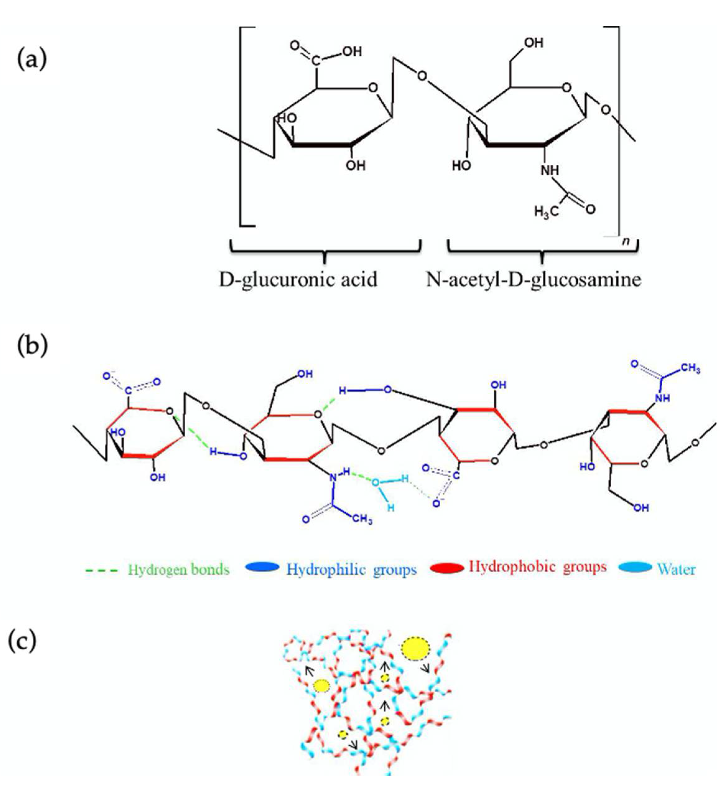
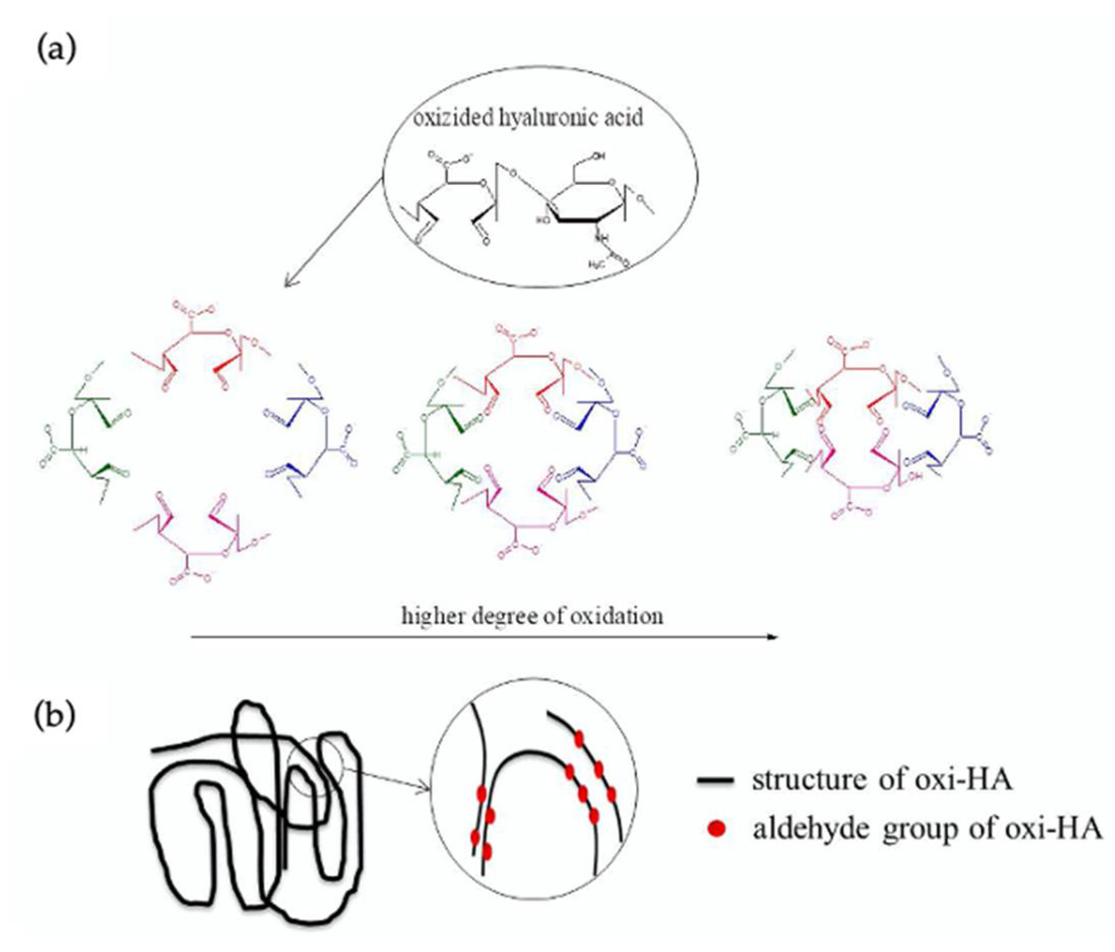
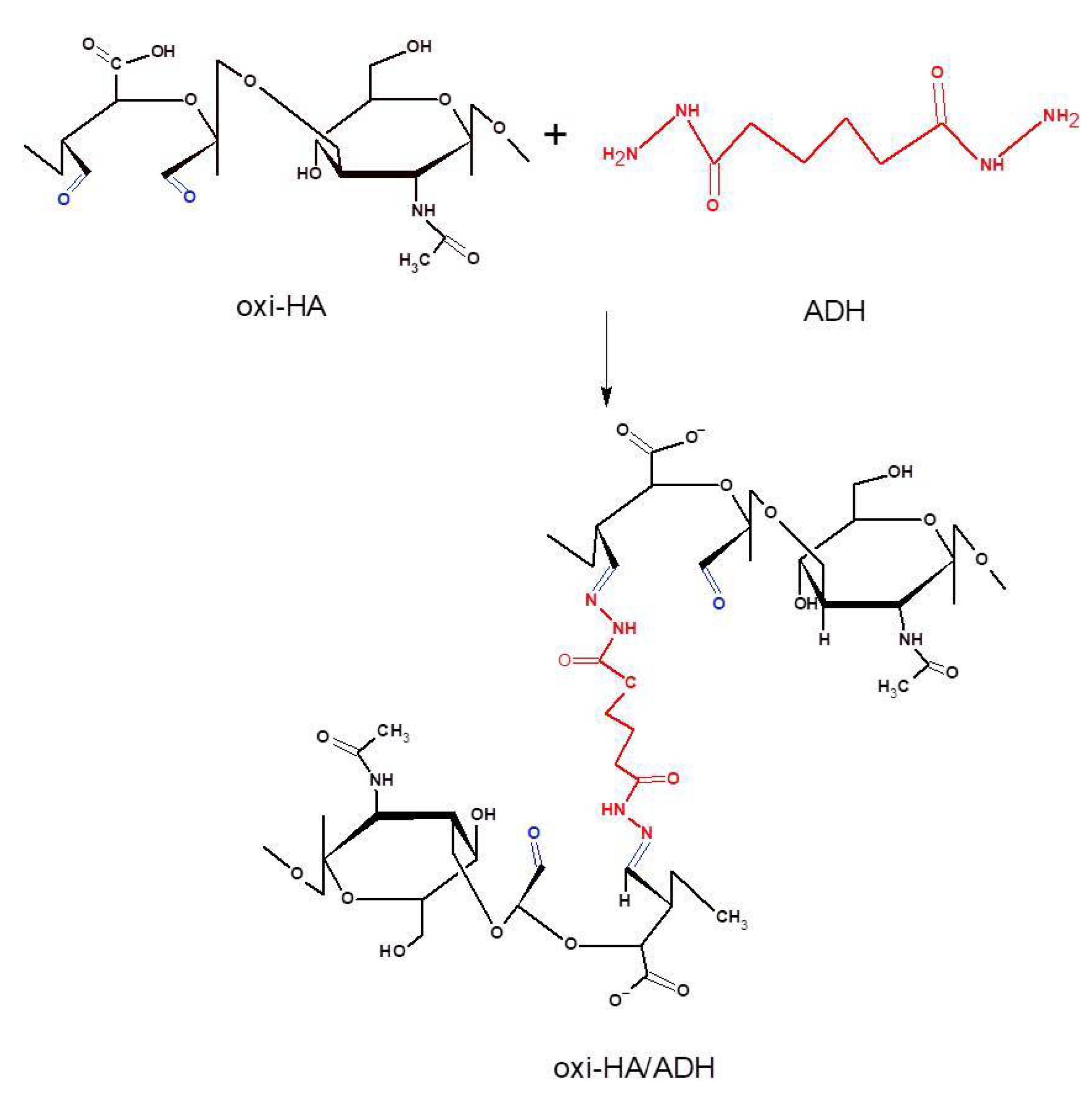
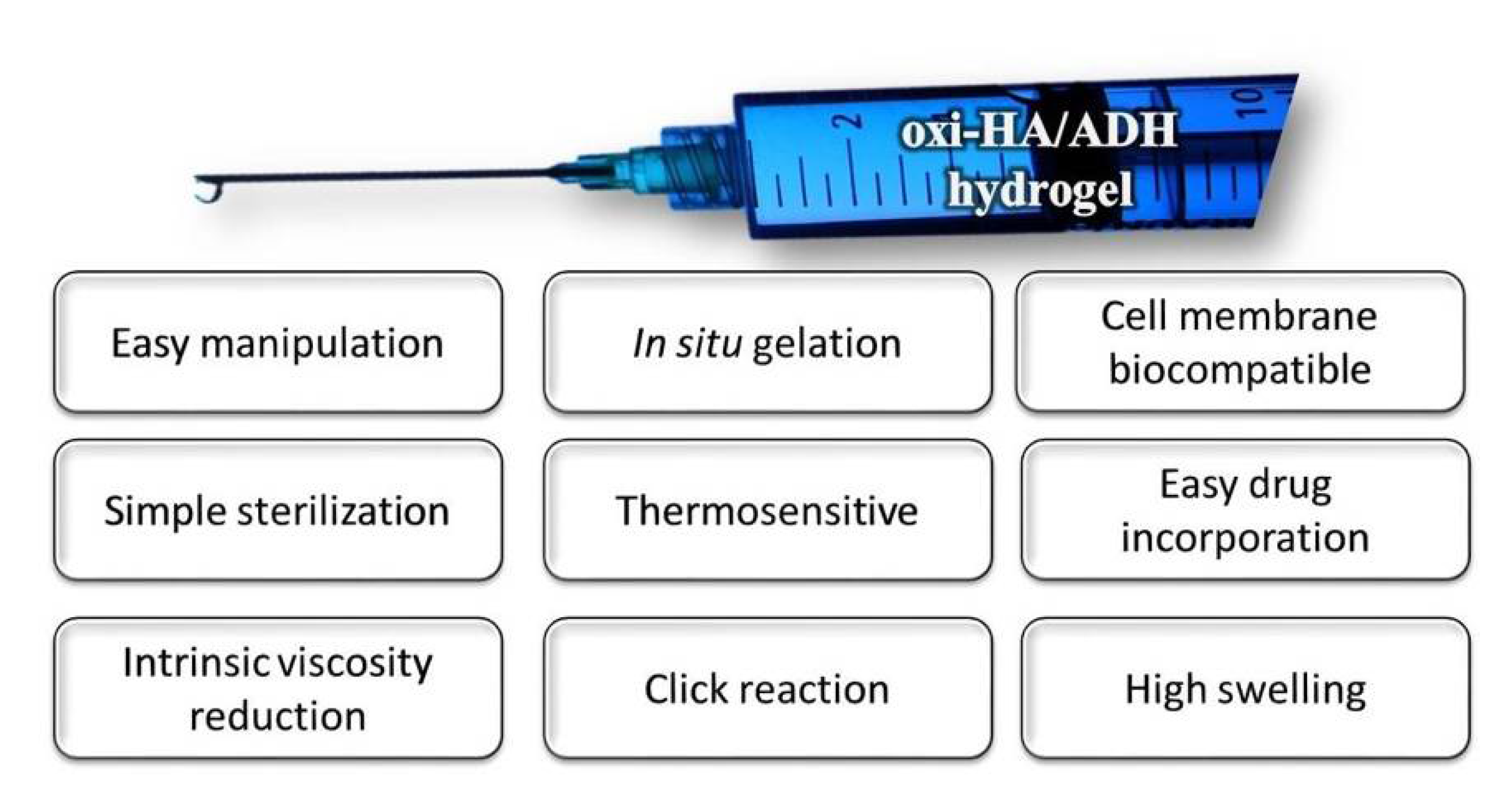
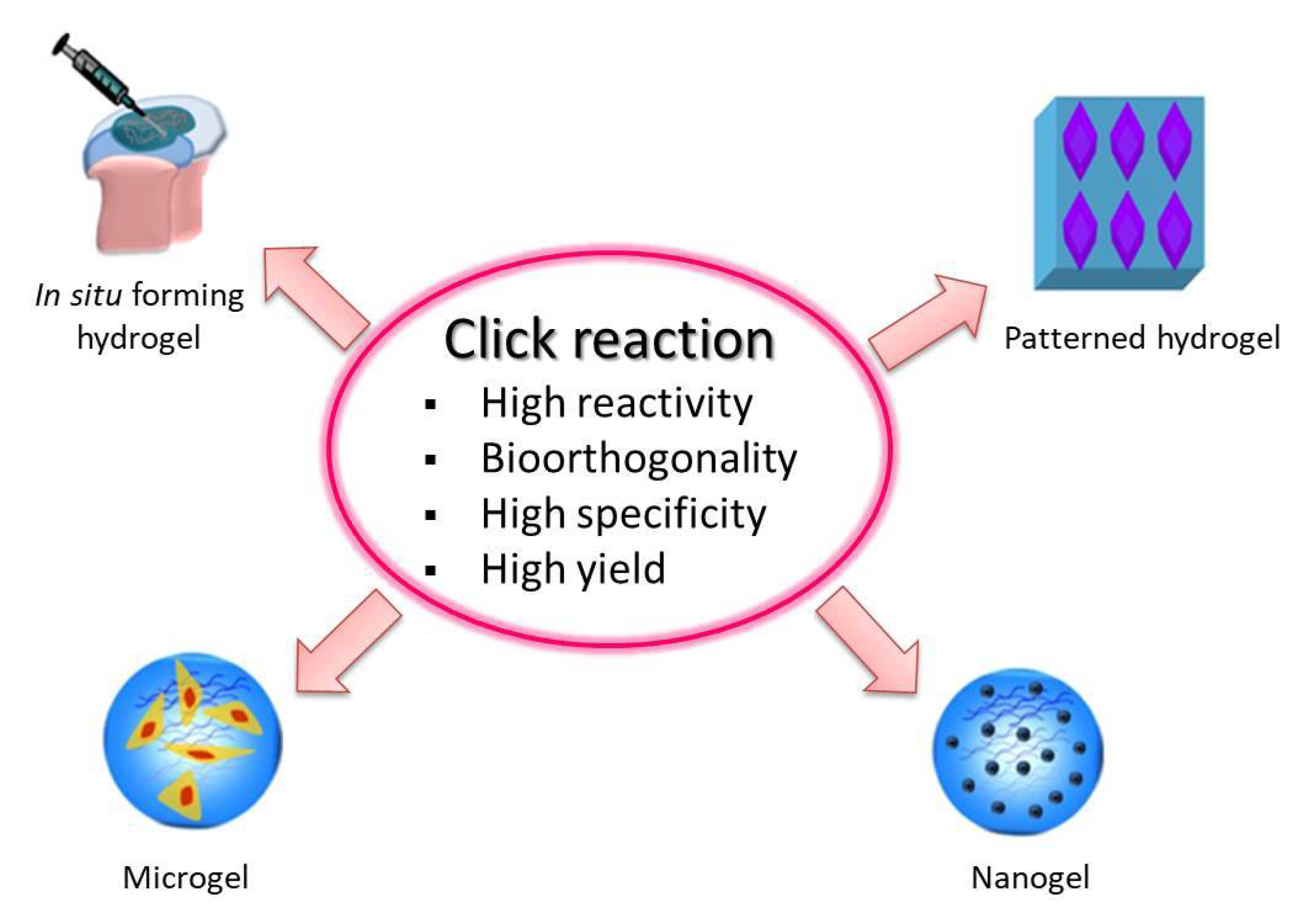
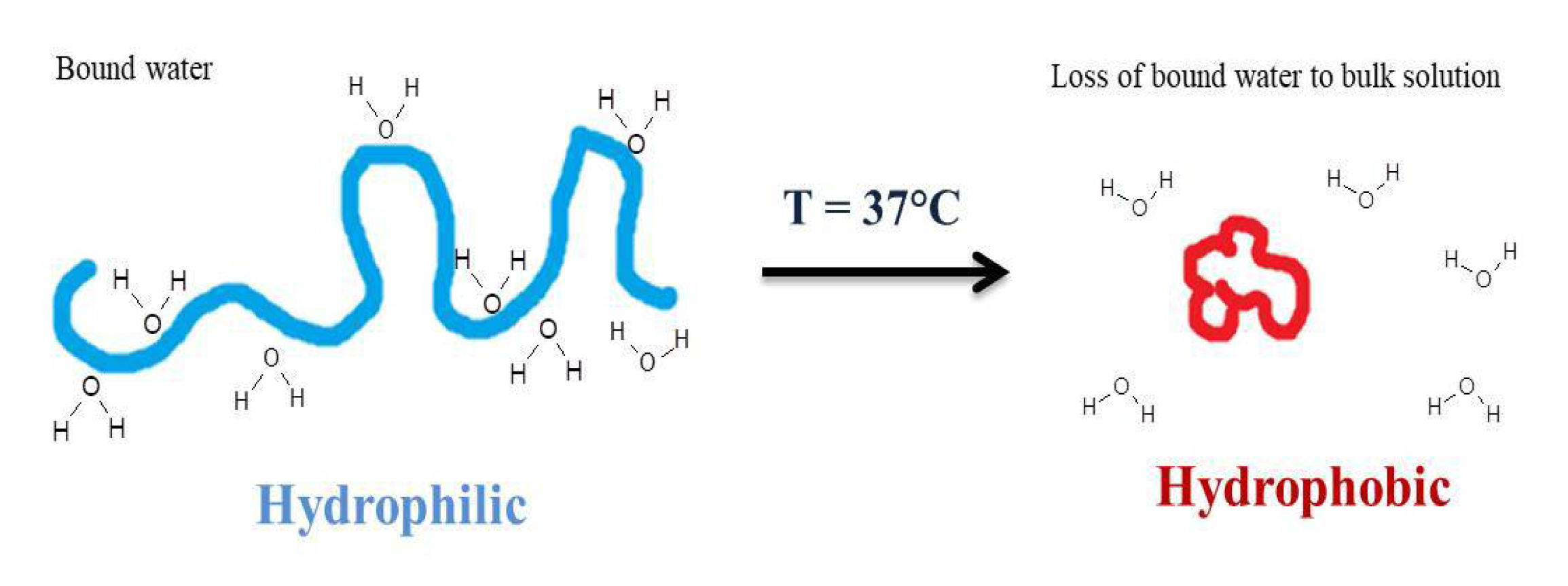
Table 1.
Applications of HA-cross-linked hydrogels in the biomedical field.
| Biomaterial | Application | Reference |
|---|---|---|
| HA 1-collagen matrix | cartilage tissue repair and neural regeneration | [40,41] |
| HA-gelatin hydrogels | bone regeneration and chondrogenic differentiation | [42,43,44,45] |
| HA-CHI 2 composite | cartilage TE | [36,46,47] |
| oxi-HA-resveratrol hydrogel | cell carrier for chondrocytes | [48,49] |
| PGS-PCL-Me-HA | heart valve TE | [50] |
| Alginate-HA | myocardial TE | [51] |
| thiolated-HA hydrogels | Adipose-substitutes TE | [52,53,54] |
| collagen-PEG-grafted-HA | intradermal drug delivery | [55] |
| oxi-HA-gelatin microspheres | drug delivery | [56] |
| HA-g-poly-(HEMA) | lung TE | [57] |
| HA-ADH 3-EDCI 4 | drug delivery | [58,59] |
| HA-DVS 5 | viscosupplement | [60,61] |
| HA-BDDE 6 | viscosupplement | [62,63,64] |
| HA-PRP 7 | regenerative medicine | [65,66,67] |
1 hyaluronic acid, 2 chitosan, 3 adipic dihydrazide,4 1-ethyl-3-(3-dimethyl aminopropyl)carbodiimide, 5 divinyl sulfone, 6 1,4-butanediol diglycidyl ether, 7 platelet-rich plasma, TE- tissue engineering.
Publisher’s Note: MDPI stays neutral with regard to jurisdictional claims in published maps and institutional affiliations. |
© 2021 by the authors. Licensee MDPI, Basel, Switzerland. This article is an open access article distributed under the terms and conditions of the Creative Commons Attribution (CC BY) license (https://creativecommons.org/licenses/by/4.0/).
Share and Cite
MDPI and ACS Style
França, C.G.; Villalva, D.G.; Santana, M.H.A. Oxi-HA/ADH Hydrogels: A Novel Approach in Tissue Engineering and Regenerative Medicine. Polysaccharides 2021, 2, 477-496. https://0-doi-org.brum.beds.ac.uk/10.3390/polysaccharides2020029
AMA Style
França CG, Villalva DG, Santana MHA. Oxi-HA/ADH Hydrogels: A Novel Approach in Tissue Engineering and Regenerative Medicine. Polysaccharides. 2021; 2(2):477-496. https://0-doi-org.brum.beds.ac.uk/10.3390/polysaccharides2020029
Chicago/Turabian StyleFrança, Carla Giometti, Denise Gradella Villalva, and Maria Helena Andrade Santana. 2021. "Oxi-HA/ADH Hydrogels: A Novel Approach in Tissue Engineering and Regenerative Medicine" Polysaccharides 2, no. 2: 477-496. https://0-doi-org.brum.beds.ac.uk/10.3390/polysaccharides2020029