Plasmacytoid Variant of Urothelial Carcinoma: Poor Prognostic Variant with High Expression of CDH1 Mutation


Abstract
:1. Introduction
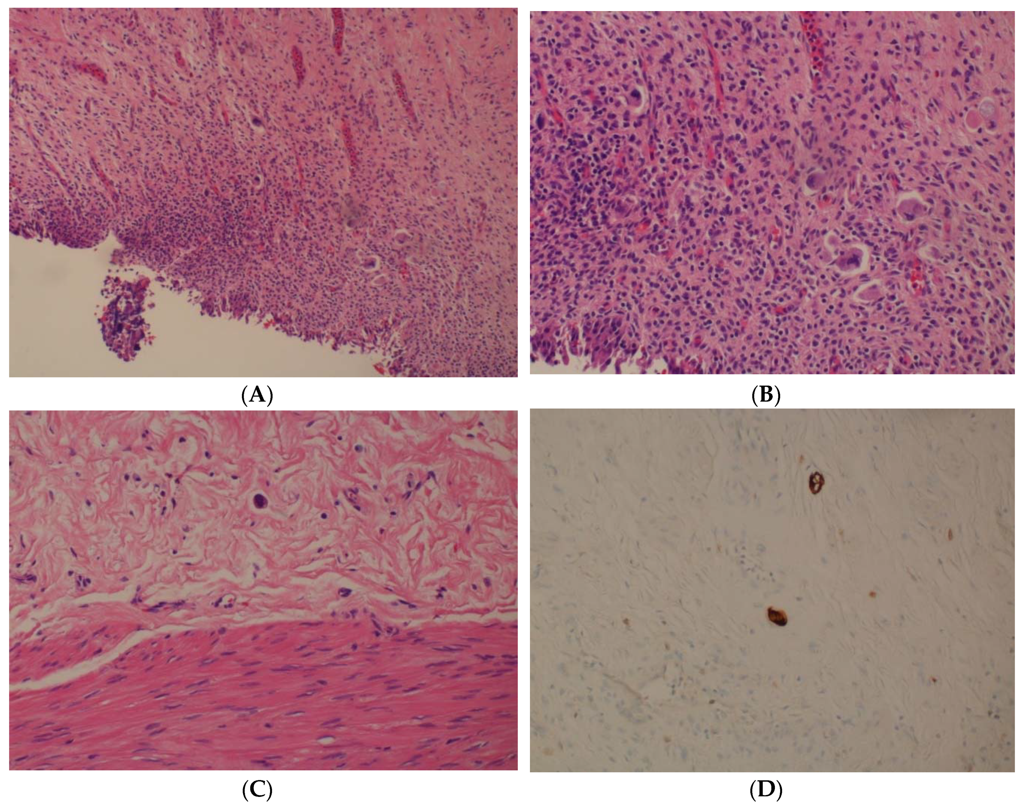
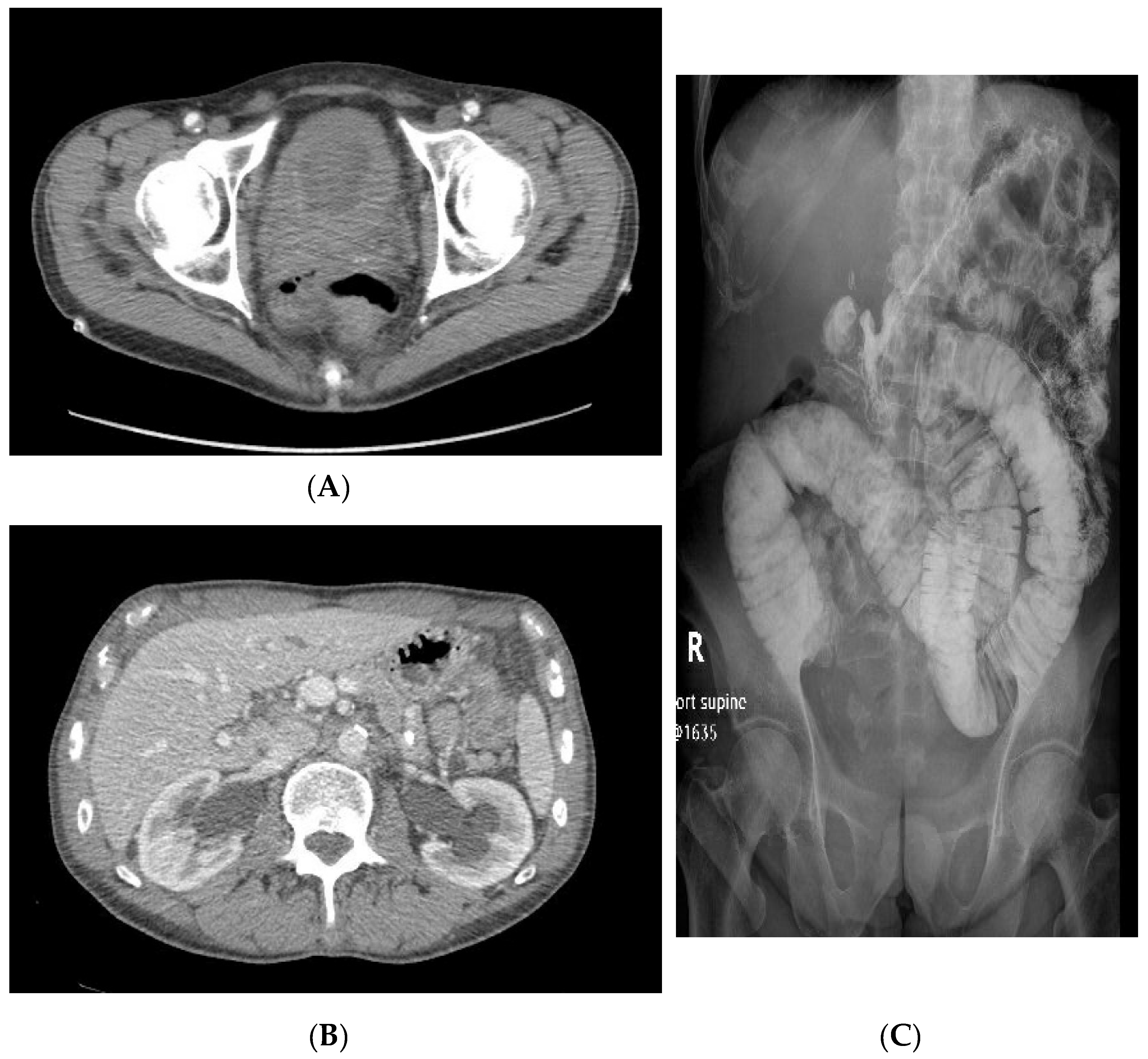
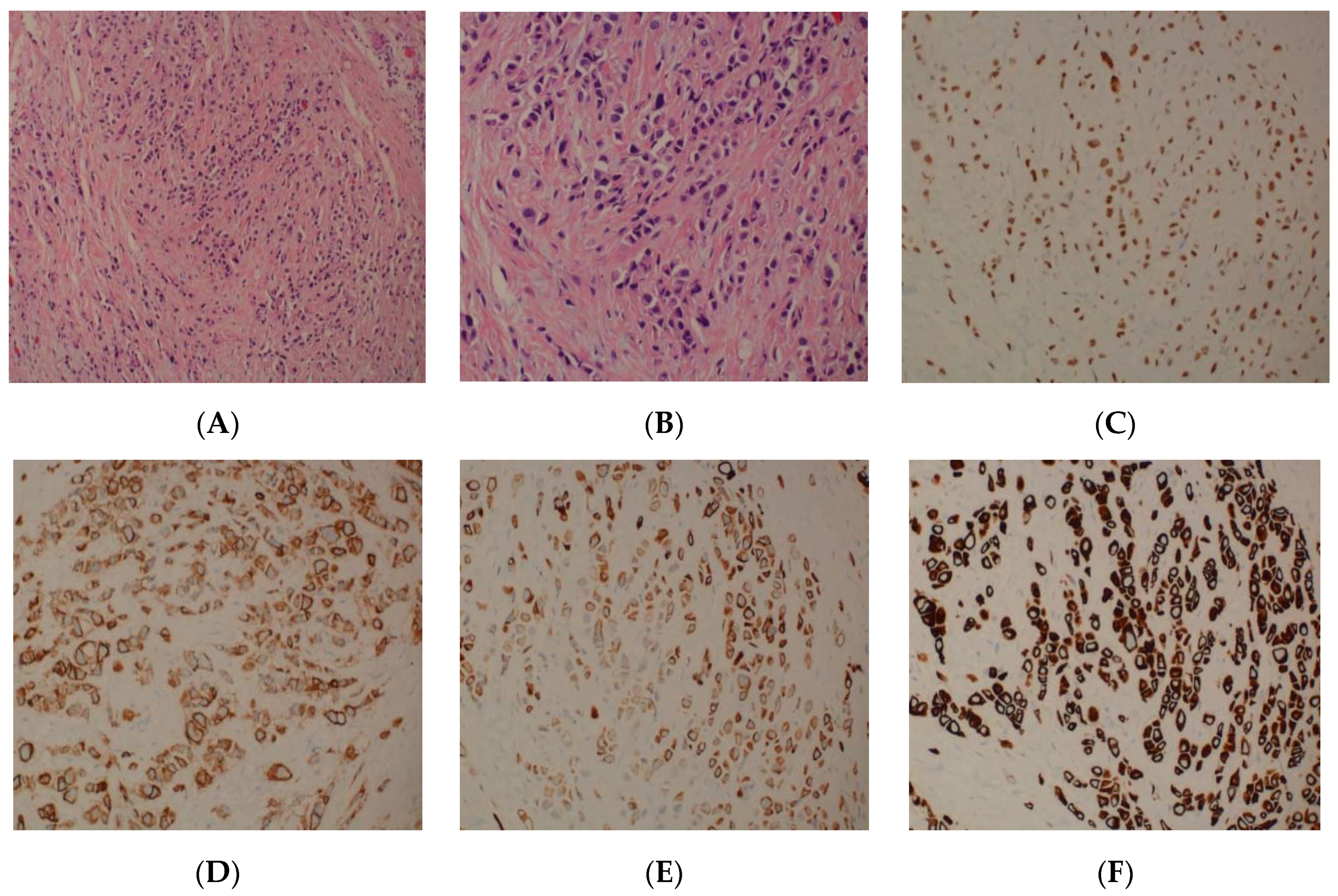
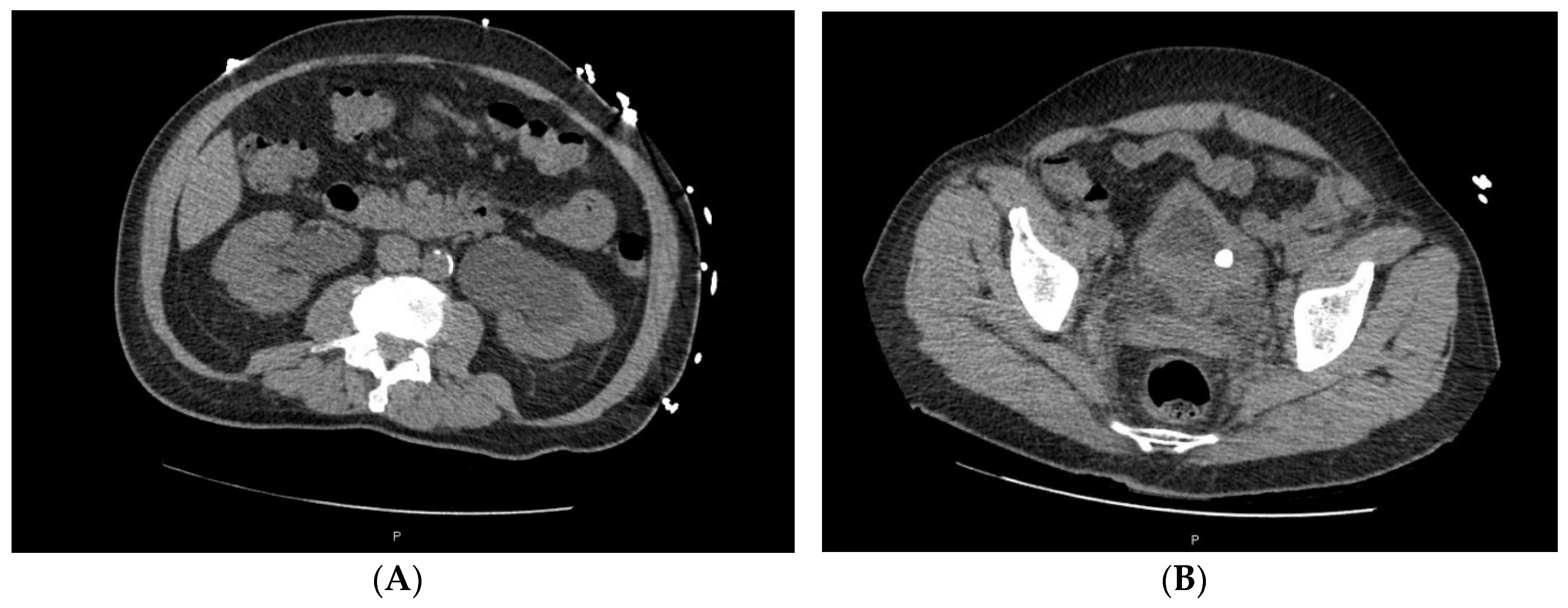
2. Case Series
3. Discussion
4. Conclusions
Author Contributions
Funding
Informed Consent Statement
Acknowledgments
Conflicts of Interest
References
- Li, Q.; Assel, M.; Benfante, N.E.; Pietzak, E.J.; Herr, H.W.; Donat, M.; Cha, E.K.; Donahue, T.F.; Bochner, B.H.; Dalbagni, G. The Impact of Plasmacytoid Variant Histology on the Survival of Patients with Urothelial Carcinoma of Bladder after Radical Cystectomy. Eur. Urol. Focus 2019, 5, 104–108. [Google Scholar] [CrossRef] [PubMed]
- Cockerill, P.A.; Cheville, J.C.; Boorjian, S.A.; Blackburne, A.; Thapa, P.; Tarrell, R.F.; Frank, I. Outcomes Following Radical Cystectomy for Plasmacytoid Urothelial Carcinoma: Defining the Need for Improved Local Cancer Control. Urology 2017, 102, 143–147. [Google Scholar] [CrossRef] [PubMed]
- Humphrey, P.A.; Moch, H.; Cubilla, A.L.; Ulbright, T.M.; Reuter, V.E. The 2016 WHO Classification of Tumours of the Urinary System and Male Genital Organs-Part B: Prostate and Bladder Tumours. Eur. Urol. 2016, 70, 106–119. [Google Scholar] [CrossRef] [PubMed] [Green Version]
- Monn, M.F.; Kaimakliotis, H.Z.; Pedrosa, J.A.; Cary, K.C.; Bihrle, R.; Cheng, L.; Koch, M.O. Contemporary bladder cancer: Variant histology may be a significant driver of disease. Urol. Oncol. 2015, 33, 18 e15–18 e20. [Google Scholar] [CrossRef] [PubMed]
- Kaimakliotis, H.Z.; Monn, M.F.; Cheng, L.; Masterson, T.A.; Cary, K.C.; Pedrosa, J.A.; Foster, R.S.; Koch, M.O.; Bihrle, R. Plasmacytoid bladder cancer: Variant histology with aggressive behavior and a new mode of invasion along fascial planes. Urology 2014, 83, 1112–1116. [Google Scholar] [CrossRef] [PubMed]
- Dayyani, F.; Czerniak, B.A.; Sircar, K.; Munsell, M.F.; Millikan, R.E.; Dinney, C.P.; Siefker-Radtke, A.O. Plasmacytoid urothelial carcinoma, a chemosensitive cancer with poor prognosis, and peritoneal carcinomatosis. J. Urol. 2013, 189, 1656–1661. [Google Scholar] [CrossRef] [PubMed]
- Ro, J.Y.; Shen, S.S.; Lee, H.I.; Hong, E.K.; Lee, Y.H.; Cho, N.H.; Jung, S.J.; Choi, Y.J.; Ayala, A.G. Plasmacytoid transitional cell carcinoma of urinary bladder: A clinicopathologic study of 9 cases. Am. J. Surg. Pathol. 2008, 32, 752–757. [Google Scholar] [CrossRef] [PubMed]
- Keck, B.; Stoehr, R.; Wach, S.; Rogler, A.; Hofstaedter, F.; Lehmann, J.; Montironi, R.; Sibonye, M.; Fritsche, H.M.; Lopez-Beltran, A.; et al. The plasmacytoid carcinoma of the bladder—Rare variant of aggressive urothelial carcinoma. Int. J. Cancer 2011, 129, 346–354. [Google Scholar] [CrossRef] [PubMed]
- Sood, S.; Paner, G.P. Plasmacytoid Urothelial Carcinoma: An Unusual Variant That Warrants Aggressive Management and Critical Distinction on Transurethral Resections. Arch. Pathol. Lab. Med. 2019, 143, 1562–1567. [Google Scholar] [CrossRef] [PubMed] [Green Version]
- Ramamurthy, C.; Arguello, D.; Anari, F.; Ghatalia, P.; Zibelman, M.R.; Plimack, E.R.; Burgess, E.F.; Hogan, T.F.; Dawson, N.A.; Hauke, R.J.; et al. Molecular profiling of aggressive variant urothelial carcinoma. J. Clin. Oncol. 2019, 37, 378. [Google Scholar] [CrossRef]
- Al-Ahmadie, H.A.; Iyer, G.; Lee, B.H.; Scott, S.N.; Mehra, R.; Bagrodia, A.; Jordan, E.J.; Gao, S.P.; Ramirez, R.; Cha, E.K.; et al. Frequent somatic CDH1 loss-of-function mutations in plasmacytoid variant bladder cancer. Nat. Genet. 2016, 48, 356–358. [Google Scholar] [CrossRef] [PubMed] [Green Version]
- McConkey, D.J.; Lee, S.; Choi, W.; Tran, M.; Majewski, T.; Lee, S.; Siefker-Radtke, A.; Dinney, C.; Czerniak, B. Molecular genetics of bladder cancer: Emerging mechanisms of tumor initiation and progression. Urol. Oncol. 2010, 28, 429–440. [Google Scholar] [CrossRef] [PubMed] [Green Version]
- Li, J.; Lu, S.; Lombardo, K.; Monahan, R.; Amin, A. ARID1A alteration in aggressive urothelial carcinoma and variants of urothelial carcinoma. Hum. Pathol. 2016, 55, 17–23. [Google Scholar] [CrossRef] [PubMed]
- Palsgrove, D.N.; Taheri, D.; Springer, S.U.; Cowan, M.; Guner, G.; Mendoza Rodriguez, M.A.; Del Carmen Rodriguez Pena, M.; Wang, Y.; Kinde, I.; Ricardo, B.F.P.; et al. Targeted sequencing of plasmacytoid urothelial carcinoma reveals frequent TERT promoter mutations. Hum. Pathol. 2019, 85, 1–9. [Google Scholar] [CrossRef] [PubMed]
- Ross, R.L.; McPherson, H.R.; Kettlewell, L.; Shnyder, S.D.; Hurst, C.D.; Alder, O.; Knowles, M.A. PIK3CA dependence and sensitivity to therapeutic targeting in urothelial carcinoma. BMC Cancer 2016, 16, 553. [Google Scholar] [CrossRef] [PubMed] [Green Version]
- Hayashi, T.; Tanigawa, G.; Fujita, K.; Imamura, R.; Nakazawa, S.; Yamamoto, Y.; Hosomi, M.; Shimazu, K.; Fushimi, H.; Yamaguchi, S. Two cases of plasmacytoid variant of urothelial carcinoma of urinary bladder: Systemic chemotherapy might be of benefit. Int. J. Clin. Oncol. 2011, 16, 759–762. [Google Scholar] [CrossRef] [PubMed]
- Keck, B.; Wach, S.; Stoehr, R.; Kunath, F.; Bertz, S.; Lehmann, J.; Stöckle, M.; Taubert, H.; Wullich, B.; Hartmann, A. Plasmacytoid variant of bladder cancer defines patients with poor prognosis if treated with cystectomy and adjuvant cisplatin-based chemotherapy. BMC Cancer 2013, 13, 71. [Google Scholar] [CrossRef] [PubMed] [Green Version]
- Diamantopoulos, L.N.; Khaki, A.; Grivas, P.; Gore, J.L.; Schade, G.; Hsieh, A.; Lee, J.K.; Yesefski, T.; Yu, E.Y.; Schweizer, M.; et al. Plasmacytoid Urothelial Carcinoma: Response to Chemotherapy and Oncologic Outcomes. Bladder Cancer 2019, 6, 71–81. [Google Scholar] [CrossRef] [Green Version]
- Gandhy, S.U.; Madan, R.A.; Aragon-Ching, J.B. The immunotherapy revolution in genitourinary malignancies. Immunotherapy 2020, 12, 819–831. [Google Scholar] [CrossRef] [PubMed]


Publisher’s Note: MDPI stays neutral with regard to jurisdictional claims in published maps and institutional affiliations. |
© 2021 by the authors. Licensee MDPI, Basel, Switzerland. This article is an open access article distributed under the terms and conditions of the Creative Commons Attribution (CC BY) license (http://creativecommons.org/licenses/by/4.0/).
Share and Cite
Erck, A.; Li, W.; Movahedi-Lankarani, S.; Chung, S.; Aragon-Ching, J.B. Plasmacytoid Variant of Urothelial Carcinoma: Poor Prognostic Variant with High Expression of CDH1 Mutation. Uro 2021, 1, 23-29. https://0-doi-org.brum.beds.ac.uk/10.3390/uro1010004
Erck A, Li W, Movahedi-Lankarani S, Chung S, Aragon-Ching JB. Plasmacytoid Variant of Urothelial Carcinoma: Poor Prognostic Variant with High Expression of CDH1 Mutation. Uro. 2021; 1(1):23-29. https://0-doi-org.brum.beds.ac.uk/10.3390/uro1010004
Chicago/Turabian StyleErck, Alisa, Wenping Li, Saeid Movahedi-Lankarani, Simon Chung, and Jeanny B. Aragon-Ching. 2021. "Plasmacytoid Variant of Urothelial Carcinoma: Poor Prognostic Variant with High Expression of CDH1 Mutation" Uro 1, no. 1: 23-29. https://0-doi-org.brum.beds.ac.uk/10.3390/uro1010004