Article

Dietary Choline Deprivation Exacerbates Cardiomyopathy in Streptozotocin-Induced Diabetic Adult Rats

1
Laboratory of Pharmacology, Medical School of Athens, National and Kapodistrian University of Athens, 75 Mikras Asias, Goudi, 11527 Athens, Greece
2
Diabetes Complications Research Centre, Conway Institute, School of Medicine and Medical Sciences, University College Dublin, D04 V1W8 Dublin, Ireland
3
Department of Clinical Pharmacy, College of Pharmacy, University of Babylon, Babylon 51001, Iraq
4
Department of Clinical Laboratory Sciences, College of Pharmacy, University of Babylon, Babylon 51001, Iraq
5
Department of Pathology, Medical School of Athens, National and Kapodistrian University of Athens, 75 Mikras Asias, Goudi, 11527 Athens, Greece
*
Author to whom correspondence should be addressed.
Submission received: 16 August 2021 / Revised: 27 September 2021 / Accepted: 1 October 2021 / Published: 9 October 2021

Abstract

:
Choline (Ch) is an essential molecule of substantial importance for the optimal development and function of several biological systems. Ch deprivation has been linked with abnormal fat metabolism, insulin resistance, and myocardial dysfunction. The current study provides evidence of an exacerbation of streptozotocin-induced cardiomyopathy in adult diabetic Wistar rats by dietary Ch deprivation through the administration of a Ch-deprived diet (CDD). Twenty-four adult male Wistar rats were randomly separated into four groups: control, diabetic (DM), choline-deprived through choline-deprived diet (CD), and diabetic choline-deprived (DM + CD). After five weeks of dietary intervention, myocardium echocardiographic and histological assessments were performed. Choline-deprived diabetic rats exhibited significantly slower heart rate, significantly higher myocardial ejection velocity and left ventricle wall tension index with a concomitant significant decreased LV posterior wall thickness as compared to diabetic rats fed on a standard diet. Moreover, histopathological evidence demonstrated an exacerbation of myocardial inflammation and fibrosis associated with significant up-regulation of VEGF expression in the diabetic rat myocardium as a result of Ch deprivation. The study’s findings are of particular significance since the examined experimental approach introduces a previously uncharacterised comorbidity simulation with regards to myocardial structure and functional profiling.

1. Introduction

Choline (Ch), a water-soluble vitamin B cofactor, has been long described as an essential nutrient [1] and dietary intake recommendations have been established by the Institute of Medicine (IOM) since 1998 [2] and by the European Food Safety Authority in 2016 [3]. Ch is an intrinsic component of several important biomolecules, has a vital role in the one-carbon cycle pathway by facilitating the metabolism of methyl groups [4] but it is also involved in crucial cellular functions, including among others lipid biochemistry [4,5,6,7], and proper cardiomyocyte contractile function [8,9]. Ch deprivation, observed either in physiological (e.g., pregnancy, lactation, intense exercise) or pathological conditions (e.g., alcoholism and malnutrition), with critical impact on brain function [10,11], has attracted much consideration due to its association with various adverse health impacts that can occur across the life span i.e. nonalcoholic fatty liver diseases [7,12,13,14,15,16,17] and insulin resistance [11,18]. In previous studies, we have demonstrated that Ch deprivation can also affect heart causing myocardial inflammatory infiltration that could lead to impaired heart mechanical properties which resemble to a restrictive pattern of cardiomyopathy characterised mainly by diastolic dysfunction [8,19]. One-carbon metabolism activation seems to have a beneficial effect on the pressure overload cardiac hypertrophy and it is necessary for the maintenance of energy balance and cardiac homeostasis [9]; thus, Ch deprivation might play a crucial role in the progression of potential myocardial dysfunction. Moreover, Ch, as a precursor of the key neurotransmitter acetylcholine, is important in the protection against various cardiovascular diseases such as myocardial infarction, arrhythmias, cardiac hypertrophy and ischaemia/reperfusion injury [20].
Diabetes mellitus is also a known leading cause of progressive heart failure [21,22,23]; diabetic cardiomyopathy describes the cardiac dysfunction in the absence of overt clinical ischaemic heart disease, valvular disease, and other conventional cardiovascular risk factors, such as dyslipidaemia and hypertension. The features of diabetic cardiomyopathy include decreased diastolic compliance, interstitial fibrosis, cardiac stiffness and hypertrophy with ultimate progression to both systolic dysfunction and clinical heart failure [24]. Diabetes is also associated with dysfunction of the cardiac microvasculature [25]; vascular endothelial growth factor (VEGF-A) is one of the important factors that exerts a substantial role in the development and functional integrity of the vasculature, acting as a signaling protein [26].
Population with diabetes is dramatically increasing worldwide [24], but, similarly, the population prone to Ch deficiency is also increasing taking into consideration that 90% of the population of the United States is not receiving sufficient intake of Ch [27,28]. These two metabolic disorders (Ch deprivation and diabetes mellitus) are resembling common cases in the clinical practice; through multiple pathophysiological mechanisms, they could potentially affect myocardium structural integrity and performance.
In the current study, we have therefore, attempted to investigate, in an experimental approach, the effect of dietary Ch deprivation through administration of a Ch-deficient diet (CDD) on the heart of streptozotocin (STZ)-induced diabetic adult Wister rats. Thus, cardiac remodeling and performance were examined under the comorbidity simulation of both metabolic insults.

2. Materials and Methods

2.1. Animals

A total of 24 three-month-old adult male albino Wistar rats with a body weight of 298.6 ± 26.2 g, were supplied by the National Centre of Scientific Research (NCSR), Athens, Greece. This study protocol was approved by the Doctorate Board of the Medical School of National and Kapodistrian University of Athens after the approval of the Animal Protocols by the Department of Rural and Veterinary Policy (RVP), General sector of Rural Economy and Veterinary, Prefecture of Attica, Hellenic Republic.
Animals were divided into groups and housed under controlled environmental conditions, at a temperature of 22 ± 1 °C, and artificial light/dark cycle (12/12 h), with adequate ventilation and appropriate humidity (~55%). Food and water were provided ad libitum. The experimental animals were cared for in accordance with the principles of the Laboratory Animal Care as previously set by the European Economic Community (EEC) Council Directive 86/609/EEC [29], and aligned and amended according to the recommendation 2007/526/EU for experimental animals.

2.2. Materials

The standard diet was a choline-enriched (1.5 g/kg) diet at the expense of sucrose. CDD was produced by Mucedola (Settino Milanese, Milan, Italy) and has been purchased from Analab Ltd. (Athens, Greece). The analytical structure of CDD was the following (g/kg): sugar (413), dextrine (110), starch (110), hydrogenated vegetable oil (100), pea protein (90), soya protein isolate (60), corn oil (50), mineral mix (35), cellulose (10), vitamin-free casein (10), vitamin mix (10), l-cystine (2), in addition to fat (16%), protein ingredient (12%), ash (3.5%), fiber (2%). The highest quality available of STZ and other reagents were used and purchased from Sigma Chemicals (Darmstadt, Germany). Vascular Endothelial Growth Factor Antibody VEGF-A kit purchased from Invitrogen by Thermo Fisher Scientific, digital ultrasound system (Vivid 7 version Pro, GE Healthcare) was used.

2.3. Experimental Procedure

Animals were separated into four groups (n = 6 per group): Control, diabetic (DM), Ch-deprived (CD), and diabetic Ch-deprived (DM + CD). Diabetes was induced by a single intraperitoneal injection of STZ (50 mg/kg of body weight diluted in a 0.1 mol/L citrate solution, pH 4.5) [30]; Control and CD groups were injected with vehicle. After 72 h following STZ administration, blood was collected from the pedal vein puncture, and blood glucose was assessed using a portable glucometer (Roche Diagnostics GmbH, Mannheim, Germany); rats with blood glucose level of ≥300 mg/dL were considered diabetic, and the dietary intervention was imposed for five weeks. Survival rate was 100% throughout the entire experiment; animals were weighted and sacrificed after five weeks and (a) blood was immediately collected from the inferior vena cava and used for assessment of biochemical parameters and (b) heart was quickly removed, weighed and prepared for histological assessment.

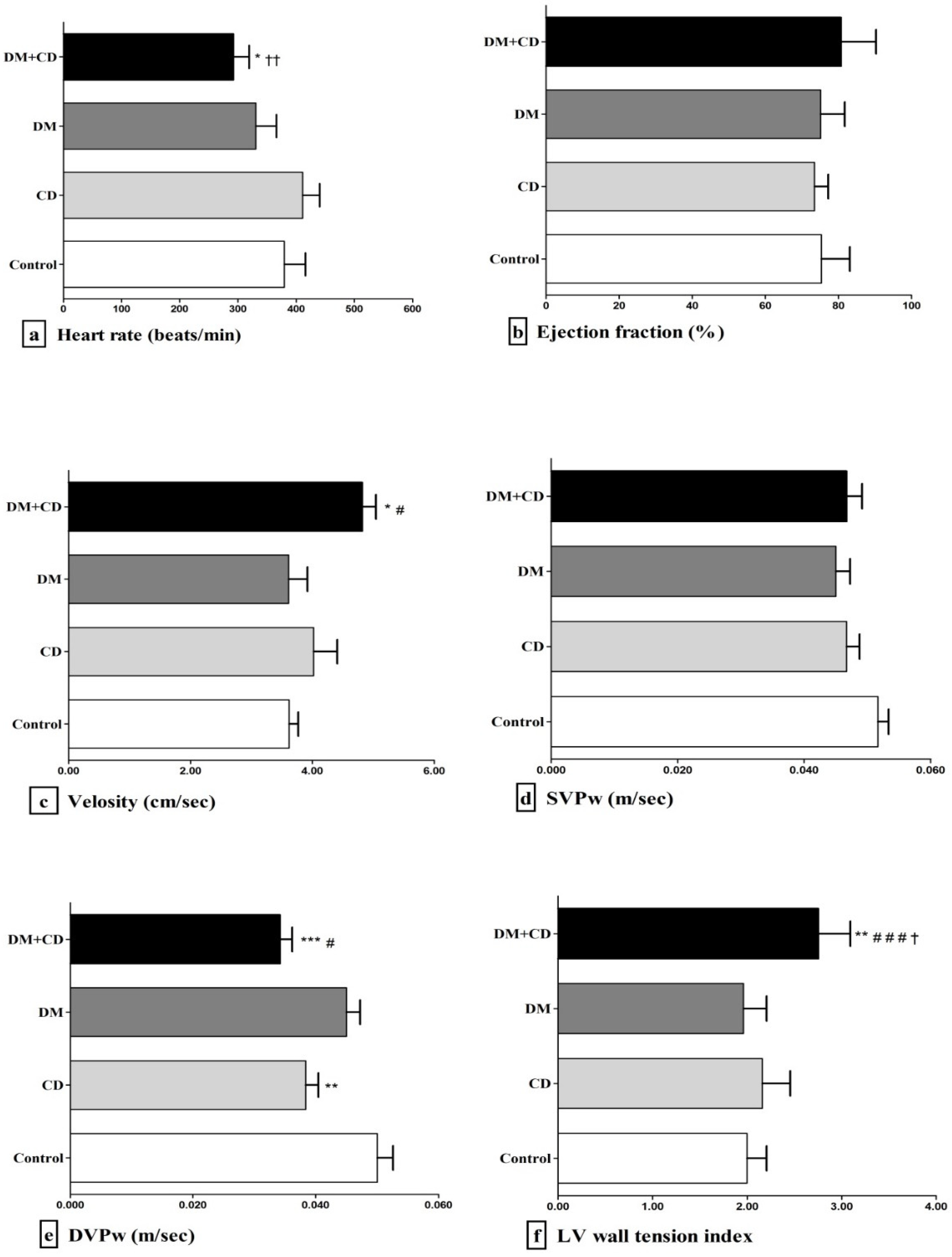
2.4. Echocardiographic Assessment

Before sacrifice, ketamine hydrochloride (100 mg/kg) was used to sedate the animals and heart function/structure was subsequently evaluated by transthoracic echocardiography as previously described in detail [31,32]. Body temperature was kept at 37 °C using heat blanket, while the animals were prone and fixed in a special apparatus. Short and long-axis images were obtained using a digital ultrasound system (Vivid 7 version Pro, GE Healthcare) with the 14.0-MHz i13L transducer. Many consecutive assessments were performed and analysed by two independent investigators. Left ventricle (LV) internal diameter at systolic phase (LVDs), LV internal diameter at the diastolic phase (LVDd), posterior wall thickness at the diastolic phase (LVPw), left atrium diameter (LA) and the ejection fraction (EF%) were measured (EF% was calculated using the Simpson equation). The fractional shortening (FS) provided by the following equation: FS = [(LVDd − LVDs)/LVDd] × 100% was used to access the global contractile LV function.
Systolic and diastolic velocity of the LV posterior wall radial displacement (SVPw and DVPw) were measured from two-dimensional guided M-mode recordings obtained at the midventricular level; SVPw and DVPw were calculated using the formula: V = ds/dt where “V” represents velocity, “s” the distance and “t” time. SVPw was used to assess the regional systolic function of the myocardium, while DVPw was used to determine the regional diastolic function of the myocardium. All echocardiographic assessments were averaged for at least 3 consecutive cardiac cycles. The LV wall tension index was calculated by the ratio of the LVDd to twice the value of LVPw [33,34] according to the following equation:
LV wall tension index = LVDd/(2 × LVPw)

2.5. Biochemical Assessment

Serum total cholesterol (TC), triglyceride (TG), low density lipoprotein (LDL), and high-density lipoprotein (HDL) levels were calculated using an automatic analyser (Hitachi, Roche modular). Fasting blood glucose levels and plasma insulin levels were assessed using the portable glucometer (Accu-Chek Roche Diagnostics GmbH, Mannheim, Germany) and the Elisaruo method of DRG ELISA, respectively.

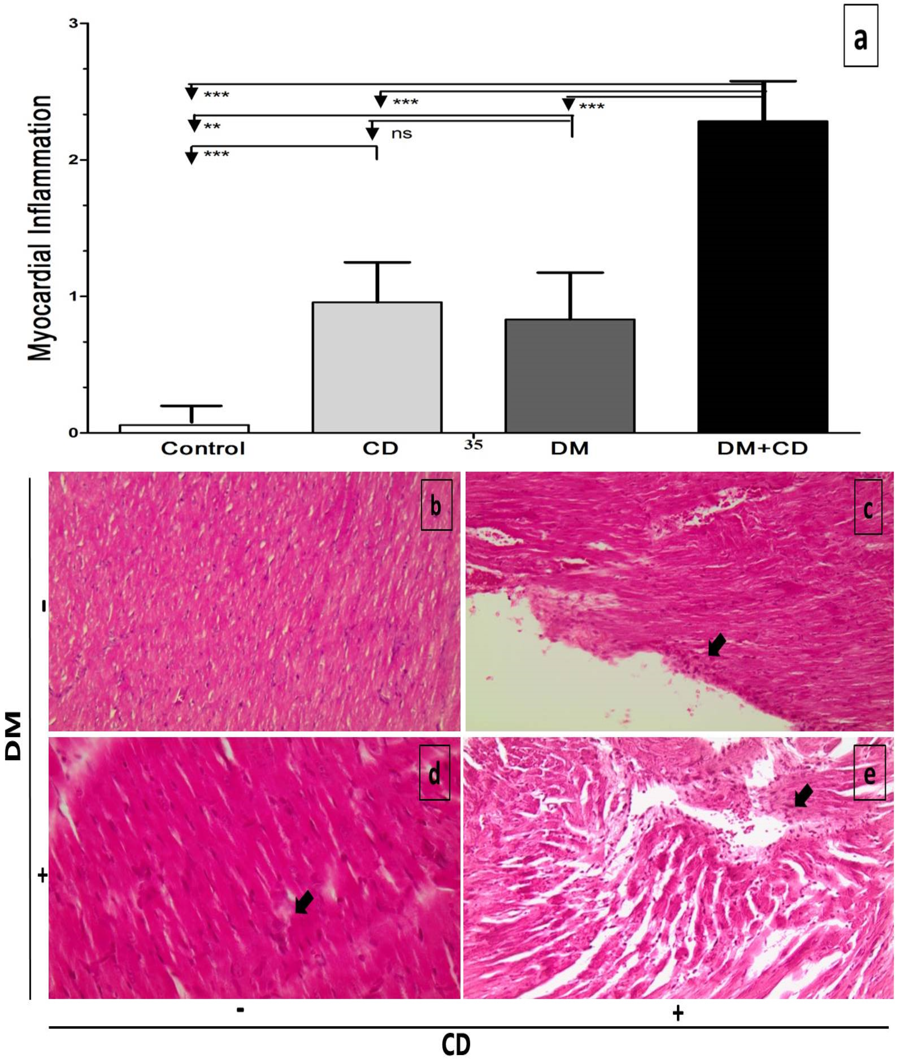
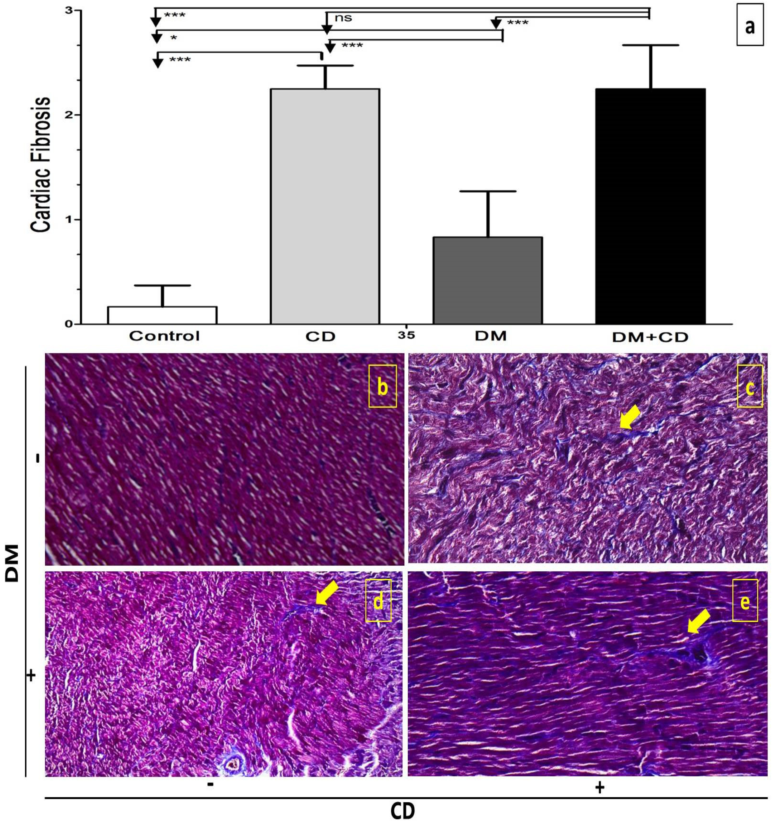
2.6. Histological Assessment

Collected heart samples were fixed in 4% (v/v) buffered formalin solution and embedded in paraffin wax using conventional techniques. Six sequential excised tissue specimens, 4 μm in thickness, were taken from each heart, at a distance approximately of 2 mm from each other. Sections were dewaxed, rinsed and stained with haematoxylin and eosin (H&E) or Masson trichrome stain, to examine and assess the myocardial infiltration with mononuclear inflammatory cells, the interstitial fibrosis and perivascular fibrosis and the interstitial oedema. The tissue specimens were randomized and given a code number for blinded assessment by two expert pathologists; cardiac inflammation and fibrosis were scored as previously described by Strilakou et al. [8].

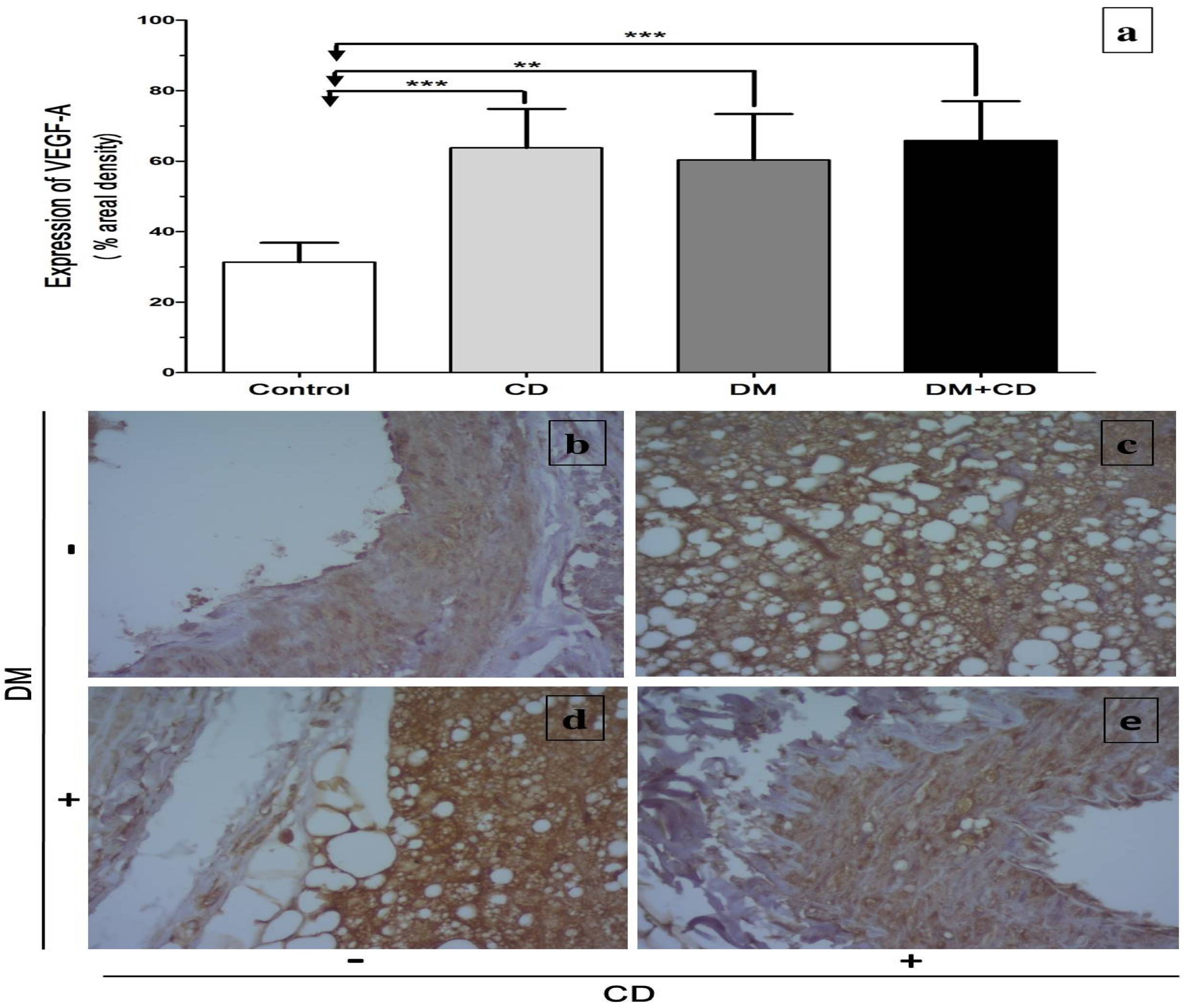
2.7. Immunohistochemical Assessment of Vascular Endothelial Factor-A165 (VEGF)

Deparaffinization, rehydration and antigen retrieval were performed by heating the slides in PTLink (Dako), using low pH solution [Envision FLEX TARGET RETRIEVAL SOLUTION Low pH (50×)]. Blocking of endogenous peroxidase was performed by incubating the slides with H2O2 3% solution for 15 min at room temperature (RT) and blocking of nonspecific staining was performed by incubating the slides with Blocking Solution (Dako REAL™ Peroxidase-Blocking Solution, code S2023) for 30 min at RT. Overnight incubation of the slides with the primary antibody followed; the antibody used was VEGF-A165 [Vascular Endothelial Growth Factor Antibody (VG1)], Invitrogen by Thermo Scientific catalogue number PA1-21796, at a dilution of 1:500. Incubation of the slides with Secondary Antibody (Dako anti-mouse immunoglobulins/ Polymer, IgG), for 30 min at RT was followed by incubation of the slides with HRP (Dako REAL™ EnVision™/HRP) for 30 min at RT (HRP recognizes the primary-secondary antiserum complex). Subsequently, the slides were incubated with diaminobenzidine (DAB) for 6 min at RT, in a dark environment (DAB reacts with HRP, resulting in the production of brown colour, visible under light microscope). Between each step the slides were washed with TBS (Tris Buffered Saline). At the end, slides were counterstained with hematoxylin, dehydrated and mounted. Evaluation of the immunohistochemical staining, assessed under light microscope in a blinded method by two independent pathologists for the VEGF stain expression in cardiac samples, was graded as follows: score 0: <15%, score 1: 16–35%, score 2: 36–65%, score 3: 65–100% according to Chen et al [35].

2.8. Statistical Analysis

Data are expressed as the mean±SD. A further analysis was performed using one-way analysis of variance (ANOVA) followed by multiple comparisons with Bonferroni’s and Tukey’s honest significant difference using GraphPad Prism 5.3 for Windows (GraphPad Software, San Diego, CA, USA). The significance level for all analyses was set at p ≤ 0.05.

3. Results

An assessment of the heart to body weight ratio at the sacrifice timepoint revealed no significant differences among the experimental groups (Table S1), and neither did the heart weight on its own.
Fasting blood glucose levels at sacrifice were significantly higher (p < 0.001) both in DM and DM + CD groups versus Control and CD groups and higher in the DM + CD group (p < 0.05) compared to DM group as well. Plasma insulin measurement revealed, as expected, a significant hypoinsulinaemia in the combination (DM + CD) and the DM groups compared to CD and control groups (p < 0.01 and p < 0.05 respectively) (Table S1).
The lipidemic profile of the nondiabetic and diabetic adult rats exposed to CDD is provided in Table 1; a significant increase in serum triglyceride was shown in DM group and a significant decrease in HDL levels in DM and CD groups, compared to control (p < 0.05).
The examination of the LV systolic velocity (SVPw) revealed no significant differences among the four examined groups (Figure 1d), but interestingly, the LV diastolic velocity (DVPw) was found significantly lower in both CD and DM + CD groups (p < 0.01 and p < 0.001 respectively) compared to control; DM + CD group exhibited a significant decrease in DVPw compared to DM (p < 0.05) (Figure 1e). The wall tension index of the LV was significantly increased in the combination group as compared to control, DM and CD rats (p < 0.01, p < 0.001, p < 0.05 respectively) (Figure 1f). LVDd was significantly increased in DM + CD rats compared to DM (p < 0.05) (Table 2) while the thickness of LVPw was significantly decreased in the DM + CD group compared to control, DM and CD group (p < 0.01, p < 0.05, p < 0.05 respectively) (Table 2). The echocardiographic measurement of the rat myocardium long axis and LVDs revealed no significant differences among the examined groups (Table 2). Finally, the left atrium (LA) diameter at long-axis slice showed significant increase in the DM + CD and CD groups compared to control (p < 0.01); and in the combination group and CD groups compared to DM (p < 0.01).
The histopathological scoring and representative light microscopy (H&E; ×400) captions of the inflammation occurring in the myocardium of nondiabetic and diabetic adult rats after a five-week exposure to CDD are provided in Figure 2. Exposure to CDD for five weeks provoked mild focal myocardial inflammation (Figure 2a,c), and so did DM (Figure 2a,d). The combined exposure to DM and CDD resulted in diffuse inflammation of the rat myocardium under the examined experimental conditions (Figure 2a,e).
The histopathological scoring and representative light microscopy (Masson’s stain; ×400) captions of the fibrosis occurring in the myocardium of nondiabetic and diabetic adult rats exposed to CDD are provided in Figure 3. Exposure to CDD for five weeks provoked diffuse myocardial fibrosis (Figure 3a,c), while DM caused mild focal myocardial fibrosis (Figure 3a,d). The combined exposure to DM and CDD resulted in more severe diffuse focal fibrosis of the rat myocardium with perivascular fibrosis, under the examined experimental conditions (Figure 3a,e).
Finally, the VEGF immunohistochemical stain expression occurring in the myocardium of nondiabetic and diabetic adult rats exposed to CDD is provided in Figure 4. All experimental groups showed a significant increase in cardiac VEGF stain expression compared to control (CD and DM + CD p < 0.001 and DM p < 0.01).

4. Discussion

Nutritional disorders can have a negative impact on cardiac metabolism and performance [36]. We have shown through the previous studies the early detrimental effect of dietary Ch deprivation on the adult healthy rat myocardium [8,19]. We herein provide novel findings that Ch deprivation impactfully aggravates the myocardial architecture and potential performance in STZ-induced diabetic adult rats. The present experimental setup revealed cardinal features of the cardiac remodeling involving at the same time inflammatory infiltration, fibrosis and stiffness along with increased LVDd and decreased LV posterior wall thickness, thus dilation of the left cavity, accompanied by slower heart rate and decreased peak diastolic velocity, but with higher LV myocardial ejection velocity and LV wall tension index.
The decreased DVPw in the diabetic setting compared to control group is consistent with the features of the early stages of development of diabetic cardiomyopathy [24]. Nevertheless, when the diabetic myocardium is concomitantly under the influence of Ch deprivation, diastolic velocity is deteriorated even more for the same time period along with cardiac fibrosis, implying increased stiffness and impaired relaxation properties of the left ventricle. Consequently, the significant increase in velocity observed in the combination group probably reflects an attempt of the Ch-deprived diabetic myocardium to counterbalance the impaired diastolic filling, to preserve the heart contractile properties. This concept is consistent with the observed preserved fractional shortening and ejection fraction under the combined insults.
In Ch-deprived diabetic design our study reveals for the first time that the structural remodeling of the LV goes beyond the established frameworks, according to which the diastolic heart failure is characterised by concentric hypertrophied myocardium [37], and shows that LV cavity with thinner wall and increased LVDd (thus potential enlargement) could also exert such functional derangement as shown by the increased LV wall tension index in the Ch-deprived myocardium. Additionally, the concurrently observed left atrium dilatation reflects the LV diastolic dysfunction and it is also probably associated with an increased atrial wall stress and stretch that could also provoke arrhythmogenesis, such as atrial fibrillation, which, in turn, jeopardises cardiac output [38].
It has been reported that autonomic nervous system (ANS) imbalance plays a crucial role in the development of heart failure [39]; decreased heart rate variability coupled with defective parasympathetic control has been shown even in the early stages of chronic heart failure. The reduced heart rate observed in the diabetic group is in line with previous studies that correlate this phenomenon with autonomic neural dysfunction and sinoatrial node impairment characterised by reduced intrinsic heart rate caused by STZ-induced diabetes [40]. Under the Ch-deprivation effect, where hyperglycaemia is exacerbated, the heart rate is decreased even more and could be the result of the severe metabolic injury not only to the pacemaker cells but also to the other nerves as well [41], promoting their denervation and the establishment of cardiovascular diabetic neuropathy [42]. The present experimental model suggests that diabetes mellitus along with dietary Ch deprivation interfere earlier with the cholinergic signaling of the heart, thus modulating cardiac remodeling [43]; the LV morphological changes might induce mechanical stretch of the cardiac muscle and further compromise the cholinergic heart signaling and the functional reservoirs in cases of pressure or volume overload, while over time they could also predispose to systolic impairment. The persistent hyperglycemia as a result of the well-established insulin resistance caused by Ch deprivation [11], and the hypoinsulinaemia of the STZ-induced diabetes in rats [44,45], might act as a significant independent contributing factor to the multifactorial cardiac fibrosis [46].
The significant inflammation observed in the choline-deprived diabetic group myocardia could be due to oxidative stress (OS) produced by exacerbation of hyperglycaemia and the excessive pro-inflammatory cytokine production [47,48,49]. However, one should not exclude the possibility of a synergistic cardiotoxic effect of CDD and diabetes that might not be restricted only to OS-mediated inflammation due to significant hyperglycaemia; previous studies of ours [8,19], and others [50,51], have suggested a complex interdependence of antioxidants (like carnitine) [52,53], and Ch, while both diabetes and Ch deprivation are known to cause serious metabolic dysregulation on multiple systems [1,7,54,55,56]. Taking into account that Ch deprivation is usually accompanied by carnitine deficiency, which has been associated by itself with dilated cardiomyopathy [57], it turns out that the setting of Ch deprivation along with diabetes introduces a new distinctive phenotype of cardiomyopathy. Ch deprivation has been repeatedly characterised as a time-dependent pathogenetic factor [14,58,59], with its toxicity to become more complex and irreversible with time. Although our study has focused on a rather short period of simultaneous Ch deprivation and diabetes mellitus exposure, this experimental time frame efficiently demonstrated myocardial injury due to the impactful effects of Ch deprivation and diabetes independently but probably not providing the full view of their potential at a longer timeframe. The herein examined experimental conditions could provide evidence of an early stage of diabetic cardiomyopathy with restrictive features [60,61,62], characterised by mild myocardial inflammation and fibrosis with preserved ejection fraction that is accompanied by impaired LV remodeling under the Ch-deprivation effect with features of dilated cardiomyopathy.
The role of VEGF, one of the most powerful and important angiogenic growth factors, in the cardiac vascularization and remodeling is indisputable; under normal conditions, the expression of VEGF is relatively low in myocardium [63]. The significant up-regulation of myocardium VEGF expression induced by Ch deprivation is in line with previous studies [64,65,66]; this paradoxical [67] although response under Ch deprivation and STZ-induced diabetes could be attributed to the mitochondrial dysfunction [15] and to the up-regulation in gene expression due to hypomethylation induced by Ch deprivation [68,69]; VEGF except for being a major regulator of cardiac angiogenesis, it also exerts cytoprotective, antioxidative and antiapoptotic effects on cardiomyocytes [70]. This concept is in line with the up-regulation of VEGF expression in the cardiac tissue of all groups in our experimental model, which reflects the imbalance in the antioxidant status that occurs when there is either Ch deprivation or diabetes mellitus alone or in combination.
Previous data of ours have demonstrated that Ch deprivation disturbs cardiac extracellular matrix homeostasis and induces a significant up-regulation of the tissue inhibitors of metalloproteinases (TIMP-2) myocardial expression accompanied by a significant down-regulation of matrix metalloproteinases (MMP-2) myocardial expression after only four weeks of dietary intervention [19]. It has been suggested that TIMP-2 inhibits angiogenesis and promotes cardiac dysfunction by decreasing the VEGF and MMP-2 expression, while it up-regulates myocardium anti-angiogenic components of myocardium that contribute to LV remodeling [71,72]. Nevertheless, studies have shown that the induced response of VEGF to pathological conditions such as OS, inflammation, hypoxia and/or ischaemia is intense and abrupt at the early phase and then fades off [73]. This finding is consistent with our experimental data showing that the up-regulation of VEGF expression in the myocardium of Ch-deprived diabetic rats does not alter significantly compared to Ch-deprived or diabetic rats, although someone would expect a synergistic effect.
It seems that Ch deprivation aggravated the collagen deposition, interstitial fibrosis and depleted the antioxidant capabilities in diabetic myocardium. According to Kim and colleagues, the mechanoregulatory mechanisms induced by the established cardiac fibrosis might also disturb and promote the myofibroblast transdifferentiation in the myocardium [74] thus exacerbating the stiffness and consequently the hemodynamic burden on the heart suggested by the increased diastolic filling resistance. The herein presented findings are of particular importance since the examined experimental setting introduces a previously unknown comorbidity simulation of two metabolic insults that seem to trigger the transition of a restrictive type of cardiomyopathy to a potential dilated type.

5. Conclusions

The Ch deprivation through the administration of a CDD, can exacerbate STZ-induced cardiac remodeling in rats, within five weeks.
STZ-induced diabetic rats exposed to Ch deprivation exhibit significantly slower heart rate, significantly higher myocardial ejection velocity and left ventricle wall tension index as compared to diabetic rats fed a Ch-supplemented diet. The echocardiographic data are compatible with the aggravation of inflammation and fibrosis in the diabetic rat myocardium as a result of Ch deprivation in association with the up-regulation of VEGF expression in myocardium. The two metabolic insults seem to trigger the transition of a restrictive type of cardiomyopathy to a potential dilated type.
Furthermore, considering the major role of OS in the development of diabetes-induced complications [75], in addition to our previous findings on the cardioprotective role of carnitine in attenuating cardiotoxicity induced by Ch deprivation [8,19], the herein presented experimental setup could (a) provide a suitable basis for the screening of several antioxidant compounds, and (b) act as an ideal model for the further exploring of the role of Ch supplementation in the course of diabetic cardiac injury.
Even though for most the studied factors the effect was clear, a limitation of the study was the short time of the experimental procedure; thus, further research is warranted to determine the progress of this cardiomyopathy over time.

6. Clinical Perspectives

Both metabolic insults (Ch deprivation and diabetes) could eventually resemble common cases in clinical practice, such as diabetes in post-menopausal women and pregnancy/lactating [76], diabetic patients with malnutrition and/or alcoholism and/or hepatic failure [77], diabetic patients with chronic renal failure [50], diabetic patients on parental nutrition and/or short bowel syndrome and/or autoimmune diseases [13], but also in critically ill patients [78]. Thus, Ch could eventually be considered to be a supplement in the prevention (or at least modification) of the course of diabetic cardiac injury and in the treatment strategies.

Supplementary Materials

The following are available online at https://0-www-mdpi-com.brum.beds.ac.uk/article/10.3390/diabetology2040017/s1, Table S1: Fasting blood glucose, insulin levels and heart to body weight ratio of nondiabetic and diabetic adult rats exposed to CDD.

Author Contributions

A.A.-H. and C.L.: conceived and designed the presented idea of the study. A.A.-H. run the experiment protocol, acquisition, interpretation of data, and achieved the original draft of the manuscript. A.S. and H.A.-H.: participated in reviewing the manuscript. E.A.: performed the histopathological procedure and assessment. I.M.: implemented the echocardiographic examination and assessment. R.A.-S.: performed the statistical analysis. W.A.-N.: performed manuscript revision and editing and C.L.: supervised the article and performed the critical revision. All authors have read and agreed to the published version of the manuscript.

Funding

This research received no external funding.

Institutional Review Board Statement

The study was conducted according to the guidelines of the Laboratory Animal Care as previously set by the European Economic Community (EEC) Council Directive 86/609/EEC and approved by the Ethical Committee of Doctorate Board of the Medical School of National and Kapodistrian University of Athens and by the Department of Rural and Veterinary Policy (RVP) [N:3067 (19/05/2014)], General sector of Rural Economy and Veterinary, Prefecture of Attica, Hellenic Republic.

Informed Consent Statement

Not applicable.

Data Availability Statement

Data are available from the corresponding author only upon a reasonable request.

Acknowledgments

The authors would like to thank Polyxeni Matzouratou and Eugene Danas for their valuable assistance.

Conflicts of Interest

The authors declare no conflict of interests. This research did not receive any specific grant from funding agencies in the public, commercial or not-for-profit sectors.

References

  1. Zeisel, S.H.; Blusztajn, J.K. Choline and human nutrition. Annu. Rev. Nutr. 1994, 14, 269–296. [Google Scholar] [CrossRef] [PubMed]
  2. Institute of Medicine Food and Nutrition Board. Choline. In Dietary Reference Intakes for Thiamin, Riboflavin, Niacin, Vitamin B6, Vitamin B12, Pantothenic Acid, Biotin, and Choline; National Academies Press: Washington, DC, USA, 1998; pp. 390–422. [Google Scholar]
  3. European Food Safety Authority. Dietary reference values for choline. EFSA J. 2016, 14, e04484. [Google Scholar]
  4. Canty, D.J.; Zeisel, S.H. Lecithin and choline in human health and disease. Nutr. Rev. 1994, 52, 327–339. [Google Scholar] [CrossRef] [PubMed]
  5. Buchman, A.L.; Dubin, M.; Jenden, D.; Moukarzel, A.; Roch, M.H.; Rice, K.; Gornbein, J.; Ament, M.E.; Eckhert, C.D. Lecithin increases plasma free choline and decreases hepatic steatosis in long-term total parenteral nutrition patients. Gastroenterology 1992, 102, 1363–1370. [Google Scholar] [CrossRef]
  6. Konstandi, M.; Segos, D.; Galanopoulou, P.; Theocharis, S.; Zarros, A.; Lang, M.A. Effects of choline-deprivation on paracetamol- or phenobarbital-induced rat liver metabolic response. J. Appl. Toxicol. 2009, 29, 101–109. [Google Scholar] [CrossRef]
  7. Zeisel, S.H. Choline: An essential nutrient for humans. Nutrition 2000, 16, 669–671. [Google Scholar] [CrossRef]
  8. Strilakou, A.A.; Lazaris, A.C.; Perelas, A.I.; Mourouzis, I.; Douzis, I.C.; Karkalousos, P.L.; Stylianaki, A.T.; Pantos, C.I.; Liapi, C.A. Heart dysfunction induced by choline-deficiency in adult rats: The protective role of l-carnitine. Eur. J. Pharmacol. 2013, 709, 20–27. [Google Scholar] [CrossRef]
  9. Padrón-Barthe, L.; Orero, M.V.; Gómez-Salinero, J.M.; Acin-Perez, R.; Cogliati, S.; López-Olañeta, M.; Ortiz-Sánchez, P.; Bonzon-Kulichenko, E.; Vázquez, J.; García-Pavía, P.; et al. Activation of Serine One-Carbon Metabolism by Calcineurin Aβ1 Reduces Myocardial Hypertrophy and Improves Ventricular Function. J. Am. Coll. Cardiol. 2018, 71, 654–667. [Google Scholar] [CrossRef] [PubMed]
  10. Lombardi, B.; Pani, P.; Schlunk, F.F. Choline-deficiency fatty liver: Impaired release of hepatic triglycerides. J. Lipid Res. 1968, 9, 437–446. [Google Scholar] [CrossRef]
  11. Veteläinen, R.; van Vliet, A.; van Gulik, T.M. Essential pathogenic and metabolic differences in steatosis induced by choline or methione-choline deficient diets in a rat model. J. Gastroenterol. Hepatol. 2007, 22, 1526–1533. [Google Scholar] [CrossRef]
  12. da Costa, K.A.; Badea, M.; Fischer, L.M.; Zeisel, S.H. Elevated serum creatine phosphokinase in choline-deficient humans: Mechanistic studies in C2C12 mouse myoblasts. Am. J. Clin. Nutr. 2004, 80, 163–170. [Google Scholar] [CrossRef] [PubMed] [Green Version]
  13. Buchman, A.L. The addition of choline to parenteral nutrition. Gastroenterology 2009, 137, S119–S128. [Google Scholar] [CrossRef]
  14. Al-Humadi, H.; Theocharis, S.; Dontas, I.; Stolakis, V.; Zarros, A.; Kyriakaki, A.; Al-Saigh, R.; Liapi, C. Hepatic injury due to combined choline-deprivation and thioacetamide administration: An experimental approach to liver diseases. Dig. Dis. Sci. 2012, 57, 3168–3177. [Google Scholar] [CrossRef]
  15. Corbin, K.D.; Zeisel, S.H. Choline metabolism provides novel insights into nonalcoholic fatty liver disease and its progression. Curr. Opin. Gastroenterol. 2012, 28, 159–165. [Google Scholar] [CrossRef] [Green Version]
  16. Guerrerio, A.L.; Colvin, R.M.; Schwartz, A.K.; Molleston, J.P.; Murray, K.F.; Diehl, A.; Mohan, P.; Schwimmer, J.B.; Lavine, J.E.; Torbenson, M.S.; et al. Choline intake in a large cohort of patients with nonalcoholic fatty liver disease. Am. J. Clin. Nutr. 2012, 95, 892–900. [Google Scholar] [CrossRef]
  17. Yu, D.; Shu, X.O.; Xiang, Y.B.; Li, H.; Yang, G.; Gao, Y.-T.; Zheng, W.; Zhang, X. Higher dietary choline intake is associated with lower risk of nonalcoholic fatty liver in normal-weight Chinese women. J. Nutr. 2014, 144, 2034–2040. [Google Scholar] [CrossRef] [PubMed] [Green Version]
  18. Wu, G.; Zhang, L.; Li, T.; Lopaschuk, G.; Vance, D.E.; Jacobs, R.L. Choline Deficiency Attenuates Body Weight Gain and Improves Glucose Tolerance in ob/ob Mice. J. Obes. 2012, 2012, 319172. [Google Scholar] [CrossRef] [PubMed]
  19. Strilakou, A.; Perelas, A.; Lazaris, A.; Papavdi, A.; Karkalousos, P.; Giannopoulou, I.; Kriebardis, A.; Panayiotides, I.; Liapi, C. Immunohistochemical determination of the extracellular matrix modulation in a rat model of choline-deprived myocardium: The effects of carnitine. Fundam. Clin. Pharmacol. 2016, 30, 47–57. [Google Scholar] [CrossRef]
  20. Liu, L.; Lu, Y.; Bi, X.; Xu, M.; Yu, X.; Xue, R.; He, X.; Zang, W. Choline ameliorates cardiovascular damage by improving vagal activity and inhibiting the inflammatory response in spontaneously hypertensive rats. Sci. Rep. 2017, 7, 42553. [Google Scholar] [CrossRef] [Green Version]
  21. Factor, S.M.; Okun, E.M.; Minase, T. Capillary microaneurysms in the human diabetic heart. N. Engl. J. Med. 1980, 302, 384–388. [Google Scholar] [CrossRef]
  22. Kawaguchi, M.; Techigawara, M.; Ishihata, T.; Asakura, T.; Saito, F.; Maehara, K.; Maruyama, Y. Comparison of ultrastructural changes on endomyocardial biopsy specimens obtained from patients with diabetes mellitus with and without hypertension. Heart Vessel. 1997, 12, 267–274. [Google Scholar] [CrossRef]
  23. De Boer, R.A.; Pinto, Y.M.; Van Veldhuisen, D.J. The imbalance between oxygen demand and supply as a potential mechanism in the pathophysiology of heart failure: The role of microvascular growth and abnormalities. Microcirculation 2003, 10, 113–126. [Google Scholar] [CrossRef]
  24. Jia, G.; Hill, M.A.; Sowers, J.R. Diabetic Cardiomyopathy: An Update of Mechanisms Contributing to This Clinical Entity. Circ. Res. 2018, 122, 624–638. [Google Scholar] [CrossRef]
  25. Touyz, R.M.; Herrmann, J. Cardiotoxicity with vascular endothelial growth factor inhibitor therapy. NPJ Precis. Oncol. 2018, 2, 13. [Google Scholar] [CrossRef] [Green Version]
  26. Ferrara, N.; Gerber, H.P. The role of vascular endothelial growth factor in angiogenesis. Acta Haematol. 2001, 106, 148–156. [Google Scholar] [CrossRef] [PubMed]
  27. Wallace, T.C.; Fulgoni, V.L. 3rd Assessment of total choline intakes in the United States. J. Am. Coll. Nutr. 2016, 35, 108–112. [Google Scholar] [CrossRef]
  28. Wallace, T.C.; Blusztajn, J.K.; Caudill, M.A.; Klatt, K.C.; Natker, E.; Zeisel, S.H.; Zelman, K.M. Choline: The Underconsumed and Underappreciated Essential Nutrient. Nutr Today 2018, 53, 240–253. [Google Scholar] [CrossRef] [PubMed]
  29. Council of the European Union. Council Directive 86/609/EEC of 24 November 1986 on the approximation of laws, regulations and administrative provisions of the Member States regarding the protection of animals used for experimental and other scientific purposes. J. Eur. Union 1986, 358, 1–28. [Google Scholar]
  30. Zarros, A.; Liapi, C.; Galanopoulou, P.; Marinou, K.; Mellios, Z.; Skandali, N.; Al-Humadi, H.; Anifantaki, F.; Gkrouzman, E.; Tsakiris, S. Effects of adult-onset streptozotocin-induced diabetes on the rat brain antioxidant status and the activities of acetylcholinesterase, (Na(+),K (+))-and Mg(2+)-ATPase: Modulation by l-cysteine. Metab. Brain Dis. 2009, 24, 337–348. [Google Scholar] [CrossRef] [PubMed]
  31. Pantos, C.; Mourouzis, I.; Markakis, K.; Dimopoulos, A.; Xinaris, C.; Kokkinos, A.D.; Panagiotou, M.; Cokkinos, D.V. Thyroid hormone attenuates cardiac remodeling and improves hemodynamics early after acute myocardial infarction in rats. Eur. J. Cardiothorac. Surg. 2007, 32, 333–339. [Google Scholar] [CrossRef]
  32. Pantos, C.; Mourouzis, I.; Markakis, K.; Tsagoulis, N.; Panagiotou, M.; Cokkinos, D.V. Long-term thyroid hormone administration reshapes left ventricular chamber and improves cardiac function after myocardial infarction in rats. Basic Res. Cardiol. 2008, 103, 308–318. [Google Scholar] [CrossRef]
  33. Grossman, W.; Jones, D.; McLaurin, L.P. Wall stress and patterns of hypertrophy in the human left ventricle. J. Clin. Investig. 1975, 56, 56–64. [Google Scholar] [CrossRef] [PubMed] [Green Version]
  34. Iliopoulou, I.; Mourouzis, I.; Lambrou, G.; Iliopoulou, D.; Koutsouris, D.; Pantos, C. Time-dependent and independent effects of thyroid hormone administration following myocardial infarction in rats. Mol. Med. Rep. 2018, 18, 864–876. [Google Scholar] [CrossRef] [Green Version]
  35. Chen, Y.X.; Nakashima, Y.; Tanaka, K.; Shiraishi, S.; Nakagawa, K.; Sueishi, K. Immunohistochemical expression of vascular endothelial growth factor/vascular permeability factor in atherosclerotic intimas of human coronary arteries. Arterioscler. Thromb. Vasc. Biol. 1999, 19, 131–139. [Google Scholar] [CrossRef] [PubMed] [Green Version]
  36. Sze, S.; Pellicori, P.; Zhang, J.; Weston, J.; Clark, A.L. Agreement and Classification Performance of Malnutrition Tools in Patients with Chronic Heart Failure. Curr. Dev. Nutr. 2020, 4, nzaa071. [Google Scholar] [CrossRef] [PubMed]
  37. Aurigemma, G.P.; Zile, M.R.; Gaasch, W.H. Response to Letter Regarding Article “Contractile Behavior of the Left Ventricle in Diastolic Heart Failure: With Emphasis on Regional Systolic Function”. Circulation 2006, 113, 3296–3304. [Google Scholar] [CrossRef]
  38. Rosenberg, M.A.; Manning, W.J. Diastolic dysfunction and risk of atrial fibrillation: A mechanistic appraisal. Circulation 2012, 126, 2353–2362. [Google Scholar] [CrossRef]
  39. Zhang, D.Y.; Anderson, A.S. The sympathetic nervous system and heart failure. Cardiol. Clin. 2014, 32, 33–45. [Google Scholar] [CrossRef] [PubMed] [Green Version]
  40. Jang, S.J.; Park, M.H.; Lee, T.K.; Choi, S.H. Healing Effect of Platelet-rich Plasma on Decellularized Tracheal Allotransplantation in Rabbits. Vivo 2018, 32, 1433–1441. [Google Scholar] [CrossRef] [Green Version]
  41. Schaan, B.D.; Dall’Ago, P.; Maeda, C.Y.; Ferlin, E.; Fernandes, T.G.; Schmid, H.; Irigoyen, M.C. Relationship between cardiovascular dysfunction and hyperglycemia in streptozotocin-induced diabetes in rats. Braz. J. Med. Biol. Res. 2004, 37, 1895–1902. [Google Scholar] [CrossRef] [Green Version]
  42. Vinik, A.I.; Maser, R.E.; Mitchell, B.D.; Freeman, R. Diabetic autonomic neuropathy. Diabetes Care 2003, 26, 1553–1579. [Google Scholar] [CrossRef] [Green Version]
  43. Roy, A.; Guatimosim, S.; Prado, V.F.; Gros, R.; Prado, M.A. Cholinergic activity as a new target in diseases of the heart. Mol. Med. 2015, 20, 527–537. [Google Scholar] [CrossRef] [Green Version]
  44. Das, A.V.; Padayatti, P.S.; Paulose, C.S. Effect of leaf extract of Aegle Marmelose (L.) Correa ex Roxb. On histological and ultrastructural changes in tissues of Streptozotocin induced diabetic rats. Indian J. Exp. Biol. 1996, 34, 341–345. [Google Scholar]
  45. Ohno, T.; Horio, F.; Tanaka, S.; Terada, M.; Namikawa, T.; Kitch, J. Fatty liver and hyperlipidemia in IDDM (insulin dependent diabetes mellitus) of Streptozotocin treated shrews. Life Sci. 2000, 66, 125–131. [Google Scholar] [CrossRef]
  46. Dobrin, J.S.; Lebeche, D. Diabetic cardiomyopathy: Signaling defects and therapeutic approaches. Expert Rev. Cardiovasc. Ther. 2010, 8, 373–391. [Google Scholar] [CrossRef] [Green Version]
  47. Guha, M.; Bai, W.; Nadler, J.L.; Natarajan, R. Molecular mechanisms of tumor necrosis factor alpha gene expression in monocytic cells via hyperglycemia-induced oxidant stress-dependent and -independent pathways. J. Biol. Chem. 2000, 275, 17728–17739. [Google Scholar] [CrossRef] [PubMed] [Green Version]
  48. Shen, Y.; Qin, J.; Bu, P. Pathways involved in interleukin-1β-mediated murine cardiomyocyte apoptosis. Tex. Heart Inst. J. 2015, 42, 109–116. [Google Scholar] [CrossRef] [PubMed] [Green Version]
  49. Münzel, T.; Gori, T.; Keaney, J.F., Jr.; Maack, C.; Daiber, A. Pathophysiological role of oxidative stress in systolic and diastolic heart failure and its therapeutic implications. Eur. Heart J. 2015, 36, 2555–2564. [Google Scholar] [CrossRef] [PubMed] [Green Version]
  50. Zeisel, S.H.; Niculescu, M.D. Modern Nutrition in Health and Disease, 10th ed.; Shils, M.E., Moshe, S., Shike, M., Ross, A.C., Caballero, B., Cousins, R.J., Eds.; Lippincott Williams & Wilkins: Philadelphia, PA, USA, 2005; p. 531. [Google Scholar]
  51. Strunz, C.M.C.; Roggerio, A.; Cruz, P.L.; Pacanaro, A.P.; Salemi, V.M.C.; Benvenuti, L.A.; Mansur, A.D.P.; Irigoyen, M.C. Down-regulation of fibroblast growth factor 2 and its co-receptors heparan sulfate proteoglycans by resveratrol underlies the improvement of cardiac dysfunction in experimental diabetes. J. Nutr. Biochem. 2017, 40, 219–227. [Google Scholar] [CrossRef] [PubMed]
  52. Dayanand, C.D.; Krishnamurthy, N.; Ashakiran, S.; Shashidhar, K.N. Carnitine: A novel health factor–an overview. Int. J. Pharm. Biomed. Res. 2011, 2, 79–89. [Google Scholar]
  53. Mingorance, C.; Rodriguez-Rodriguez, R.; Justo, M.L.; Herrera, M.D.; de Sotomayor, M.A. Pharmacological effects and clinical applications of propionyl-L-carnitine. Nutr. Rev. 2011, 69, 279–290. [Google Scholar] [CrossRef]
  54. Lin, Y.; Rajala, M.W.; Berger, J.P.; Moller, D.E.; Barzilai, N.; Scherer, P.E. Hyperglycemia-induced production of acute phase reactants in adipose tissue. J. Biol. Chem. 2001, 276, 42077–42083. [Google Scholar] [CrossRef] [Green Version]
  55. Westermann, D.; Rutschow, S.; Jäger, S.; Linderer, A.; Anker, S.; Riad, A.; Unger, T.; Schultheiss, H.-P.; Pauschinger, M.; Tschöpe, C. Contributions of inflammation and cardiac matrix metalloproteinase activity to cardiac failure in diabetic cardiomyopathy: The role of angiotensin type 1 receptor antagonism. Diabetes 2007, 56, 641–646. [Google Scholar] [CrossRef] [Green Version]
  56. Al-Humadi, H.; Zarros, A.; Kyriakaki, A.; Al-Saigh, R.; Liapi, C. Choline deprivation: An overview of the major hepatic metabolic response pathways. Scand. J. Gastroenterol. 2012, 47, 874–886. [Google Scholar] [CrossRef] [PubMed]
  57. Japp, A.G.; Gulati, A.; Cook, S.A.; Cowie, M.R.; Prasad, S.K. The Diagnosis and Evaluation of Dilated Cardiomyopathy. J. Am. Coll. Cardiol. 2016, 67, 2996–3010. [Google Scholar] [CrossRef] [PubMed]
  58. Liapi, C.; Al-Humadi, H.; Zarros, A.; Galanopoulou, P.; Stolakis, V.; Gkrouzman, E.; Mellios, Z.; Skandali, N.; Anifantaki, F.; Tsakiris, S. Combined thirty-day exposure to thioacetamide and choline-deprivation alters serum antioxidant status and crucial brain enzyme activities in adult rats. Metab. Brain Dis. 2009, 24, 441–451. [Google Scholar] [CrossRef] [PubMed]
  59. Al-Humadi, H.; Alhumadi, A.; Al-Saigh, R.; Strilakou, A.; Lazaris, A.C.; Gazouli, M.; Liapi, C. Extracellular matrix remodelling in the liver of rats subjected to dietary choline deprivation and/or thioacetamide administration. Clin. Exp. Pharmacol. Physiol. 2018, 45, 1245–1256. [Google Scholar] [CrossRef]
  60. Battiprolu, P.K.; Gillette, T.G.; Wang, Z.V.; Lavandero, S.; Hill, J.A. Diabetic Cardiomyopathy: Mechanisms and Therapeutic Targets. Drug Discov. Today Dis. Mech. 2010, 7, e135–e143. [Google Scholar] [CrossRef] [PubMed] [Green Version]
  61. Tarquini, R.; Lazzeri, C.; Pala, L.; Rotella, C.M.; Gensini, G.F. The diabetic cardiomyopathy. Acta Diabetol. 2011, 48, 173–181. [Google Scholar] [CrossRef]
  62. Miki, T.; Yuda, S.; Kouzu, H.; Miura, T. Diabetic cardiomyopathy: Pathophysiology and clinical features. Heart Fail. Rev. 2013, 18, 149–166. [Google Scholar] [CrossRef] [Green Version]
  63. Liu, D.; Song, J.; Ji, X.; Liu, Z.; Cong, M.; Hu, B. Association of genetic polymorphisms on VEGFA and VEGFR2 with risk of coronary heart disease. Medicine 2016, 95, e3413. [Google Scholar] [CrossRef] [PubMed]
  64. Mehedint, M.G.; Craciunescu, C.N.; Zeisel, S.H. Maternal dietary choline deficiency alters angiogenesis in fetal mouse hippocampus. Proc. Natl. Acad. Sci. USA 2010, 107, 12834–12839. [Google Scholar] [CrossRef] [PubMed] [Green Version]
  65. Kaji, K.; Yoshiji, H.; Kitade, M.; Ikenaka, Y.; Noguchi, R.; Shirai, Y.; Aihara, Y.; Namisaki, T.; Yoshii, J.; Yanase, K.; et al. Combination treatment of angiotensin II type I receptor blocker and new oral iron chelator attenuates progression of nonalcoholic steatohepatitis in rats. Am. J. Physiol. Gastrointest. Liver Physiol. 2011, 300, G1094–G1104. [Google Scholar] [CrossRef] [PubMed] [Green Version]
  66. Takeuchi-Yorimoto, A.; Noto, T.; Yamada, A.; Miyamae, Y.; Oishi, Y.; Matsumoto, M. Persistent fibrosis in the liver of choline-deficient and iron-supplemented l-amino acid-defined diet-induced nonalcoholic steatohepatitis rat due to continuing oxidative stress after choline supplementation. Toxicol. Appl. Pharmacol. 2013, 268, 264–277. [Google Scholar] [CrossRef]
  67. Albright, C.D.; Siwek, D.F.; Craciunescu, C.N.; Mar, M.-H.; Kowall, N.W.; Williams, C.L.; Zeisel, S.H. Choline availability during embryonic development alters the localization of calretinin in developing and aging mouse hippocampus. Nutr. Neurosci. 2003, 6, 129–134. [Google Scholar] [CrossRef] [PubMed]
  68. Kim, J.A.; Wei, Y.; Sowers, J.R. Role of mitochondrial dysfunction in insulin resistance. Circ. Res. 2008, 102, 401–414. [Google Scholar] [CrossRef]
  69. Niculescu, M.D.; Craciunescu, C.N.; Zeisel, S.H. Dietary choline deficiency alters global and gene-specific DNA methylation in the developing hippocampus of mouse fetal brains. FASEB J. 2006, 20, 43–49. [Google Scholar] [CrossRef] [Green Version]
  70. Gogiraju, R.; Bochenek, M.L.; Schäfer, K. Angiogenic Endothelial Cell Signaling in Cardiac Hypertrophy and Heart Failure. Front. Cardiovasc. Med. 2019, 6, 20. [Google Scholar] [CrossRef] [Green Version]
  71. Givvimani, S.; Tyagi, N.; Sen, U.; Mishra, P.K.; Qipshidze, N.; Munjal, C.; Vacek, J.C.; Abe, O.A.; Tyagi, S.C. MMP-2/TIMP-2/TIMP-4 versus MMP-9/TIMP-3 in transition from compensatory hypertrophy and angiogenesis to decompensatory heart failure. Arch. Physiol. Biochem. 2010, 116, 63–72. [Google Scholar] [CrossRef]
  72. Givvimani, S.; Kundu, S.; Narayanan, N.; Armaghan, F.; Qipshidze, N.; Pushpakumar, S.; Vacek, T.P.; Tyagi, S.C. TIMP-2 mutant decreases MMP-2 activity and augments pressure overload induced LV dysfunction and heart failure. Arch. Physiol. Biochem. 2013, 119, 65–74. [Google Scholar] [CrossRef]
  73. Zhao, T.; Zhao, W.; Chen, Y.; Ahokas, R.A.; Sun, Y. Vascular endothelial growth factor (VEGF)-A: Role on cardiac angiogenesis following myocardial infarction. Microvasc. Res. 2010, 80, 188–194. [Google Scholar] [CrossRef] [Green Version]
  74. Kim, P.; Chu, N.; Davis, J.; Kim, D.H. Mechanoregulation of Myofibroblast Fate and Cardiac Fibrosis. Adv. Biosyst. 2018, 2, 1700172. [Google Scholar] [CrossRef] [PubMed]
  75. Baynes, J.W. Role of oxidative stress in development of complications in diabetes. Diabetes 1991, 40, 405–412. [Google Scholar] [CrossRef] [PubMed]
  76. Fisher, L.M.; da Costa, K.A.; Kwock, L.; Galanko, J.; Zeisel, S.H. Dietary choline requirements of women: Effects of estrogen and genetic variation. Am. J. Clin. Nutr. 2010, 92, 1113–1119. [Google Scholar] [CrossRef] [PubMed] [Green Version]
  77. Zeisel, S.H.; da Costa, K.A. Choline: An essential nutrient for public health. Nutr. Rev. 2009, 67, 615–623. [Google Scholar] [CrossRef] [PubMed] [Green Version]
  78. Krinsley, J.S. Effect of an intensive glucose management protocol on the mortality of critically ill adult patients. Mayo Clin. Proc. 2004, 79, 992–1000. [Google Scholar] [CrossRef] [Green Version]
Figure 1. Functional myocardial profile of nondiabetic and diabetic adult rats after a five-week exposure to CDD: heart rate (a), ejection fraction (b), velocity (c), SVPw (d), DVPw (e) and LV wall tension index (f). After a five-week exposure of nondiabetic and diabetic adult rats to CDD. Data refer as mean ± SD. *: p < 0.05, **: p < 0.01, ***: p < 0.001 as compared to Control; #: p < 0.05, ###: p < 0.001 compared to DM; : p < 0.05, ††: p < 0.01 compared to CD. Control: rats receiving standard diet and water; CDD: choline-deficient diet; CD: choline-deprived group; DM: diabetic group; DM + CD: diabetic rats exposed to CDD; LV: left ventricle; SVPw: systolic velocity of left ventricular posterior wall; DVPw: diastolic velocity of left ventricular posterior wall.
Diabetology 02 00017 g001
Figure 2. Histopathological scoring (a) and representative light microscopy (H&E; ×400) captions (be) of the inflammation occurring in the myocardium of nondiabetic and diabetic adult rats after a five-week exposure to CDD. Control: rats receiving standard diet and water (b), CD: Choline-deprived group (c); DM: diabetic group (d); DM + CD: diabetic rats exposed to CDD (e). Data refer as percentage (%) of inflammation per optical view after a five-week exposure of nondiabetic and diabetic adult rats to CDD; H&E: eosin and haematoxylin; CDD: choline-deprived diet. Data refer as mean ± SD. **: p < 0.01, ***: p < 0.001 compared to other groups. ns, not significant.
Diabetology 02 00017 g002
Figure 3. Histopathological scoring (a) and representative light microscopy (Masson’s stain; ×400) captions (be) of the fibrosis occurring in the myocardium of nondiabetic and diabetic adult rat to CDD. Control: rats receiving standard diet and water (b), CD: Choline-deprived group (c); DM: diabetic group (d); DM + CD: diabetic rats exposed to CDD (e). Data refer as percentage (%) of fibrosis per optical view of nondiabetic and diabetic adult rats after a five-week exposure to CDD. Data refer as mean ± SD. *: p < 0.05, ***: p < 0.001 as compared to other groups. CDD: choline-deficient diet. ns, not significant.
Diabetology 02 00017 g003
Figure 4. Myocardial expression of VEGF-A165 as measured by IHC in myocardium of nondiabetic and diabetic rats after a five-week exposure to CDD. IHC scoring (a) and representative light microscopy (VEGF-A165 stain expression; ×200), captions (be) expression of VEGF-A165 occurring in myocardium as percentage (%) per optical view. Control: rats receiving standard diet and water (b); CD: choline-deprived group (c), DM: diabetic group (d); CD + DM: diabetic rats exposed to CCD (e); IHC: immunohistochemistry; VEGF: vascular endothelial factor. Data refer as mean ± SD. **: p < 0.01, ***: p < 0.001. CDD: choline-deficient diet.
Diabetology 02 00017 g004
Table 1. Lipidaemic profile of nondiabetic and diabetic adult rats exposed to CDD.
GroupsTC (mg/dL)TG (mg/dL)HDL (mg/dL)LDL (mg/dL)
Control75.17 ± 8.8461.17 ± 36.5552.83 ± 7.9410.10 ± 15.18
DM72.20 ± 12.38137.00 ± 47.93 *35.80 ± 5.22 *9.00 ± 8.28
CD59.50 ± 12.3472.00 ± 22.6738.33 ± 11.48 *6.77 ± 4.57
DM + CD69.33 ± 12.0486.00 ± 64.5046.50 ± 9.815.63 ± 6.13
Data refers as mean ± SD of serum levels after a five-week exposure of nondiabetic and diabetic adult rats to CDD. Statistical significance: *: p < 0.05 compared to control. Where no sign, no significant difference among the groups. CDD: choline-deprived diet; CD: choline-deprived group; DM: diabetic group; DM + CD: diabetic rats exposed to CDD; HDL: high-density lipoprotein; LDL: low density lipoprotein; TC: total cholesterol; TG: triglycerides.Regarding echocardiographic findings, diabetic rats exposed to CDD exhibited significantly slower heart rate as compared to control (p < 0.05) and to CD group (p < 0.01) (Figure 1a). The assessment of the ejection fraction (EF%) and fractional shortening (FS) showed no significant differences among the examined groups (Figure 1b) (Table 2); the assessment of the LV myocardial ejection velocity of the posterior wall was significantly higher in DM + CD group compared to control and DM (p < 0.05) (Figure 1c).
Table 2. Structural myocardial profile of nondiabetic and diabetic adult rats exposed to CDD.
GroupHeart Weight
(g)
Long Axis
(cm)
LVDd
(cm)
LVDs
(cm)
LVPw
(cm)
FS
(%)
LA
(cm)
Control1.258 ± 0.2031.67 ± 0.020.68 ± 0.060.35 ± 0.090.18 ± 0.0149 ± 11.170.29 ± 0.02
DM0.995 ± 0.0931.66 ± 0.020.64 ± 0.030.38 ± 0.070.16 ± 0.0244 ± 7.770.31 ± 0.02
CD1.134 ± 0.1411.70 ± 0.090.68 ± 0.060.38 ± 0.070.17 ± 0.0239.83 ± 8.750.39 ± 0.04 **,##
DM + CD1.001 ± 0.0991.63 ± 0.040.70 ± 0.03 #0.37 ± 0.080.13 ± 0.01 **,†,#44.66 ± 13.300.40 ± 0.03 **,##
Data refers as mean ± SD of measurements of nondiabetic and diabetic adult rat after a five-week exposure s to CDD. Statistical significance: **: p < 0.01 compared to control; #: p < 0.05, ##: p < 0.01 compared to DM; : p < 0.05 compared to CD. Where no sign, no significant differences among the groups. CDD: choline-deficient diet; CD: choline-deprived group; DM: diabetic group; DM + CD: diabetic rats exposed to CDD; FS: fractional shortening; LVDd: end diastolic diameter of the left ventricle at a short axis slice; LVDs: end systolic diameter of the left ventricle at a short axis slice; LVPw: thickness of the posterior wall of the left ventricle; LA: left atrium diameter at long-axis slice.
Publisher’s Note: MDPI stays neutral with regard to jurisdictional claims in published maps and institutional affiliations.

