
Protective Effect of Cyclolepis genistoides Aqueous Extract against Cellular Oxidative Stress †


1
Instituto de Investigaciones Bioquímicas de Bahía Blanca (INIBIBB)—Consejo Nacional de Investigaciones Científicas y Técnicas (CONICET), Camino La Carrindanga km 7, Bahía Blanca 8000, Argentina
2
Departamento de Química—Universidad Nacional del Sur (UNS), Avenida Alem 1253, Bahía Blanca 8000, Argentina
3
Departamento de Biología, Bioquímica y Farmacia—(UNS), San Juan 670, Bahía Blanca 8000, Argentina
4
Instituto de Química del Sur (INQUISUR-CONICET) Avenida Alem 1253, Bahía Blanca 8000, Argentina
*
Author to whom correspondence should be addressed.
†
In Proceedings of the 24th International Electronic Conference on Synthetic Organic Chemistry,
15 November–15 December 2020; Available online: https://ecsoc-24.sciforum.net/.
Chem. Proc. 2021, 3(1), 104; https://0-doi-org.brum.beds.ac.uk/10.3390/ecsoc-24-08344
Published: 14 November 2020
(This article belongs to the Proceedings of The 24th International Electronic Conference on Synthetic Organic Chemistry)
Abstract
:The development of neuroprotective agents constitutes one of the most promising strategies to treat neurodegenerative disorders, such as Parkinson’s disease (PD). Oxidative stress (OS) is a major contributor to the death of dopaminergic neurons in PD. In the present work, we investigated the effect of aqueous extracts of three Argentinian medicinal plants, Agalinis genistifolia (Cham. & Schltdl.) D’Arcy, Cyclolepis genistoides Gillies ex D. Don and Margyricarpus pinnatus (Lam.) Kuntze, on cellular models of metal-induced OS. These species have been traditionally used to treat PD-related symptoms, such as paralysis for A. genistifolia and inflammation for C. genistoides and M. pinnatus. To evaluate the potential neuroprotective activity of the aqueous extracts, we used the human neuroblastoma cell line IMR-32 exposed to ferric ammonium citrate (FAC) as an OS inducer. Whereas cells exposed to FAC exhibited increased levels of reactive oxygen species (ROS) after the treatment with A. genistifolia and M. pinnatus (50 µg/mL extract), the exposure to C. genistoides extract at 20 µg/mL showed a reduction in ROS levels. In line with this finding, we found that C. genistoides treatment decreased lipid peroxidation under the same experimental conditions (20 µg/mL). Furthermore, the induction of ROS production by manganese in IMR-32 cells and by FAC in N27 rat dopaminergic cells was attenuated by the exposure to C. genistoides extract. Our results suggest that the aqueous extract of C. genistoides has potential as a source of neuroprotective agents that can target OS, a hallmark of neuronal death in PD.
1. Introduction
Oxidative stress (OS) is a hallmark of several neurodegenerative disorders, including Parkinson’s disease (PD). OS is one of the well-described causes of the massive death of dopaminergic neurons in PD, together with the formation of neurotoxic oligomers of the protein α-synuclein [1,2]. Oxidative damage has been detected in different brain regions, mainly in the substancia nigra, and peripheral tissues of PD patients, leading to the production of reactive oxygen species (ROS), increased lipid peroxidation, and the oxidation of proteins and nucleic acids [3,4]. An imbalance between ROS production and the cellular antioxidant capacity is responsible for this pathological condition.
Worldwide, there are numerous efforts ongoing to discover disease-modifying therapies that could change PD’s beginning or progression. In spite of this, current treatments are symptomatic and point towards neurotransmitter replacement by levodopa and dopamine agonists [5]. Neuroprotective agents constitute a promising therapy to treat PD, and especially the protection against OS could prevent the death of dopaminergic neurons. Phytochemicals are able to protect against OS-induced damage by free radical scavenging capacity or through the enhancement of cellular antioxidant defenses [6]. In the present study, we investigated the potential neuroprotective action against OS of aqueous extracts of three Argentinian medicinal plants.
2. Materials and Methods
2.1. Plant Material Extraction
A. genistifolia, C. genistoides and M. pinnatus were collected during the flowering period (September to December 2019) in Bahía Blanca, Argentina. Voucher specimens were identified by Dr. Maria Gabriela Murray and deposited in the Herbarium of the Universidad Nacional del Sur (BBB) in Bahía Blanca, Argentina. Aerial parts were dried at room temperature for at least two weeks and cut into small pieces. A total of 50 g of dried plant material of each sample was extracted in 600 mL of distilled water by heating for 45 min at 50–60 °C with continuous stirring. After filtration, aqueous extracts were lyophilized and kept at −80 °C until use. Prior to each assay, they were dissolved in sterile distilled water.
2.2. Cell Culture
Cell lines were obtained from ATCC. IMR-32 and N27 cells were grown in DMEM-high glucose medium or RPMI 1640 medium, respectively, supplemented with 10% (v/v) fetal bovine serum, 100 U/mL penicillin, 100 μg/mL streptomycin and 0.25 μg/mL amphotericin B in a humidified atmosphere of 5% CO2 at 37 °C.
2.3. MTT Reduction Assay
To determine the concentration of each extract to be used in the assays, cell viability was assessed using the MTT reduction assay. Briefly, cells were seeded (1 × 10⁴ cells/well) into 96-well plates. After 24 h, they were treated with aqueous extracts at different concentrations (100 µg/mL, 50 µg/mL, 20 µg/mL, 5 µg/mL, and 0.1 µg/mL) for 24 h. They were then incubated with MTT at a final concentration of 0.5 mg/mL for 2 h at 37 °C in a 5% CO2 atmosphere. The MTT-containing medium was removed and the formazan crystals were dissolved with 200 μL of 20% sodium dodecyl sulfate (pH 4.7). The MTT reduction was measured spectrophotometrically at 570 nm [7]. Results are expressed as a percentage of the control. The lowest cytotoxic concentration was chosen for subsequent assays to analyze protection against oxidative stress.
2.4. Experimental Treatments
All treatments were performed in serum-free medium with 80–90% confluence. After medium was removed and replaced with serum-free medium, cells were exposed to the insults to induce oxidative stress (500 µM ferric ammonium citrate -FAC-) or 100 µM Mn) for 30 min. They were then treated with the aqueous extracts at the desired final concentration for 24 h (50 µg/mL A. genistifolia and M. pinnatus extracts, and 20 µg/mL C. genistoides extract). Controls received vehicle alone.
2.5. Determination of Cellular Oxidant Levels
To evaluate cellular oxidant levels, the probe dichloro-dihydro-fluorescein diacetate (DCFH-DA) was used [8]. After treatments, the media were replaced by medium containing 10 μM DCFH-DA. After 30 min of incubation at 37 °C, the medium was removed and cells were rinsed three times with phosphate-buffered saline (PBS). Subsequently, they were lysed in a buffer containing PBS and 1% Nonidet P-40 (NP-40). Fluorescence in the lysates (λex = 495, λem = 530 nm) was measured. Results are expressed as arbitrary units (AU) per mg of protein.
2.6. Determination of Lipid Peroxidation
Lipid peroxidation was analyzed by the thiobarbituric acid reactive substance (TBARS) assay [9]. Following the corresponding treatments, cells were scraped off into 200 μL of ice-cold water. Lysates were mixed with 0.5 mL of 30% trichloroacetic acid, and 50 µL of 5 N HCl and 0.5 mL of 0.75% thiobarbituric acid were then added. The mixtures were subsequently heated at 100 °C for 15 min in a boiling water bath, and centrifuged at 1000× g for 10 min. TBARSs were measured spectrophotometrically in the supernatant at 535 nm. Results are expressed as a percentage of the control.
2.7. Statistical Analysis
Data represent the mean value ± SD of at least three independent experiments. Statistical significance was determined by either Student’s t-test or one-way ANOVA followed by a Tukey’s test. p-values lower than 0.05 were considered statistically significant; *, ** or *** represent p < 0.05, p < 0.01 and p < 0.001, respectively.
3. Results and Discussion
The present study was focused on the evaluation of the protective effect against OS of three Argentinian medicinal plants traditionally used to treat PD-related symptoms. Extracts of A. genistifolia (used to treat paralysis) and C. genistoides and M. pinnatus (both used to treat inflammation) were prepared by water decoction to keep the traditional practices [10].
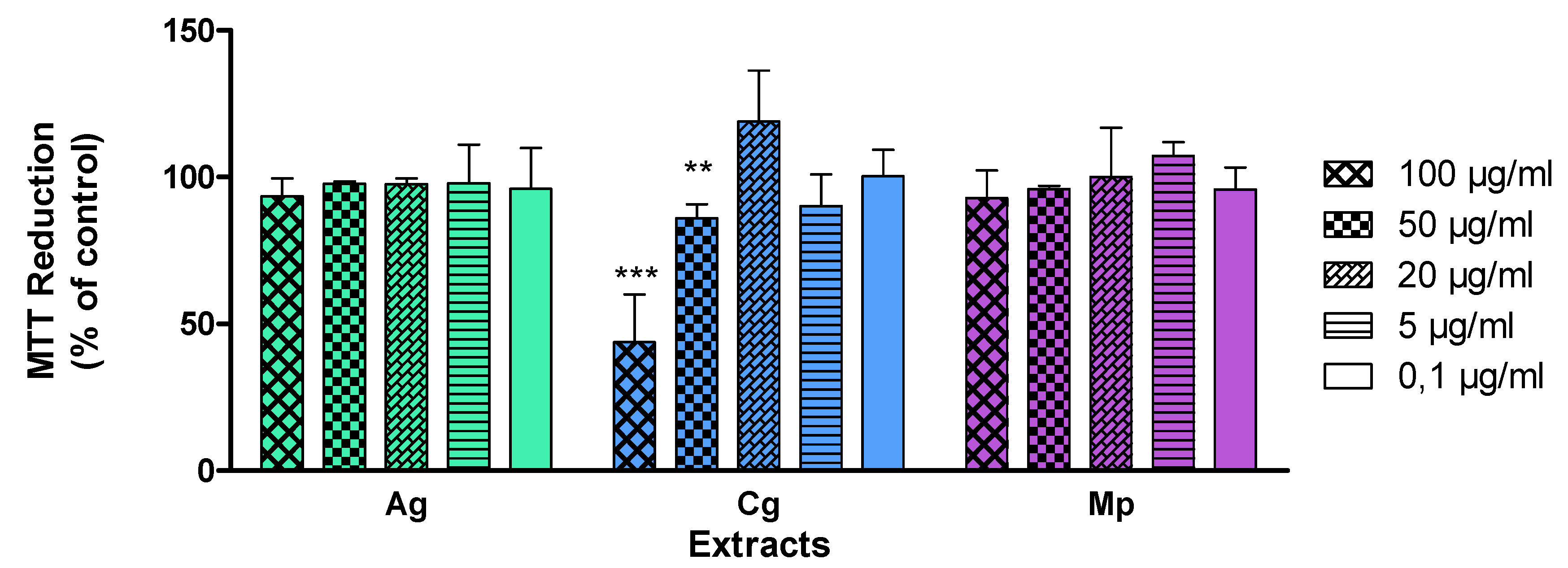
To fix a non-cytotoxic concentration of each extract for the assays, cell viability was evaluated using the IMR-32 human neuroblastoma cell line. Cells were treated with a wide range of concentrations from 0.1 µg/mL to 100 µg/mL and viability was assessed by the MTT reduction assay (Figure 1). Based on these results, the selected concentrations were 50 µg/mL for the aqueous extracts of A. genistifolia (Ag) and M pinnatus (Mp), and 20 µg/mL for C. genistoides aqueous extract (Cg).
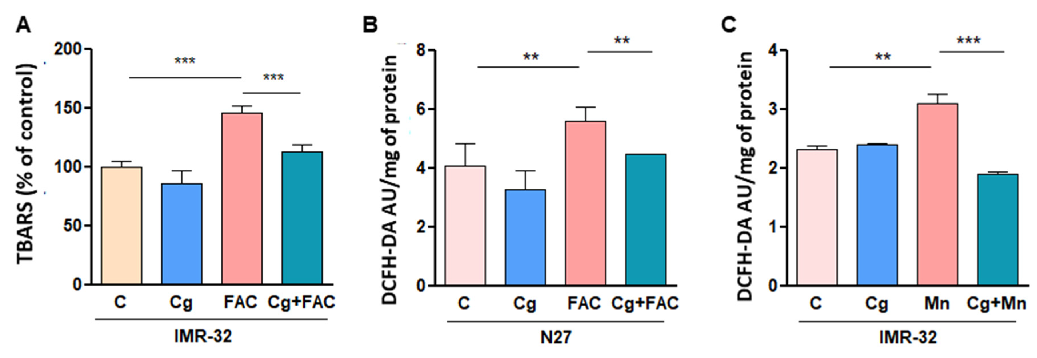
Our group has broad experience in the study of OS in the context of different neurodegenerative diseases based on cellular models, particularly in the field of iron-induced OS [8,11,12,13]. Here, ROS production was induced by FAC treatment in IMR-32 cells in order to analyze the neuroprotective effect of the extracts. To this end, assays with the DCFH-DA probe were performed to measure cellular oxidant levels in the presence of Ag, Cg and Mp after FAC exposure (Figure 2). While ROS levels were not altered by Cg treatment, they were increased in the presence of Ag and Mp. Our most interesting finding was the effect of Cg treatment against FAC-induced OS: ROS were reduced to control levels by the action of Cg. In line with these results, lipid peroxidation promoted by FAC exposure was diminished by Cg treatment (Figure 3A).
To corroborate the protective action against OS of Cg, we evaluated its effect using another neuronal cell line and a different OS inducer. N27 rat dopaminergic cells showed a reduction in ROS levels in the presence of Cg after FAC exposure (Figure 3B). Furthermore, when Mn was used to induce OS, ROS production was diminished after IMR-32 cells were exposed to Cg (Figure 3C).
These findings are in line with previous studies supporting the potential of phytotherapy in PD. Several phytochemicals obtained from Chinese herbs have been studied for their antioxidant capacity to mitigate OS and rescue the neuronal cell death in in vitro and in vivo PD models [14]. Other medicinal plants and their active ingredients presented positive effects in animal models of PD, such as the strengthening of the cellular of antioxidant capacity [15]. Furthermore, medicinal plants have been tested for their beneficial effects in OS conditions related to PD, such as Camelia sinensis and Whitania somnifera. C. sinensis showed neuroprotective action in 6-hydroxydopamine and 1-methyl-4-phenyl-1,2,3,6-tetrahydropyridine (MPTP) animal models of PD, its mechanism of action being the reversion of OS alterations [16]. Interestingly, W. somnifera root extract lessens oxidative markers and astrocyte activation in the Maneb–Paraquat PD model, concomitantly with an enhancement in locomotor activity [17].
4. Conclusions
Our findings suggest a protective activity against OS of the aqueous extract of C. genistoides. These data address this plant species as a promising candidate to continue our search for neuroprotective agents. Future studies will be carried out to determine the chemical composition of the aqueous extract of C. genistoides and its mechanism of action.
Funding
This study was supported by grants from Agencia Nacional de Promoción Científica y Tecnológica [PICT2013-0987, PICT2017-0224] to G. Salvador, and the Universidad Nacional del Sur [PGI24B292] to G. Salvador.
Institutional Review Board Statement
Not applicable.
Informed Consent Statement
Not applicable.
Data Availability Statement
Data is contained within the article.
Acknowledgments
N. Alza, A. Murray, and G. Salvador are research members of the Consejo Nacional de Investigaciones Científicas y Técnicas (CONICET). Oriana Benzi Juncos is a student fellow of the Universidad Nacional del Sur.
Conflicts of Interest
The authors declare no conflict of interest.
References
- Ingelsson, M. Alpha-synuclein oligomers-neurotoxic molecules in Parkinson’s disease and other Lewy body disorders. Front. Neurosci. 2016, 10, 408. [Google Scholar] [CrossRef] [PubMed]
- Lashuel, H.A.; Overk, C.R.; Oueslati, A.; Masliah, E. The many faces of α-synuclein: From structure and toxicity to therapeutic target. Nat. Rev. Neurosci. 2013, 14, 38–48. [Google Scholar] [CrossRef] [PubMed]
- Jiang, T.; Sun, Q.; Chen, S. Oxidative stress: A major pathogenesis and potential therapeutic target of antioxidative agents in Parkinson’s disease and Alzheimer’s disease. Prog. Neurobiol. 2016, 147, 1–19. [Google Scholar] [CrossRef] [PubMed]
- Sanders, L.H.; Greenamyren, J.T. Oxidative damage to macromolecules in human Parkinson disease and the rotenone model. Free Radic. Biol. Med. 2013, 62, 111–120. [Google Scholar] [CrossRef] [PubMed]
- McFarthing, K.; Buff, S.; Rafaloff, G.; Dominey, T.; Wyse, R.K.; Stott, S.R.W. Parkinson’s Disease Drug Therapies in the Clinical Trial Pipeline: 2020. J. Parkinsons Dis. 2020, 10, 757–774. [Google Scholar] [CrossRef] [PubMed]
- Hannan, M.A.; Dash, R.; Sohag, A.A.M.; Haque, M.N.; Moon, I.S. Neuroprotection Against Oxidative Stress: Phytochemicals Targeting TrkB Signaling and the Nrf2-ARE Antioxidant System. Front. Mol. Neurosci. 2020, 13, 116. [Google Scholar] [CrossRef] [PubMed]
- Alza, N.P.; Murray, A.P.; Salvador, G.A. Cativic acid-caffeic acid hybrid exerts cytotoxic effects and induces apoptotic death in human neuroblastoma cells. Naunyn Schmiedebergs Arch. Pharmacol. 2017, 390, 1229–1238. [Google Scholar] [CrossRef] [PubMed]
- Sánchez Campos, S.; Alza, N.P.; Salvador, G.A. Lipid metabolism alterations in the neuronal response to A53T α-synuclein and Fe-induced injury. Arch. Biochem. Biophys. 2018, 655, 43–54. [Google Scholar] [CrossRef] [PubMed]
- Uranga, R.M.; Alza, N.P.; Conde, M.A.; Antollini, S.S.; Salvador, G.A. Phosphoinositides: Two-Path Signaling in Neuronal Response to Oligomeric Amyloid β Peptide. Mol. Neurobiol. 2017, 54, 3236–3252. [Google Scholar] [CrossRef] [PubMed]
- de Rus Jacquet, A.; Tambe, M.A.; Ma, S.Y.; McCabe, G.P.; Vest, J.H.C.; Rochet, J.C. Pikuni-Blackfeet traditional medicine: Neuroprotective activities of medicinal plants used to treat Parkinson’s disease-related symptoms. J. Ethnopharmacol. 2017, 206, 393–407. [Google Scholar] [CrossRef] [PubMed]
- Uranga, R.M.; Salvador, G.A. Unraveling the Burden of Iron in Neurodegeneration: Intersections with Amyloid Beta Peptide Pathology. Oxidative Med. Cell. Longev. 2018, 2018, 2850341. [Google Scholar] [CrossRef] [PubMed]
- Sánchez Campos, S.; Rodríguez Diez, G.; Oresti, G.M.; Salvador, G.A. Dopaminergic neurons respond to iron-induced oxidative stress by modulating lipid acylation and deacylation cycles. PLoS ONE 2015, 10, e0130726. [Google Scholar] [CrossRef] [PubMed]
- Salvador, G.A. Iron in neuronal function and dysfunction. BioFactors 2010, 13, 103–110. [Google Scholar] [CrossRef] [PubMed]
- Ding, Y.; Xin, C.; Zhang, C.-W.; Lim, K.-L.; Zhang, H.; Fu, Z.; Li, L.; Huang, W. Natural Molecules From Chinese Herbs Protecting Against Parkinson’s Disease via Anti-oxidative Stress. Front. Aging Neurosci. 2018, 10, 246. [Google Scholar] [CrossRef] [PubMed]
- Rabiei, Z.; Solati, K.; Amini-Khoei, H. Phytotherapy in treatment of Parkinson’s disease: A review. Pharm. Biol. 2019, 57, 355–362. [Google Scholar] [CrossRef] [PubMed]
- Bitu Pinto, N.; da Silva Alexandre, B.; Tavares Neves, K.R.; Silva, A.; Lea, L.; Viana, G. Neuroprotective Properties of the Standardized Extract from Camellia sinensis (Green Tea) and Its Main Bioactive Components, Epicatechin and Epigallocatechin Gallate, in the 6-OHDA Model of Parkinson’s Disease. Evid. Based Complement. Altern. Med. 2015, 2015, 161092. [Google Scholar] [CrossRef] [PubMed]
- Prakash, J.; Yadav, S.K.; Chouhan, S.; Singh, S.P. Neuroprotective role of Withania somnifera root extract in maneb-paraquat induced mouse model of parkinsonism. Neurochem. Res. 2013, 38, 972–980. [Google Scholar] [CrossRef] [PubMed]
Figure 1.
Evaluation of cytotoxicity of aqueous extracts of A. genistifolia (Ag), C. genistoides (Cg) and M pinnatus (Mp). IMR-32 cell viability was determined by the MTT reduction assay to fix the concentration for assays. Bars represent means ± SD. ** p < 0.01, *** p < 0.001 with respect to the control conditions.

Figure 2.
Effect of aqueous extracts of A. genistifolia (Ag), C. genistoides (Cg) and M pinnatus (Mp) on reactive oxygen species (ROS) production induced by ferric ammonium citrate (FAC). The dichloro-dihydro-fluorescein diacetate (DCFH-DA) probe was used to determine ROS levels in IMR-32 cells. Bars represent means ± SD. ** p < 0.01, *** p < 0.001 with respect to the control conditions.

Figure 3.
Neuroprotective effect of the aqueous extract of C. genistoides (Cg) against oxidative stress (OS). (A) Evaluation of lipid peroxidation in the presence of Cg after FAC exposure. Thiobarbituric acid reactive substance (TBARS) assay was performed in IMR-32 cell line. (B) Determination of FAC-induced ROS generation in N27 cells treated with Cg using the DCFH-DA probe. (C) Measurement of ROS levels in IMR-32 cells exposed to Mn as OS inducer in the presence of Cg. Bars represent means ± SD. ** p < 0.01, *** p < 0.001 with respect to the control conditions.

Publisher’s Note: MDPI stays neutral with regard to jurisdictional claims in published maps and institutional affiliations. |
© 2020 by the authors. Licensee MDPI, Basel, Switzerland. This article is an open access article distributed under the terms and conditions of the Creative Commons Attribution (CC BY) license (https://creativecommons.org/licenses/by/4.0/).
Share and Cite
MDPI and ACS Style
Alza, N.; Juncos, O.B.; Murray, A.; Salvador, G. Protective Effect of Cyclolepis genistoides Aqueous Extract against Cellular Oxidative Stress. Chem. Proc. 2021, 3, 104. https://0-doi-org.brum.beds.ac.uk/10.3390/ecsoc-24-08344
AMA Style
Alza N, Juncos OB, Murray A, Salvador G. Protective Effect of Cyclolepis genistoides Aqueous Extract against Cellular Oxidative Stress. Chemistry Proceedings. 2021; 3(1):104. https://0-doi-org.brum.beds.ac.uk/10.3390/ecsoc-24-08344
Chicago/Turabian StyleAlza, Natalia, Oriana Benzi Juncos, Ana Murray, and Gabriela Salvador. 2021. "Protective Effect of Cyclolepis genistoides Aqueous Extract against Cellular Oxidative Stress" Chemistry Proceedings 3, no. 1: 104. https://0-doi-org.brum.beds.ac.uk/10.3390/ecsoc-24-08344