Hybrid Oxidation of Titanium Substrates for Biomedical Applications †


Department of Innovation and Safety Systems, Czestochowa University of Technology (CUT), 42-200 Czestochowa, Poland
†
Presented at the 2nd Coatings and Interfaces Web Conference, 15–31 May 2020; Available online: https://ciwc2020.sciforum.net/.
Mater. Proc. 2020, 2(1), 8; https://0-doi-org.brum.beds.ac.uk/10.3390/CIWC2020-06845
Published: 15 May 2020
(This article belongs to the Proceedings of 2nd Coatings and Interfaces Web Conference (CIWC-2 2020))
Abstract
:Titanium oxidation for biomedical applications is still a challenge in obtaining favorable mechanical and physicochemical properties of thin oxide layers, as well as the required high bioactivity. Interesting techniques for TiO2 layer formation are electrochemical, plasma and diffusive methods. Each method aims to create a thin oxide layer characterized by thermal stability and re-passivation in the presence of a simulated body fluid SBF environment. However, an important aspect here is also the phase composition of oxide layers, essential for osseointegration. Accordingly, the research carried out aims to produce such a titanium substrate, where the surface zone is a Tiα(O) solid solution formed with fluidized bed (FB) diffusion process (640 °C, 8 h) and the top layer is TiO2 produced by physical vapour deposition PVD—magnetron sputtering. The effects of such hybrid oxidation on titanium surface properties were investigated with scanning electron microscopy SEM/scanning transmission electron microscopy STEM/ Raman spectroscopy RS and nanoindentation tests. The results showed that hybrid oxidation made it possible to generate a favorable synergetic effect between FB and PVD oxide layers and to reduce the stresses at their interface. In turn, a variable share of TiO2 phases (rutile + anatase mixture) obtained at the titanium surface allowed for the significant enhancement of hydroxyapatite compound growth, which was confirmed by a 14-day Kokubo test.
1. Introduction
Titanium oxide thin layers are still the subject of numerous studies due to their highly interesting properties for biomedicine and implantology, especially for third-generation biomaterials, which are produced to stimulate specific cell responses and tissue regeneration [1,2,3]. In fact, thin TiO2 oxide layers are obtained with the use of several methods, such as: anodizing, laser treatment PLD, physical methods PVD and diffusion methods, which result in reduced thickness and poor adhesion, which depends on many factors, including surface preparation for the oxidation, the phase composition of surface oxides and the substrate’s chemical and strength properties [4,5,6,7]. Nevertheless, each method aims to create a passive oxide coating, which is characterized by homogeneity, low thermal conductivity, chemical stability and the ability to re-passivate after being defected in the presence of a corrosive environment. The mechanism of titanium oxidation differs from the oxidation of other metals. This is due to stability of the material: Tiα—stable up to 882 °C and Tiβ—stable over 882 °C. At room temperature, on the Ti surface, a thin (5–15 nm) passive nanolayer is formed, where, in turn, the oxidation of titanium at a high temperature (>400 °C) leads to the formation of the crystalline layer with a TiO/inter-Ti2O3 layer and a TiO2 layer (rutile or anatase) zone structure. The oxide coating formed at room temperature is stable and adheres well to the substrate, but is too thin. In turn, at high temperatures, titanium oxidizes rapidly and forms thick oxide layer which is often porous and poorly bonded (anchored) to the substrate, and thus delaminates and cracks [8,9,10,11]. An important role of the oxide layer on the titanium surface, in addition to the aforementioned properties, is to provide the required osseointegration process kinetics by forcing the biochemical activity of the layers leading to accelerated interaction with the body’s tissues [12,13,14,15,16]. Initially, it was thought that the titanium substrates are inert to the body. However, when in direct contact with the tissues of organisms, titanium can release Ti ions into the body’s environment, which causes the occurrence of edema and inflammation, generates health problems for patients and ultimately the rejection of the implant. The biocompatibility and bioactivity of titanium are directly related to the physicochemical properties of the substrate surface. To improve the bioactivity of titanium substrates, the best-known solutions are single-stage surface treatment and the production of multilayers [17,18,19,20]. However, surface methods, due to the conditions of rapid chemical interaction between the atmosphere and substrate, have very limited influence on oxygen diffusion processes towards the substrate surface layer and the formation of a Tiα(O) diffusion layer with good strength properties. Thus, it is difficult to obtain substrates with the following arrangement: I. Tiα substrate/II. Tiα(O) solid solution/III. thin TiO2 oxide layer, with both stable oxide phases at the surface, a low hardness gradient between the matrix and layer and a reduced state of stress (compressive stresses required) at the interface [21,22,23,24]. Accordingly, highly bioactive titanium materials (i.e., third-generation metallic biomaterials) might be produced by the adequate functionalization of the thin oxide layers (tailored phase composition morphology and adhesion to the substrate) together with the control of the substrate surface stress state and structure. Therefore, the research carried out by the author aims to develop such a titanium substrate, where on the diffusion oxide layer (Tiα(O) solid solution), a homogenous, tight and smooth thin TiO2 layer is formed by surface treatment, i.e., PVD magnetron sputtering. Such a hybrid method uses the advantages of continuous substrate activation and defect by the influence of a fluidized bed aeromechanical factor and non-equilibrium PVD surface oxidation. There is the expectation that a combination of TiO2 layers will ensure a synergistic effect in the improvement of the titanium substrate’s biofunctional properties.
2. Materials and Methods
The substrates used for hybrid oxidation were made of Tiα single-phase commercially pure titanium manufactured by Kobe Steel LTD in accordance with ASTM 8348, with the chemical composition presented in Table 1.
Before hybrid oxidation, the substrates were mechanically activated by blasting with a mixture of Al2O3 + ZrO2 + Ti. Diffusive oxidation was carried out in a fluidized bed (FB) reactor with Al2O3 grain material at 640 °C for 8 h in an air atmosphere. After the FB treatment, substrates were cooled down in air. A further oxidation process was conducted with PVD magnetron sputtering using a TiO2 target, with a pressure of 3 × 10₋2 mbar, an Ar (99.95%) atmosphere, constant power mode P = 350 W, target–substrate distance 60 mm and deposition time 20 min. The thin TiO2 oxide layer structure and interface were analyzed by a scanning electron microscopy (SEM) (FEI E-SEM XL30 microscope) and scanning transmission electron microscopy (STEM) (FEI S/TEM TITAN 80-300) method. The surface morphology of the substrates was evaluated by confocal laser scanning microscopy (CLSM) (LEXT OLS4000 microscope OLYMPUS, Tokyo, Japan). Phase analysis of the TiO2 layers was conducted by Raman spectroscopy (RS) (LabRAM HR micro-Raman spectrometer equipped with a CCD detector HORIBA Scientific France SAS), under an excitation wavelength of 532 nm and an intensity of ca. 10 mW. The acquisition time was set at 30 s. The precise determination of the oxide layers’ hardness, Young’s modulus and elastic and plastic energy was realized with nanoindentation mechanical tests (NanoTest Vantage Micro Materials Quantum Design Europe, United Kingdom). The bioactivity response of the titanium substrates was evaluated by a 14-day Kokubo test using c-SBF2 solution. It was found that the hybrid oxidation method (FB+PVD) led to the formation of tight, homogeneous thin TiO2 layers, which highly improves the bioactivity of the titanium surface in the aspect of biomedical applications.
3. Results and Discussion
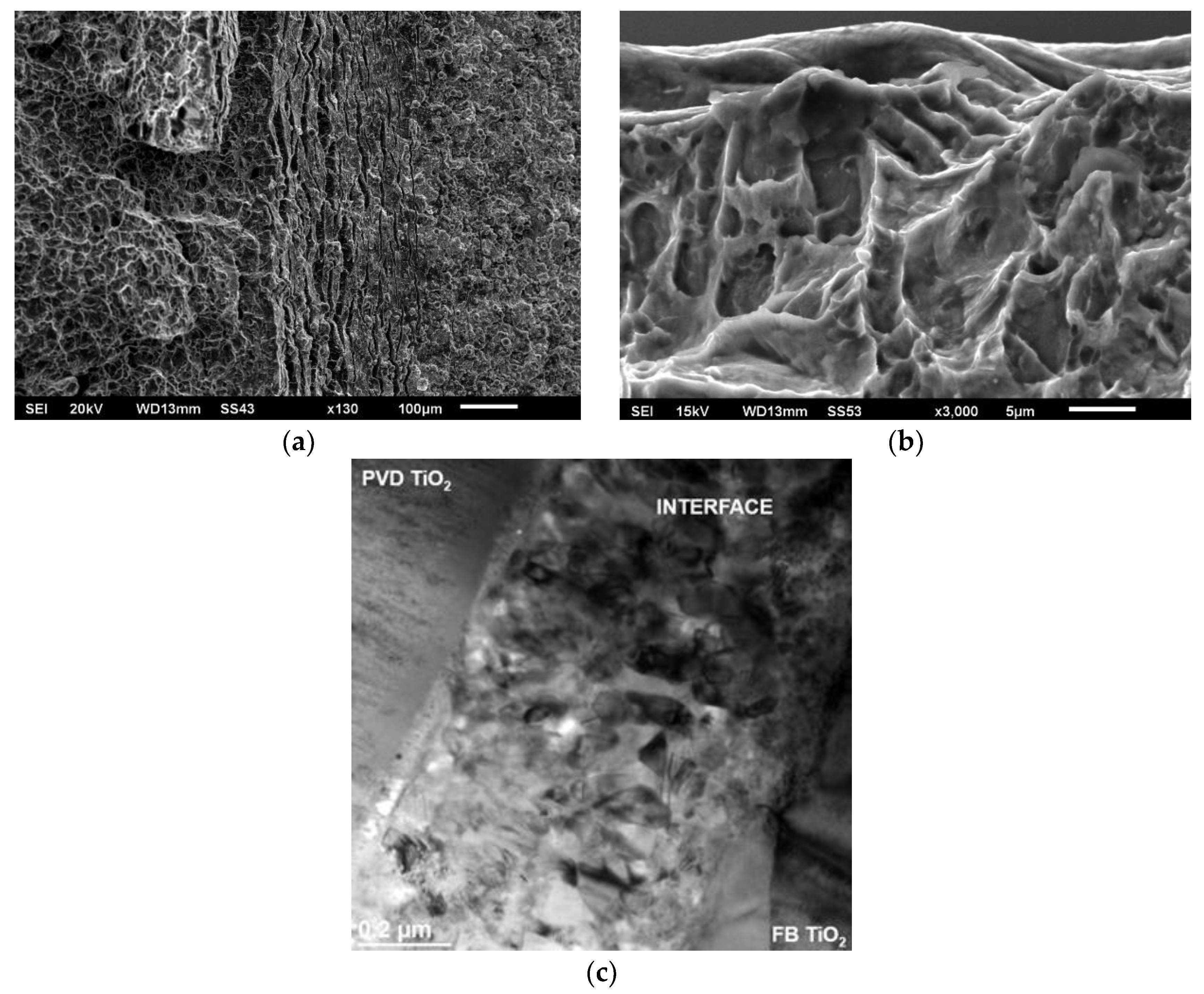
Titanium oxidation, realized by a two-stage hybrid process (FB + PVD), allowed for the production of strong substrates with rutile and anatase TiO2 thin layers at the top surface. The first stage of the oxidation process was conducted in a fluidized bed (FB), which allowed for the production of a Tiα(O) diffusion layer with a thickness of 11 µm and a ca. 2 µm nano-porous TiO2 oxide layer. The saturation of titanium with oxygen atoms leads to the strengthening of the substrate matrix and improves its hardness. Furthermore, a fine-grained diffusion zone under a nano-porous oxide layer aimed to reduce the stress gradient between the matrix and the TiO2 layer. The second stage of the oxidation process was PVD magnetron sputtering, which resulted in the deposition of a thin TiO2 oxide layer with a thickness of ca. 0.8–1 µm. The plasma interaction with the FB substrate involved the continuous bombardment of nano-porous TiO2 and enhanced local heat transfer to control chemical reactions (physisorption) when forming thin TiO2 PVD layers. Hybrid oxidation also produced a stable and fine FB TiO2/PVD TiO2 interface with a thickness of ca. 600–620 nm (Figure 1).
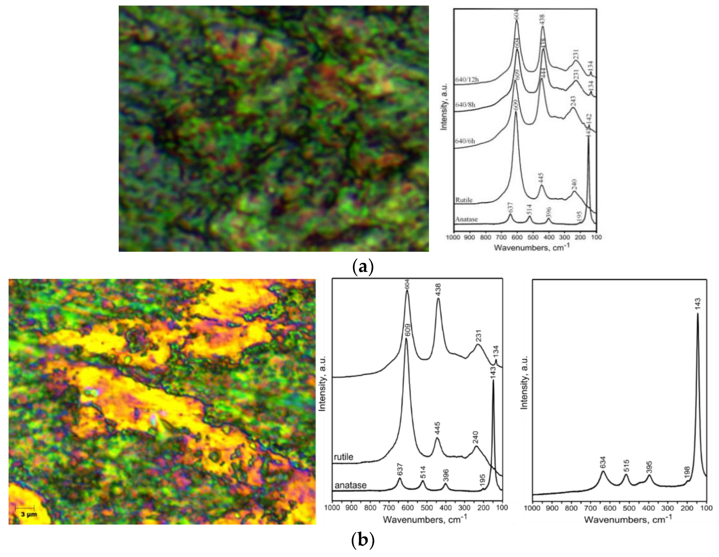
At the interface zone, there were visible areas of nano-pores, which were free gaps for further anchoring the TiO2 layers deposited by the PVD method. The next step of the research was substrate surface morphology and phase analysis conducted by confocal laser scanning microscopy and Raman spectroscopy (Figure 2).
The Raman spectra obtained for titanium after FB oxidation showed the presence of the strongest peaks coming from the rutile phase. Hardly noticeable bands located at wave numbers of 143–146 cm−1 were obtained for the anatase phase. However, hybrid oxidation FB + PVD showed the presence of visible TiO2 rutile (wave numbers: 604 cm−1, 438 cm−1 and 231 cm−1) and anatase bands in the wave numbers of 143 cm−1, 395 and 515 cm−1. In addition, it was also observed that the bands shifted towards lower wave numbers, suggesting the occurrence of compressive stresses in the TiO2 thin layer. The rutile phase of TiO2 layers plays an important role in inducing the apatite deposition as a result of the crystal lattice matching between rutile and apatite. In fact, there are some literature data which show biomedical properties of anatase, including the author’s previous works [25,26]. The author tried to define and precisely indicate the favorable phase share of the rutile and anatase titanium oxide mixture at the surface, which promises to have a great influence on the bioactive behavior of the substrates. Such a phase gradient (between rutile and anatase) has a great influence on the osteogenesis and bioactivity of titanium substrates. The next step of the research was the nanomechanical investigation of the PVD thin TiO2 oxide layer. The results showed the favorable strength properties of the layers. A series of indentations (at nano- and micro-scales) was performed on pure titanium (raw substrates) and the specimens after hybrid oxidation. The results of the nanoindentation tests are shown in Table 2.
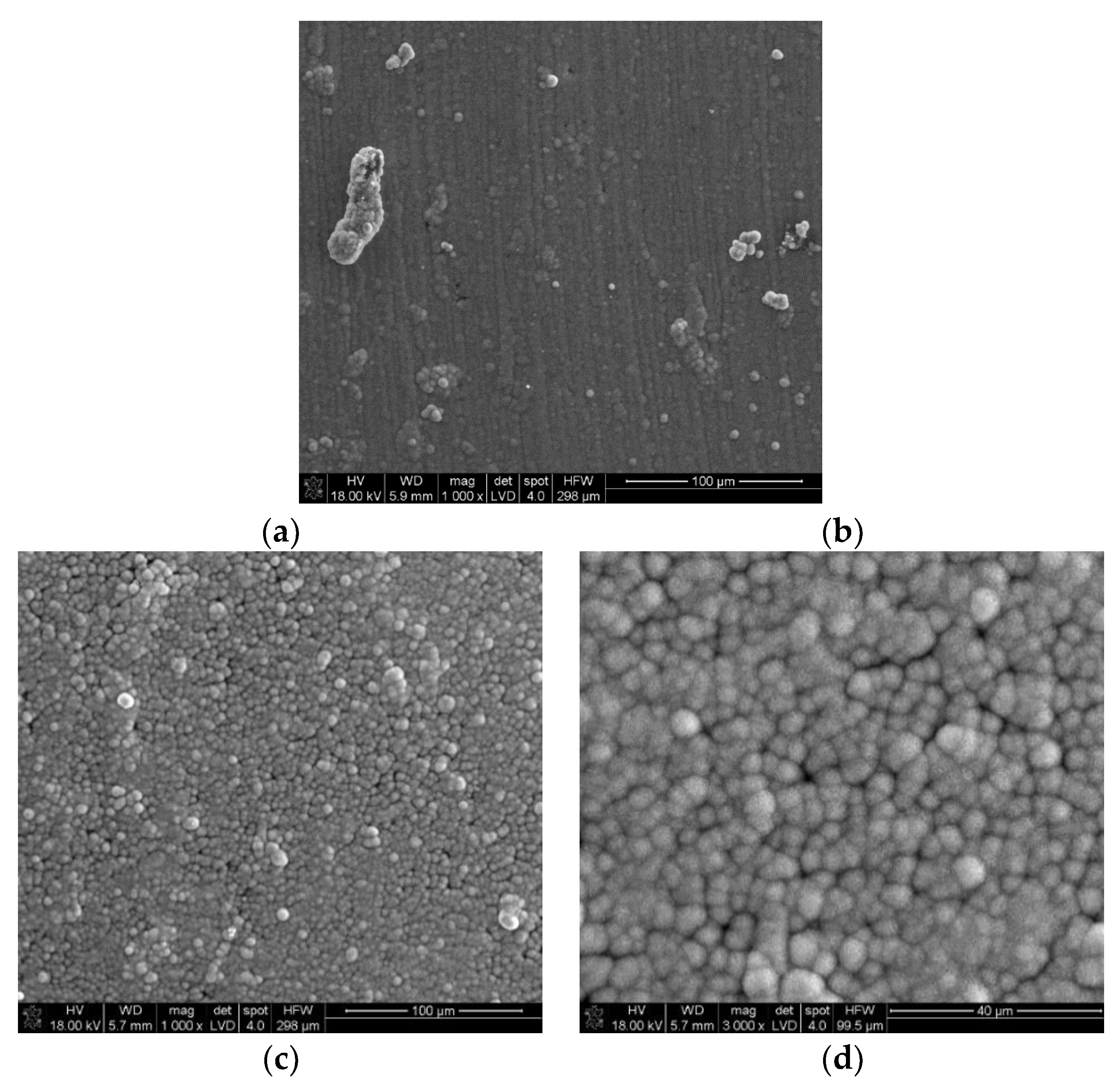
Results allowed for finding a correlation between the mechanical parameters measured at the nano- and micro-scales for the substrates. Special attention was devoted to the mechanical properties of the FB + PVD interface, which plays a crucial role in the integrity of the whole hybrid system. The nanoindentation hardness and Young’s modulus measured for the FB + PVD TiO2 were H = 15.21 GPa and E = 261 GPa, which were slightly higher values than the results of sputtered TiO2 layers reported in the literature [27]. The nanoindentation results confirmed that hybrid oxidation affects the improvement of titanium surface hardness and strength. From the point of view of the application of the obtained substrates as biomaterials, it is necessary to determine their bioactivity. Such important results were obtained after the 14-day Kokubo test in simulated body fluid SBF [28,29] (Figure 3).
The intensive growth of globular hydroxyapatite compounds was visible at the surface of FB + PVD TiO2 thin rutile/anatase layers. Such an improvement in biochemical activity was reached both through stabilization and the reduction of stresses at the FB TiO2/PVD TiO2 interface, and the tailoring of the TiO2 phase composition at the surface. The Kokubo test results confirmed that the hybrid oxidation significantly enhances the bioactivity and allows for the biofunctional modification of titanium substrates.
4. Conclusions
- Diffusion oxidation in a fluidized bed (FB) leads to the formation of a highly defected Tiα(O) diffusion zone with good strength properties and nano-porous TiO2. Such a system plays a role as a foundation for the subsequent deposition of thin TiO2 layers by PVD magnetron sputtering.
- The hybrid oxidation treatment applies two types of surface activation, I: mechanical, as an impact of an aeromechanical factor in FB; II: sputtering, with simultaneous oxidation by PVD. Activation increases the number of active centers, and enhances oxygen mass transport to finally form homogenous thin TiO2 layers. The layers are characterized by a high level of homogeneity and resistance to cracking and delayering.
- In hybrid oxidation, the interface between nano-porous FB TiO2 and PVD TiO2 has a favorable state of stress and further influences the formation of a bioactive rutile and anatase mixture, which improves the rate of osseointegration.
- The presented hybrid oxidation is a promising surface treatment for biomedical applications, indicating the directions of forming bioactive layers on titanium substrates. The solution corresponds with the new trends in biomaterials and surface engineering to combine different processing techniques in order to improve implants and medical devices.
5. Patents
Patent no PL 221053: Method for modifying the surface layer of titanium alloy implants. P. Podsiad, J.J. Jasinski, J. Jasinski, R. Czyz.
Funding
This research was funded by the National Science Centre, Poland.
Conflicts of Interest
The author declares no conflict of interest.
References
- Rack, H.J.; Qazi, J.I. Titanium alloys for biomedical applications. Mater. Sci. Eng. C 2006, 26, 1269–1277. [Google Scholar] [CrossRef]
- Kokubo, T.; Kim, H.M.; Kawashita, M.; Nakamura, T. Bioactive metals: preparation and properties. J. Mater. Sci. Mater. Med. 2004, 15, 99–107. [Google Scholar] [CrossRef] [PubMed]
- Rahimi, N.; Pax, R.A.; Mac, A.; Gray, E. Review of functional titanium oxides. I: TiO2 and its modifications. Prog. Solid State Chem. 2016, 44, 86–105. [Google Scholar] [CrossRef]
- Zhou, B.; Jiang, X.; Shen, R.; Rogachev, A.V. Preparation and characterization of TiO2 thin film by thermal oxidation of sputtered Ti film. Mater. Sci. Semicond. Process. 2013, 16, 513–519. [Google Scholar] [CrossRef]
- Radmanesh, M.; Kiani, A. Bioactivity enhancement of titanium induced by Nd:Yag laser pulses. J. Appl. Biomater. Funct. Mater. 2016, 14, 70–77. [Google Scholar]
- Wu, B.; Yu, Y.; Wu, J.; Shchelkanov, I.; Ruzic, D.N.; Huang, N.; Len, Y.X. Tailoring of titanium thin film properties in high power pulsed magnetron sputtering. Vacuum 2018, 150, 144–154. [Google Scholar] [CrossRef]
- Heinrichs, J.; Jarmar, T.; Wiklund, U.; Engqvist, H. Physical Vapour Deposition and Bioactivity of Crystalline Titanium Dioxide Thin Films. Trends Biomater. Artif. Organs 2008, 22, 104–110. [Google Scholar]
- Shannon, R.D.; Pask, J.A. Kinetics of the anatase-rutile transformation. J. Am. Ceram. Soc. 1965, 48, 391–398. [Google Scholar] [CrossRef]
- Aniołek, K. The influence of thermal oxidation parameters on the growth of oxide layers on titanium. Vacuum 2017, 144, 94–100. [Google Scholar] [CrossRef]
- Satoh, N.; Nakashima, T.; Yamamoto, K. Metastability of anatase: size dependent and irreversible anatase-rutile phase transition in atomic-level precise titania. Sci. Rep. 2013, 3, 1959. [Google Scholar] [CrossRef] [PubMed]
- Pradhan, S.S.; Sahoo, S.; Pradhan, S.K. Influence of annealing temperature on the structural, mechanical and wetting property of TiO2 films deposited by RF magnetron sputtering. Thin Solid Films 2010, 518, 6904–6908. [Google Scholar] [CrossRef]
- Ochsenbein, A.; Chai, F.; Winter, S.; Traisnel, M.; Breme, J.; Hildebrand, H.F. Osteoblast responses to different oxide coatings produced by the sol-gel process on titanium substrates. Acta Biomater. 2008, 4, 1506–1517. [Google Scholar] [CrossRef] [PubMed]
- Barfeie, A.; Wilson, J.; Rees, J. Implant surface characteristics and their effect on osseointegration. Br. Dent. J. 2015, 218, E9. [Google Scholar] [CrossRef]
- Niinomi, M.; Nakai, M.; Hieda, J. Development of new metallic alloys for biomedical applications. Acta Biomater. 2012, 8, 38883–38903. [Google Scholar] [CrossRef]
- Forsgren, J.; Svahn, F.; Jarmar, T.; Engqvist, H. Formation and adhesion of biomimetic hydroxyapatite deposited on titanium substrates. Acta Biomater. 2007, 3, 980–984. [Google Scholar] [CrossRef]
- Rosales-Leal, J.I.; Rodríguez-Valverde, M.A.; Mazzaglia, G.; Ramon-Torregrosa, P.J.; Diaz-Rodriguez, L.; Garcia-Martinez, O.; Vallecillo-Capilla, M.; Ruiz, C.; Cabrerizo-Vilchez, M.A. Effect of roughness, wettability and morphology of engineered titanium surfaces on osteoblast-like cell adhesion. Colloids Surf. A Physicochem. Eng. Asp. 2010, 365, 222–229. [Google Scholar] [CrossRef]
- Yamaguchi, S.; Nath, S.; Sugawara, Y.; Divakarla, K.; Das, T.; Manos, J.; Chrzanowski, W.; Matsushita, T.; Kokubo, T. Two-in-one biointerfaces—Antimicrobial and bioactive nanoporous gallium titanate layers for titanium implants. Nanomaterials 2017, 7, 229. [Google Scholar] [CrossRef]
- Ding, Z.; Hu, X.; Yue, P.L.; Lu, G.Q.; Greenfield, P.F. Synthesis of anatase TiO2 supported on porous solids by chemical vapor deposition. Catal. Today 2001, 68, 173–182. [Google Scholar] [CrossRef]
- Sabetrasekh, R.; Tiainen, H.; Lyngstadaas, S.P.; Reseland, J.; Haugen, H. A novel ultra-porous titanium dioxide ceramic with excellent biocompatibility. J. Biomater. Appl. 2011, 25, 559–580. [Google Scholar] [CrossRef]
- Sengottuvelan, A.; Balasubramanian, P.; Will, J.; Boccaccini, A.R. Bioactivation of titanium dioxide scaffolds by ALP-functionalization. Bioact. Mater. 2017, 2, 108–115. [Google Scholar] [CrossRef]
- Li, D.; Ferguson, S.J.; Beutler, T.; Cochran, D.L.; Siting, C.; Hirt, H.P.; Buser, D.J. Biomechanical comparison of the sandblasted and acid-etched and the machined and acid-etched titanium surface for dental implants. J. Biomed. Mater. Res. 2002, 60, 325–332. [Google Scholar] [CrossRef] [PubMed]
- Lubas, M.; Sitarz, M.; Jasinski, J.J.; Jelen, P.; Klita, L.; Podsiad, P.; Jasinski, J. Fabrication and characterization of oxygen–Diffused titanium using spectroscopy method. Spectrochim. Acta A 2014, 133, 883–886. [Google Scholar] [CrossRef]
- Sarvadii, S.Y.; Gatin, A.K.; Kharitonov, V.A.; Dokhlikova, N.V.; Ozerin, S.A.; Grishin, M.V.; Shub, B.R. Oxidation of Thin Titanium Films: Determination of the Chemical Composition of the Oxide and the Oxygen Diffusion Factor. Crystals 2020, 10, 117. [Google Scholar] [CrossRef]
- Toptan, F.; Alves, A.C.; Pinto, A.M.P.; Ponthiaux, P. Tribocorrosion behavior of bio-functionalized highly porous titanium. J. Mech. Behav. Biomed. 2017, 69, 144–152. [Google Scholar] [CrossRef] [PubMed]
- He, J.; Zhou, W.; Zhou, X.; Zhong, X.; Zhang, X.; Wan, P.; Zhu, B.; Chen, W. The anatase phase of nanotopography titania plays an important role on osteoblast cell morphology and proliferation. J. Mater. Sci. Mater. Med. 2008, 19, 3465–3472. [Google Scholar] [CrossRef]
- Jasinski, J.J.; Lubas, M.; Kurpaska, L.; Napadlek, W.; Sitarz, M. Functionalization of Ti99.2 substrates surface by hybrid treatment investigated with spectroscopic methods. J. Mol. Struct. 2018, 1164, 412–419. [Google Scholar] [CrossRef]
- Pang, M.; Bahr, D. Thin-film fracture during nanoindentation of a titanium oxide film–titanium system. J. Mater. Res. 2001, 16, 2634–2643. [Google Scholar] [CrossRef]
- Kokubo, T.; Takadama, H. How useful is SBF in predicting in vivo bone bioactivity. Biomaterials 2006, 27, 2907–2915. [Google Scholar] [CrossRef] [PubMed]
- Oyane, A.; Onuma, K.; Ito, A.; Kim, H.M.; Kokubo, T.; Nakamura, T. Formation and growth of clusters in conventional and new kinds of simulated body fluids. J. Biomed. Mater. Res. 2003, 64, 339–348. [Google Scholar] [CrossRef]
Figure 1.
SEM/STEM images of the titanium substrate microstructure and interface after hybrid oxidation (a) fluidized bed FB 640 °C/8 h, (b) fluidized bed FB + PVD magnetron sputtering and (c) FB TiO2/PVD TiO2 interface.
Figure 1.
SEM/STEM images of the titanium substrate microstructure and interface after hybrid oxidation (a) fluidized bed FB 640 °C/8 h, (b) fluidized bed FB + PVD magnetron sputtering and (c) FB TiO2/PVD TiO2 interface.

Figure 2.
Confocal laser scanning microscopy image and Raman spectra of the titanium surface after hybrid oxidation, (a) FB 640 ℃/8 h and (b) FB + PVD magnetron sputtering.
Figure 2.
Confocal laser scanning microscopy image and Raman spectra of the titanium surface after hybrid oxidation, (a) FB 640 ℃/8 h and (b) FB + PVD magnetron sputtering.

Figure 3.
SEM image of the effect of hydroxyapatite on the growth of titanium substrates after hybrid oxidation—14-day simulated body fluid SBF Kokubo test, (a) FB 640 ℃/8 h and (b) FB + PVD rutile + anatase phase.
Figure 3.
SEM image of the effect of hydroxyapatite on the growth of titanium substrates after hybrid oxidation—14-day simulated body fluid SBF Kokubo test, (a) FB 640 ℃/8 h and (b) FB + PVD rutile + anatase phase.


Table 1.
The chemical composition of commercially pure titanium used for hybrid oxidation (in accordance with ASTM 8348) (mass %).
Table 1.
The chemical composition of commercially pure titanium used for hybrid oxidation (in accordance with ASTM 8348) (mass %).
| Material | Chemical Composition | |||||
|---|---|---|---|---|---|---|
| KOBE Steel LTD Titanium Grade 2 (ASTM 8348) | O | N | C | H | Fe | Ti |
| 0.20 | 0.03 | 0.10 | 0.015 | 0.30 | rest | |
Table 2.
Nanoindentation test results of titanium substrates before and after hybrid oxidation (FB + PVD).
Table 2.
Nanoindentation test results of titanium substrates before and after hybrid oxidation (FB + PVD).
| Substrate Type | Hardness, H (GPa) | Reduced Young’s Modulus, ER (GPa) | Calculated Young’s Modulus, E (GPa) | Maximum Depth (nm) | Plastic Depth (nm) | |||||
|---|---|---|---|---|---|---|---|---|---|---|
| Titanium Grade 2 (ASTM 8348) | Value | SD | Value | SD | Value | SD | Value | SD | Value | SD |
| 9.33 | 4.14 | 160.00 | 60.30 | 148.34 | 55.91 | 204.08 | 57.41 | 167.17 | 54.36 | |
| Titanium after hybrid oxidation FB + PVD | Value | SD | Value | SD | Value | SD | Value | SD | Value | SD |
| 15.21 | 6.04 | 281.83 | 87.79 | 261.28 | 81.39 | 144.90 | 28.87 | 119.20 | 27.37 | |
© 2020 by the author. Licensee MDPI, Basel, Switzerland. This article is an open access article distributed under the terms and conditions of the Creative Commons Attribution (CC BY) license (http://creativecommons.org/licenses/by/4.0/).
Share and Cite
MDPI and ACS Style
Jasinski, J.J. Hybrid Oxidation of Titanium Substrates for Biomedical Applications. Mater. Proc. 2020, 2, 8. https://0-doi-org.brum.beds.ac.uk/10.3390/CIWC2020-06845
AMA Style
Jasinski JJ. Hybrid Oxidation of Titanium Substrates for Biomedical Applications. Materials Proceedings. 2020; 2(1):8. https://0-doi-org.brum.beds.ac.uk/10.3390/CIWC2020-06845
Chicago/Turabian StyleJasinski, Jaroslaw Jan. 2020. "Hybrid Oxidation of Titanium Substrates for Biomedical Applications" Materials Proceedings 2, no. 1: 8. https://0-doi-org.brum.beds.ac.uk/10.3390/CIWC2020-06845