Review

Reprogramming of Central Carbon Metabolism in Myeloid Cells upon Innate Immune Receptor Stimulation

Centre International de Recherche en Infectiologie (CIRI), Univ Lyon, Inserm, U1111, Université Claude Bernard Lyon 1, CNRS, UMR5308, ENS de Lyon, F-69007 Lyon, France
*
Author to whom correspondence should be addressed.
Submission received: 18 November 2020 / Revised: 27 December 2020 / Accepted: 4 January 2021 / Published: 11 January 2021

Abstract

:
Immunometabolism is a relatively new field of research that aims at understanding interconnections between the immune system and cellular metabolism. This is now well-documented for innate immune cells of the myeloid lineage such as macrophages and myeloid dendritic cells (DCs) when they engage their differentiation or activation programs. Several studies have shown that stimulation of DCs or macrophages by the binding of pathogen-associated molecular patterns (PAMPs) to pattern recognition receptors (PRRs) leads to increased glycolytic activity and rewiring of central carbon metabolism. These metabolic modulations are essential to support and settle immunological functions by providing energy and immunoregulatory metabolites. As the understanding of molecular mechanisms progressed, significant differences between cell types and species have also been discovered. Pathways leading to the regulation of central carbon metabolism in macrophages and DCs by PRR signaling and consequences on cellular functions are reviewed here.

1. Introduction

It is now widely accepted that the metabolic status and effector functions of immune cells are intimately connected [1]. Indeed, immune cells must adjust their cellular metabolism depending on their microenvironment and the stimulatory or inhibitory signals they receive through their multiple receptors. According to their differentiation or activation program, immune cells may change their need for nutrients and the manner they metabolize them. In addition, recent studies have revealed that certain metabolites have potent immunomodulatory properties that were underestimated until recently. For example, the end product of the metabolic breakdown of purine nucleotides, uric acid, plays the role of alarmin by activating the inflammatory response, whereas kynurenine, a metabolite of tryptophan degradation, is a potent immunosuppressive metabolite. The word “immunometabolism” refers to these complex interactions between metabolites, metabolic pathways and immunity, and covers an extremely dynamic field of research. Among immune cells, macrophages and dendritic cells (DCs) play a pivotal role at the interface between innate and adaptive immunity. Main functions include endocytosis and phagocytosis of pathogens, secretion of antimicrobial, inflammatory and immunoregulatory factors, and presentation of antigens to lymphoid cells. Both macrophages and DCs are from the myeloid lineage and express a large panel of pattern recognition receptors (PRRs) such as toll-like receptors (TLRs) and RIG-like receptors (RLRs), allowing the detection of pathogen-associated molecular patterns (PAMPs) and damage-associated molecular patterns (DAMPs). When PRRs are engaged by their cognate ligands, macrophages and DCs are activated and undergo in-depth metabolic reprogramming. These modulations are required for these cells to fulfill their immunological functions [2,3]. This review summarizes recent findings on the regulation of central carbon metabolism in macrophages and DCs upon activation by PRRs and how this can lead to the production of specific immunomodulatory metabolites by these cells.

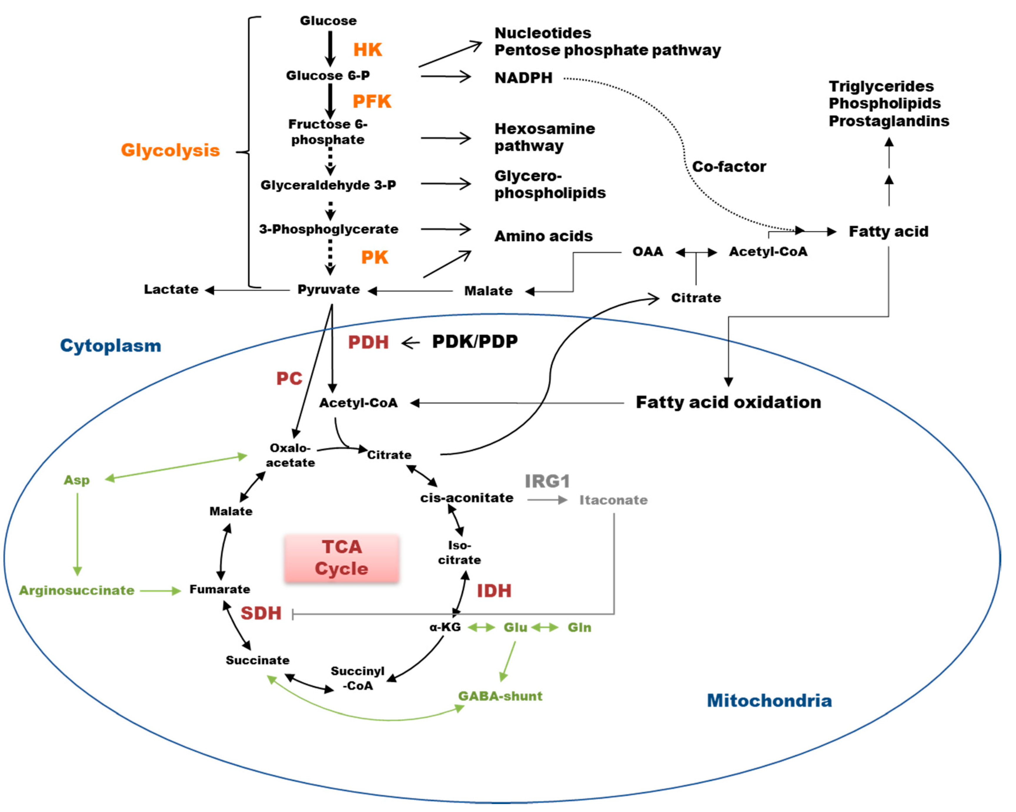
2. Glycolysis in Cell Bioenergetics

Glucose catabolism is a major source of energy for the cell. Glycolysis is the metabolic pathway converting glucose to pyruvate by ten enzymatic reactions (Figure 1), producing 2 moles of ATP together with 2 moles of NADH per mole of glucose. Pyruvate is then converted either into lactate that is secreted or into oxaloacetate (OAA) or acetyl-CoA when entering the mitochondrial matrix to feed the tricarboxylic acid (TCA) cycle (Figure 1). When oxygen is available, glycolysis is connected to the mitochondrial respiratory chain and enables oxidative phosphorylation (OXPHOS). Transport of electrons through the proteins of the respiratory chain generates a gradient of proton necessary for ATP synthesis. When glycolysis and OXPHOS are coupled, up to 36 moles of ATP per mole of glucose can be produced. Under hypoxic conditions, OXPHOS reactions are impaired, and therefore, glycolytic activity is increased to fulfill the needs in energy [4]. Because it is the fastest way to produce ATP, glycolysis is also rapidly enhanced when the demand for ATP suddenly increases, and mitochondrial activity is insufficient [5]. Finally, intermediate metabolites of glycolysis are precursors for the biosynthesis of pentose phosphates, hexosamines, glycerophospholipids and amino acids, so that glycolysis can fuel various anabolic pathways whenever required [2] (Figure 1). Hexokinase (HK), phosphofructokinase (PFK) and pyruvate kinase (PK) are the three rate-limiting enzymes regulating the glycolytic flux (Figure 1). HK controls the entry of glucose into the pathway by producing glucose-6-phosphate. Depending on cell type and tissue, this first step of glycolysis can be catalyzed by one of the 4 isoenzymes produced by a set of paralogous genes named HK1, HK2, HK3, and HK4 (or GCK for glucokinase). HK1 and HK3 are ubiquitously expressed, while HK4 expression is essentially restricted to the liver and pancreas. Interestingly, the high-affinity HK2 enzyme is mainly expressed in tissues having specific needs in energy, including tumor cells. Both HK1 and HK2 can bind to the voltage-dependent anion channel VDAC1 at mitochondria-endoplasmic reticulum junctions or mitochondria associated membranes (MAMs). This localization is associated with a higher HK activity, likely due to a better coupling to the production of ATP [6].

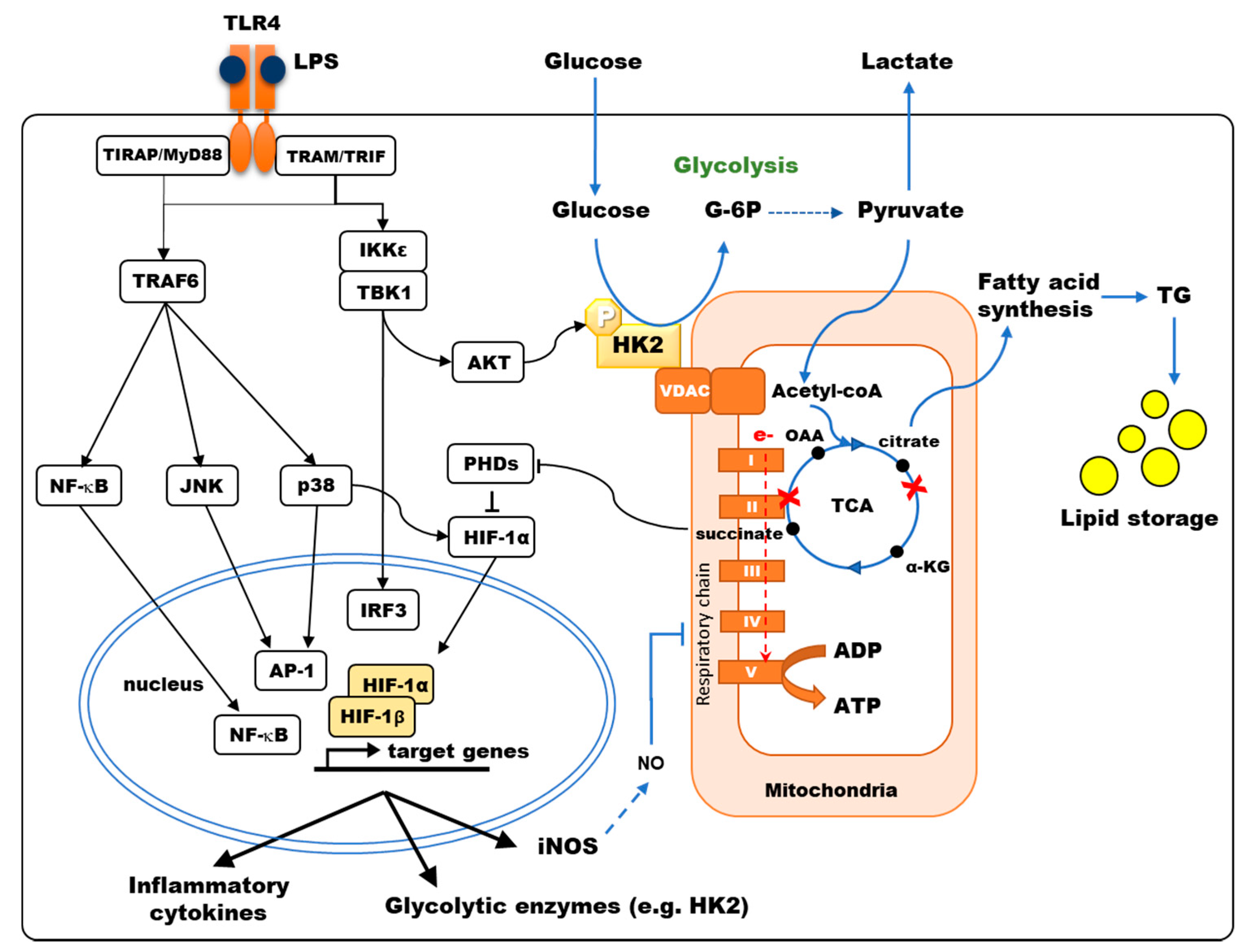
3. PRR Stimulation Triggers a Glycolytic Reprogramming of Immune Cells

In both immune and non-immune cells, PRRs participate in immune surveillance by detecting PAMPs and DAMPs. TLRs form a family of transmembrane receptors, including 10 members in humans that localize at the cell surface or in endocytic vesicles with their sensing domain exposed to extracellular PAMPs. Among TLRs, TLR4, which is mainly expressed on monocytes, DCs, and macrophages, is the sensor for lipopolysaccharide (LPS), the Gram-negative bacterial endotoxin, and DAMPs such as HMGB1. Upon stimulation, TLRs recruit adapter molecules and initiate a cascade of signaling events, activating transcription factors such as NF-κB, AP-1, IRF3 and IRF7 that regulate the expression of many genes involved in innate immunity and metabolic reprogramming [2]. Metabolic regulations that were reported in macrophages and myeloid DCs upon TLR engagement are summarized in Table 1. In particular, HK2 is induced by TLR stimulation in innate immune cells such as human monocyte-derived DCs (MoDCs) [7]. The stimulation of TLRs in general and TLR4 in particular, results in increased glycolytic activity in DCs and macrophages (Table 1), a process essential to support their immunological functions [3,4,7,8,9,10,11,12,13,14,15]. Indeed, 2-deoxyglucose (2-DG), a potent inhibitor of HK, strongly hampers the entire activation process, reducing the secretion of cytokines, motility properties and the expression of costimulatory molecules that characterize mature DCs [7,9,10,12]. In murine macrophages and DCs, TLR4 stimulation by LPS is also associated with the inhibition of OXPHOS activity despite the presence of oxygen in the microenvironment [3,8,9,10,16]. This metabolic switch from OXPHOS to glycolysis in the presence of oxygen—or aerobic glycolysis—is also a frequent feature of tumor cells and is known as the Warburg effect. In human plasmacytoid DCs (pDCs) from the lymphoid lineage, TLR7 and TLR9 activation also stimulate the glycolytic activity, supporting the production of type-I interferon (IFN) [17,18]. This metabolic adaptation of macrophages, DCs and pDCs, is likely to support the synthesis of metabolites that are essential to immune functions, to satisfy energy needs for cell activation, and to allow immune cells to be functional even when localized in an oxygen-deprived environment (Figure 1).
As opposed to TLRs, the RLR sensors RIG-I (retinoic acid-inducible gene I, also known as DDX58) and MDA5 (melanoma differentiation-associated protein 5) mainly detect viral intracellular nucleic acids and recruit a signaling adaptor called mitochondrial antiviral-signaling (MAVS) upon activation. MAVS signalosome drives IRF3 and NF-κB activation by TBK1 and IKKε, resulting in type I IFN and inflammatory cytokines production [49]. Influenza virus infection of bone-marrow-derived dendritic cells (BMDCs) increases glycolytic activity similarly to TLR stimulation but also increases basal respiration in contrast to TLR3, 4 and 8 stimulation [50]. The specific stimulation of RIG-I also results in enhanced glycolytic activity in human MoDCs, allowing type-I IFN synthesis [51]. Reciprocally, several recent reports have established that glucose metabolism regulates cell signaling downstream of RLRs [51,52]. Interestingly, the hexosamine biosynthetic pathway producing UDP-GlcNAc promotes the O-GlcNAcylation of MAVS, thus increasing RIG-I and MDA5 signaling [51]. Moreover, HK2 knockdown has been shown to promote the antiviral response induced by RIG-I stimulation as HK2 interaction with MAVS restrains RIG-I-induced IFN-β secretion [52,53]. Altogether, these observations clearly establish functional links between the immune signaling pathways activated downstream of PRRs and modulations of the glycolytic pathway.

4. Different Metabolic Reprogramming According to Cell Type, Species and Origin

As our knowledge improves, it unveils the diversity of metabolic programs running in different cell types and situations. The immunometabolism of macrophages has been extensively studied, revealing much diversity. Macrophages can be classified according to their functional polarization ranging from pro-inflammatory M1 macrophages to anti-inflammatory M2 macrophages, and this process of polarization has been associated with metabolic reprogramming [44]. In general, M1 polarization of murine macrophages is associated with a metabolism dependent on aerobic glycolysis, whereas in M2 macrophages, the glycolytic activity remains unchanged, and metabolism mainly relies on fatty acid oxidation (FAO) [2,54]. Glycogen metabolism is also crucial for several immune cell types, including macrophages [55]. Induction of glycogen synthesis has been reported together with its degradation in inflammatory macrophages [56]. Glucose-6-phosphate (G6P) derived from glycogenolysis is primarily used to fuel glycolysis to produce ATP. Recent work has shown that it also plays an important role in polarizing M1 macrophages by feeding the pentose phosphate pathway (PPP) [56], increasing the amount of intracellular NADPH supporting inflammatory macrophages survival. The glycogen metabolic intermediate UDP-glucose (UDPG) also promotes STAT1 induction and phosphorylation via the purinergic receptor P2Y14 in macrophages [55].
Macrophages exhibit different metabolic behaviors when activated by LPS whether they were differentiated from human monocytes or mouse bone marrow (BMDMs) or were isolated from the peritoneal cavity [20,57], highlighting differences related to both species and origin of cells. In mice, BMDMs respond to LPS with a typical Warburg shift of their metabolism, whereas the response of peritoneal macrophages is characterized by the induction of both glycolysis and mitochondrial respiration. Even the macrophages of the human upper and lower respiratory tract are metabolically distinct. Upper tract macrophages mainly rely on glycolysis, while bronchoalveolar macrophages are more dependent on mitochondrial respiration [24]. Although less studied, this diversity in metabolic response is also found in DCs. For example, human monocytes stimulated by TLR4 undergo little modulation of glycolytic activity while DCs derived from human monocytes react by a glycolytic burst [7,11]. Furthermore, tolerogenic DCs show a metabolic signature characterized by high glycolytic capacity and high mitochondrial activity fueled by FAO [27]. Therefore, glycolytic reprogramming is highly dependent on myeloid cell type and the subpopulation they belong to.

5. Mechanisms Controlling Glycolytic Reprogramming in Myeloid Cells

The molecular mechanisms controlling glycolytic reprogramming upon TLR stimulation have been partially uncovered and differ according to cell types. In murine BMDCs stimulated through TLR4 by LPS, the early increase in glycolysis is controlled by activation of TBK1, IKKε and AKT kinases, favoring mitochondrial translocation of HK2 [9] (Figure 2). Relocalization of HK2 to the mitochondria, occurring within the first hour of LPS stimulation, enhances HK activity, thus participating in the early glycolytic burst, fueling the TCA cycle and fatty acids (FA) synthesis. Glycogen metabolism supports early glycolytic reprogramming required for DC immune responses [13,14]. The modulation of pyruvate kinase M2 (PKM2) activity, another key enzyme of glycolysis, is required to induce inflammatory responses in macrophages. Deacetylation of PKM2 by class II histone deacetylases enhances its activity and thus promotes LPS-inducible interleukin (IL)-1β production in human and mouse macrophages [15]. Upon TLR4 stimulation, the expression of inducible nitric oxide synthase (iNOS) is also upregulated in mouse macrophages and DCs. This enzyme catalyzes the production of nitrogen oxide (NO) from L-arginine. NO, that diffuses in the microenvironment, then interferes with TCA cycle functioning and inhibits mitochondrial electron transport chain complexes, thus reducing O2 consumption and ATP production by OXPHOS [32]. The late increase in glycolytic metabolism observed in these cells may be a survival mechanism to maintain ATP production despite OXPHOS inhibition [4]. Importantly, human macrophages, which do not produce significant NO, present unaltered mitochondrial respiration upon LPS stimulation [58,59].
Hypoxia-inducible factor 1-alpha (HIF-1α) is a master transcriptional regulator of glycolytic enzymes, including HK and PKM2 [60]. Under normoxia, HIF-1α degradation is induced by prolyl-hydroxylase domain enzymes (PHDs) that hydroxylate proline and asparagine residues. The interaction of the von Hippel–Lindau (VHL) factor with hydroxylated residues of HIF-1α recruits another component of an E3 ubiquitin–ligase that targets this factor to the proteasome for degradation [29]. PHD requires O2 and α-ketoglutarate (α-KG) to hydroxylate HIF-1α and generates CO2 and succinate as byproducts of this reaction. Hence, PHD activity is controlled by intracellular concentrations of O2 and α-KG as substrates but is also regulated by succinate, which is acting as a competitive inhibitor of these enzymes. Hypoxia inhibits PHDs, resulting in HIF-1α accumulation [61]. Nevertheless, in normoxic conditions, alternative mechanisms also result in HIF-1α accumulation upon LPS stimulation. PHD inhibition depends on reactive oxygen species (ROS) production and succinate accumulation, thus increasing HIF-1α stability [57,62]. In murine macrophages and DCs stimulated by LPS, the signaling of mammalian target of rapamycin complex 1 (mTORC1), whose activation depends on available nutrients including glucose, is sustained, and HIF-1α is upregulated, thus increasing glycolysis and triggering iNOS expression with consequences on OXPHOS as aforementioned [16,39,48]. Moon et al. show that Raptor/mTORC1 complex is involved in the regulation of HK1 expression and glycolysis that regulates NLRP3 inflammasome activation [39]. This glucose-sensitive signal transduction circuit coordinates DC metabolism and function to limit DC-stimulated T-cell responses [48]. In glucose-deprived cells, this mTORC1/HIF-1α/iNOS pathway is impaired, thus impacting both DC metabolism and immunological functions [35]. In these cells, NF-κB- and ERK-dependent transcriptional events, which are induced upon TLR engagement, are also required to trigger HIF-1α accumulation [63,64]. mTORC2 also enhances glycolytic metabolism by activating AKT and promoting MYC transcription activation [65]. In addition, PKM2 can associate and regulate HIF-1α activity with consequences on IL-1β induction in LPS-activated macrophages [30]. We have shown that quite differently, the increased expression of HIF-1α observed in human MoDCs stimulated by TLR4 depends on p38-MAPK activation [7]. HIF-1α then increases HK2 levels in both human MoDCs and mouse BMDCs, resulting in higher HK activity and glycolytic flux [7,9]. In human MoDCs, cytokine secretion triggered by TLR4 stimulation depends on this p38-MAPK/HIF-1α/HK2 pathway, while other pathways are controlling the induction of maturation markers such as MHC-class II, CD40, and CD86 [7]. Although TLR1/2 or TLR2/6 stimulation also results in a glycolytic burst in human MoDCs, the molecular mechanisms involved are not dependent on p38-MAPK activation of HIF-1α [7]. Beyond its impact on glycolysis, this upregulation of HK2 has consequences on apoptosis and autophagy due to the nonenzymatic functions of this protein. Overexpression and mitochondrial association of HK2 confer protection to apoptotic or necrotic stimuli in different cell types by several mechanisms [66]. Moreover, in response to glucose starvation, HK2 binds and inhibits mTORC1, thus facilitating autophagy [66]. Although established in non-immune cells, this mechanism could contribute to protecting DCs or macrophages from cellular damage, providing energy by recycling intracellular components, but also contribute to internalized pathogen processing, antigen presentation and immune activation.

6. TCA Rewiring

Metabolic reprogramming by TLR stimulation is not limited to enhanced glycolysis but also results in reconfiguration of the TCA cycle, which contributes to macrophage and DC activation [67]. TLR4 activation results in the inhibition of pyruvate dehydrogenase (PDH) in murine macrophages, thus limiting the metabolization of pyruvate into acetyl-CoA, which is normally fueling the TCA cycle (Figure 1) [64]. This restriction of pyruvate entry into the TCA is a late event occurring after LPS + IFNɣ stimulation [36]. LPS stimulation of murine macrophages also leads to reduced expression of several enzymes of the TCA cycle, therefore modifying the balance of flux for different metabolites [64]. For example, decreased expression of iso-citrate dehydrogenase (IDH) results in citrate accumulation in M1 macrophages [35]. When macrophages are stimulated by LPS, IRG1 is upregulated, favoring the production of itaconate from citrate via aconitate. Itaconate, by reducing the production of IL-1β, IL-12, IL-6 and ROS triggered by LPS stimulation, exhibits anti-inflammatory effects [37,38]. It was recently shown that itaconate could induce electrophilic stress, promoting Nrf2-dependent transcription and altering cellular response to TLR via the inhibition of IκBζ/ATF3-mediated inflammation, reducing IL-6 and IL-12 secretion [68]. Membrane permeable derivatives of itaconate have revealed potent anti-inflammatory activities that could limit excessive innate immunity induced by human pathogenic viruses, including SARS-CoV-2 [69]. Itaconate is also an inhibitor of succinate dehydrogenase (SDH; complex-II of the electron transport chain). SDH inhibition by itaconate blocks the TCA cycle resulting in succinate accumulation, thus linking citrate and succinate increase. As opposed to itaconate, succinate is acting as a pro-inflammatory signal in murine macrophages. When the electron transfer chain is inhibited downstream of complex-II, succinate oxidation provides electrons to complex I (reverse electron transport). This generates ROS leading to increased expression of pro-IL-1β and activation of the NLRP3 inflammasome for processing pro-IL-1β into IL-1β [31]. Consequently, the inhibition of complex I by metformin prevents the production of ROS and IL-1β secretion by LPS-stimulated cells [54]. Moreover, succinate accumulation inhibits prolyl-hydroxylases (PHDs), thus promoting HIF-1α accumulation and inducing the expression of glycolytic enzymes [60] and inflammatory cytokines such as IL-1β [29] without affecting TNFα secretion [29,31]. Thus, itaconate and succinate may have balanced effects on inflammation depending on the kinetics of production and the half-life of these metabolites.
Because of this broken TCA cycle, replenishment of intermediary metabolites is necessary. In particular, α-KG is generated from glutamine by glutamine dehydrogenase and from succinate via the γ-aminobutyric acid (GABA) shunt (Figure 1). Succinate is also a secreted metabolite that is signaling in an autocrine and paracrine manner. Succinate is a chemotactic factor enhancing the activation of DCs stimulated with TLR ligands [70]. In addition, engagement of its membrane receptor SUCNR1 (also known as GPR91) triggers IL-1β production [71]. In pathological conditions, such as rheumatoid arthritis and obesity, cell signaling downstream of SUCNR1 was shown to promote inflammation in myeloid cells [71,72]. However, SUCNR1 can also favor the M2 polarization of macrophages, thus limiting inflammation in obesity [73].

7. Lipid Metabolism

Citrate efflux from the mitochondria is the main source of carbon for FA synthesis and generates inflammatory free radicals (Figure 2). In murine M1 macrophages, reduced IDH expression inhibits the TCA cycle by preventing citrate degradation. As a result, citrate efflux to the cytosol by the specific transporter SLC25A1 is increased [35]. LPS stimulation in macrophages and BMDCs has been shown to induce the expression of this transporter, further increasing citrate efflux [9,63]. Cytosolic citrate is then processed into oxaloacetate and acetyl-CoA, which fuels Wakil’s helix for FA elongation. The conversion of oxaloacetate into malate and then pyruvate is producing NADPH, which can be used for the production of NO by iNOS and O2- by NADPH oxidase [54,63]. Indeed, it has been shown that ROS and NO production in LPS-stimulated human macrophages depends on the expression of the citrate efflux transporter SLC25A1 [63].
When induced by LPS, citrate efflux from the TCA cycle promotes FA elongation through both acetyl-CoA synthesis and increased levels of NADPH that is an essential cofactor. Produced FA can activate NLRP3, thus promoting inflammation activation. Indeed, fatty acid synthetase (FASN) contributes to NLRP3 activation in LPS-stimulated murine macrophages via AKT activation [74]. When FA synthesis is reduced, IL-1β production is decreased, and the inflammatory response is reduced. In activated macrophages and DCs, FA synthesis is also required for ER/Golgi expansion that supports the secretion of cytokines [3,54,64]. As a result, BMDC maturation triggered by LPS is impaired by FA synthesis inhibition or by silencing of the mitochondrial citrate transporter SLC25A1 [9]. In the liver, immunogenic DCs display high content in intracellular lipids [62]. The uptake and storage of FA in lipid droplets is increased when macrophages are activated by TLR4 stimulation due to combined enhanced expression of acyl-CoA synthetase (ACSL1) and acyl-transferases such as diacylglycerol acyltransferase-2 (DGAT2) [21] or glycerol-3-phosphate acyltransferase 3 (GPAT3) [46]. Finally, synthesis of the specific FA arachidonic acid also supports the production of prostaglandins that are important regulators of inflammation [63].
Oxidation of FA in mitochondria produces acetyl-CoA, generating NADH and FADH and fueling the TCA cycle. Upon glucose starvation, FAO induction maintains ATP, NADH, and NADPH levels in monocytes [75]. FAO is also controlled by the energy sensor AMP-activated protein kinase (AMPK) [76], which is activated by low levels of ATP to block anabolic pathways and increase catabolic pathways such as FAO. In M1 macrophages, FAO is low and further suppressed by TLR4 stimulation. Quite similarly, AMPK is downregulated by TLR4 in MoDCs. As a result, FAO is decreased, whereas IL-12 secretion and CD86 membrane expression are enhanced [10]. This functional link between AMPK and FAO activity is supported by observations showing that upon TLR4 stimulation, macrophages and DCs from AMPK-α1 knocked-out mice produce higher levels of IL-6 and TNFα [77]. In contrast, IL-4 stimulates STAT6, thus activating AMPK, resulting in increased FAO in M2 macrophages. Moreover, immunotolerance during the late steps of sepsis is associated with increased levels of FAO [78]. Altogether, this supports the notion that a low level of FAO contributes to inflammation in macrophages and DCs.

8. Conclusions and Perspectives

In macrophages or DCs, PRR stimulation results in metabolic reprogramming, which is required for their functional maturation. Reciprocally, metabolic sensors interfere with cell signaling downstream of PRRs. This implies that the metabolic status of innate immune cells has an important impact on their function. Detailed molecular mechanisms involved in metabolic regulations have been better documented in mouse macrophages and DCs than primary human cells. Nevertheless, important differences have been identified in the mechanisms involved in the fine-tuning of cell metabolism, according to cell types and species. This calls for more detailed investigations to decipher the diversity of molecular mechanisms involved in the crosstalk between cellular metabolism and immunological functions. Moreover, future studies will need to analyze metabolic reprogramming as a dynamic process, considering modifications in metabolic fluxes rather than endpoint steady states.
Improving our knowledge of the crosstalk between cellular metabolism and the innate immune response will certainly help to design immunomodulatory drugs for future therapies against cancer, autoimmunity, inflammatory and infectious diseases. Pharmacological compounds targeting metabolic pathways are considered promising therapeutic tools to control aberrant metabolism in cancer cells. They could also be useful to modulate the quality and efficiency of innate immune responses to fight tumors. In particular, tumor-associated macrophages (TAMs) that are often abundant in solid tumors and mostly favor cancer progression through the production of immunosuppressive factors [79] could be targeted with metabolic modulators to achieve macrophage reprogramming and restore their antitumoral activity. Viruses especially highjack the cellular metabolism of host cells to generate energy, biosynthesis intermediates and lipid platforms that are necessary for their replication. Therefore, manipulation of cell metabolism using metabolite analogs or enzyme modulators could be used to control infections by playing on both pathogen replication and host defense. For example, targeting macrophages and DCs with metabolic modulators could prevent the overproduction of inflammatory cytokines that are often deleterious during acute viral infections such as flu or COVID-19. In conclusion, targeting metabolic pathways in macrophages and DCs should benefit patients with different pathological conditions, and this emerging field of research deserves in-depth investigations in the near future.

Funding

This work was supported by the Institut National de la Santé et de la Recherche Médicale (INSERM), the Center National de Recherche Scientifique (CNRS), the Université de Lyon and the Fondation pour la Recherche Médicale (FRM), grant number DEQ20160334893.

Conflicts of Interest

The authors declare no conflict of interest.

References

  1. Mathis, D.; Shoelson, S.E. Immunometabolism: An emerging frontier. Nat. Rev. Immunol. 2011, 11, 81. [Google Scholar] [CrossRef] [PubMed] [Green Version]
  2. Loftus, R.M.; Finlay, D.K. Immunometabolism: Cellular Metabolism Turns Immune Regulator. J. Biol. Chem. 2016, 291, 1–10. [Google Scholar] [CrossRef] [PubMed] [Green Version]
  3. O’Neill, L.A.; Pearce, E.J. Immunometabolism governs dendritic cell and macrophage function. J. Exp. Med. 2016, 213, 15–23. [Google Scholar] [CrossRef]
  4. Everts, B.; Amiel, E.; van der Windt, G.J.W.; Freitas, T.C.; Chott, R.; Yarasheski, K.E.; Pearce, E.L.; Pearce, E.J. Commitment to glycolysis sustains survival of NO-producing inflammatory dendritic cells. Blood 2012, 120, 1422–1431. [Google Scholar] [CrossRef] [PubMed] [Green Version]
  5. Melkonian, E.A.; Schury, M.P. Biochemistry, Anaerobic Glycolysis. In StatPearls; StatPearls Publishing: Treasure Island, FL, USA, 2020. [Google Scholar]
  6. Roberts, D.J.; Miyamoto, S. Hexokinase II integrates energy metabolism and cellular protection: Akting on mitochondria and TORCing to autophagy. Cell Death Differ. 2015, 22, 248–257. [Google Scholar] [CrossRef] [Green Version]
  7. Perrin-Cocon, L.; Aublin-Gex, A.; Diaz, O.; Ramiere, C.; Peri, F.; Andre, P.; Lotteau, V. Toll-like Receptor 4-Induced Glycolytic Burst in Human Monocyte-Derived Dendritic Cells Results from p38-Dependent Stabilization of HIF-1alpha and Increased Hexokinase II Expression. J. Immunol. 2018, 201, 1510–1521. [Google Scholar] [CrossRef] [Green Version]
  8. Jantsch, J.; Chakravortty, D.; Turza, N.; Prechtel, A.T.; Buchholz, B.; Gerlach, R.G.; Volke, M.; Glasner, J.; Warnecke, C.; Wiesener, M.S.; et al. Hypoxia and hypoxia-inducible factor-1 alpha modulate lipopolysaccharide-induced dendritic cell activation and function. J. Immunol. 2008, 180, 4697–4705. [Google Scholar] [CrossRef]
  9. Everts, B.; Amiel, E.; Huang, S.C.; Smith, A.M.; Chang, C.H.; Lam, W.Y.; Redmann, V.; Freitas, T.C.; Blagih, J.; van der Windt, G.J.; et al. TLR-driven early glycolytic reprogramming via the kinases TBK1-IKKvarepsilon supports the anabolic demands of dendritic cell activation. Nat. Immunol. 2014, 15, 323–332. [Google Scholar] [CrossRef] [Green Version]
  10. Krawczyk, C.M.; Holowka, T.; Sun, J.; Blagih, J.; Amiel, E.; DeBerardinis, R.J.; Cross, J.R.; Jung, E.; Thompson, C.B.; Jones, R.G.; et al. Toll-like receptor-induced changes in glycolytic metabolism regulate dendritic cell activation. Blood 2010, 115, 4742–4749. [Google Scholar] [CrossRef] [Green Version]
  11. Perrin-Cocon, L.; Aublin-Gex, A.; Sestito, S.E.; Shirey, K.A.; Patel, M.C.; Andre, P.; Blanco, J.C.; Vogel, S.N.; Peri, F.; Lotteau, V. TLR4 antagonist FP7 inhibits LPS-induced cytokine production and glycolytic reprogramming in dendritic cells, and protects mice from lethal influenza infection. Sci. Rep. 2017, 7, 40791. [Google Scholar] [CrossRef]
  12. Guak, H.; Al Habyan, S.; Ma, E.H.; Aldossary, H.; Al-Masri, M.; Won, S.Y.; Ying, T.; Fixman, E.D.; Jones, R.G.; McCaffrey, L.M.; et al. Glycolytic metabolism is essential for CCR7 oligomerization and dendritic cell migration. Nat. Commun. 2018, 9, 2463. [Google Scholar] [CrossRef] [PubMed] [Green Version]
  13. Thwe, P.M.; Pelgrom, L.; Cooper, R.; Beauchamp, S.; Reisz, J.A.; D’Alessandro, A.; Everts, B.; Amiel, E. Cell-Intrinsic Glycogen Metabolism Supports Early Glycolytic Reprogramming Required for Dendritic Cell Immune Responses. Cell Metab. 2017, 26, 558–567.e5. [Google Scholar] [CrossRef] [Green Version]
  14. Curtis, K.D.; Smith, P.R.; Despres, H.W.; Snyder, J.P.; Hogan, T.C.; Rodriguez, P.D.; Amiel, E. Glycogen Metabolism Supports Early Glycolytic Reprogramming and Activation in Dendritic Cells in Response to Both TLR and Syk-Dependent CLR Agonists. Cells 2020, 9, 715. [Google Scholar] [CrossRef] [Green Version]
  15. Das Gupta, K.; Shakespear, M.R.; Curson, J.E.B.; Murthy, A.M.V.; Iyer, A.; Hodson, M.P.; Ramnath, D.; Tillu, V.A.; von Pein, J.B.; Reid, R.C.; et al. Class IIa Histone Deacetylases Drive Toll-like Receptor-Inducible Glycolysis and Macrophage Inflammatory Responses via Pyruvate Kinase M2. Cell Rep. 2020, 30, 2712–2728.e8. [Google Scholar] [CrossRef] [PubMed] [Green Version]
  16. Amiel, E.; Everts, B.; Fritz, D.; Beauchamp, S.; Ge, B.; Pearce, E.L.; Pearce, E.J. Mechanistic Target of Rapamycin Inhibition Extends Cellular Lifespan in Dendritic Cells by Preserving Mitochondrial Function. J. Immunol. 2014, 193, 2821–2830. [Google Scholar] [CrossRef] [PubMed]
  17. Bajwa, G.; DeBerardinis, R.J.; Shao, B.; Hall, B.; Farrar, J.D.; Gill, M.A. Cutting Edge: Critical Role of Glycolysis in Human Plasmacytoid Dendritic Cell Antiviral Responses. J. Immunol. 2016, 196, 2004–2009. [Google Scholar] [CrossRef] [PubMed] [Green Version]
  18. Fekete, T.; Sütö, M.I.; Bencze, D.; Mázló, A.; Szabo, A.; Biro, T.; Bacsi, A.; Pazmandi, K. Human Plasmacytoid and Monocyte-Derived Dendritic Cells Display Distinct Metabolic Profile Upon RIG-I Activation. Front. Immunol. 2018, 9, 3070. [Google Scholar] [CrossRef]
  19. Murugina, N.E.; Budikhina, A.S.; Dagil, Y.A.; Maximchik, P.V.; Balyasova, L.S.; Murugin, V.V.; Melnikov, M.V.; Sharova, V.S.; Nikolaeva, A.M.; Chkadua, G.Z.; et al. Glycolytic reprogramming of macrophages activated by NOD1 and TLR4 agonists: No association with proinflammatory cytokine production in normoxia. J. Biol. Chem. 2020, 295, 3099–3114. [Google Scholar] [CrossRef]
  20. Vijayan, V.; Pradhan, P.; Braud, L.; Fuchs, H.R.; Gueler, F.; Motterlini, R.; Foresti, R.; Immenschuh, S. Human and murine macrophages exhibit differential metabolic responses to lipopolysaccharide—A divergent role for glycolysis. Redox Biol. 2019, 22, 101147. [Google Scholar] [CrossRef]
  21. Huang, Y.L.; Morales-Rosado, J.; Ray, J.; Myers, T.G.; Kho, T.; Lu, M.; Munford, R.S. Toll-like receptor agonists promote prolonged triglyceride storage in macrophages. J. Biol. Chem. 2014, 289, 3001–3012. [Google Scholar] [CrossRef] [Green Version]
  22. Chiba, S.; Hisamatsu, T.; Suzuki, H.; Mori, K.; Kitazume, M.T.; Shimamura, K.; Mizuno, S.; Nakamoto, N.; Matsuoka, K.; Naganuma, M.; et al. Glycolysis regulates LPS-induced cytokine production in M2 polarized human macrophages. Immunol. Lett. 2017, 183, 17–23. [Google Scholar] [CrossRef] [PubMed]
  23. Ménégaut, L.; Thomas, C.; Jalil, A.; Julla, J.B.; Magnani, C.; Ceroi, A.; Basmaciyan, L.; Dumont, A.; Le Goff, W.; Mathew, M.J.; et al. Interplay between Liver X Receptor and Hypoxia Inducible Factor 1α Potentiates Interleukin-1β Production in Human Macrophages. Cell Rep. 2020, 31, 107665. [Google Scholar] [CrossRef] [PubMed]
  24. Lavrich, K.S.; Speen, A.M.; Ghio, A.J.; Bromberg, P.A.; Samet, J.M.; Alexis, N.E. Macrophages from the upper and lower human respiratory tract are metabolically distinct. Am. J. Physiol. Cell. Mol. Physiol. 2018, 315, L752–L764. [Google Scholar] [CrossRef] [PubMed] [Green Version]
  25. Basit, F.; de Vries, I.J.M. Dendritic Cells Require PINK1-Mediated Phosphorylation of BCKDE1α to Promote Fatty Acid Oxidation for Immune Function. Front. Immunol. 2019, 10, 2386. [Google Scholar] [CrossRef] [PubMed] [Green Version]
  26. Dáňová, K.; Klapetková, A.; Kayserová, J.; Šedivá, A.; Špíšek, R.; Jelínková, L.P. NF-κB, p38 MAPK, ERK1/2, mTOR, STAT3 and increased glycolysis regulate stability of paricalcitol/dexamethasone-generated tolerogenic dendritic cells in the inflammatory environment. Oncotarget 2015, 6, 14123–14138. [Google Scholar] [CrossRef]
  27. Malinarich, F.; Duan, K.; Hamid, R.A.; Bijin, A.; Lin, W.X.; Poidinger, M.; Fairhurst, A.M.; Connolly, J.E. High mitochondrial respiration and glycolytic capacity represent a metabolic phenotype of human tolerogenic dendritic cells. J. Immunol. 2015, 194, 5174–5186. [Google Scholar] [CrossRef] [Green Version]
  28. Basit, F.; Mathan, T.; Sancho, D.; de Vries, I.J.M. Human Dendritic Cell Subsets Undergo Distinct Metabolic Reprogramming for Immune Response. Front. Immunol. 2018, 9, 2489. [Google Scholar] [CrossRef] [Green Version]
  29. Tannahill, G.M.; Curtis, A.M.; Adamik, J.; Palsson-McDermott, E.M.; McGettrick, A.F.; Goel, G.; Frezza, C.; Bernard, N.J.; Kelly, B.; Foley, N.H.; et al. Succinate is an inflammatory signal that induces IL-1beta through HIF-1alpha. Nature 2013, 496, 238–242. [Google Scholar] [CrossRef]
  30. Palsson-McDermott, E.M.; Curtis, A.M.; Goel, G.; Lauterbach, M.A.; Sheedy, F.J.; Gleeson, L.E.; van den Bosch, M.W.; Quinn, S.R.; Domingo-Fernandez, R.; Johnston, D.G.; et al. Pyruvate kinase M2 regulates Hif-1alpha activity and IL-1beta induction and is a critical determinant of the warburg effect in LPS-activated macrophages. Cell Metab. 2015, 21, 65–80. [Google Scholar] [CrossRef] [Green Version]
  31. Mills, E.L.; Kelly, B.; Logan, A.; Costa, A.S.H.; Varma, M.; Bryant, C.E.; Tourlomousis, P.; Dabritz, J.H.M.; Gottlieb, E.; Latorre, I.; et al. Succinate Dehydrogenase Supports Metabolic Repurposing of Mitochondria to Drive Inflammatory Macrophages. Cell 2016, 167, 457–470. [Google Scholar] [CrossRef] [Green Version]
  32. Palmieri, E.M.; Gonzalez-Cotto, M.; Baseler, W.A.; Davies, L.C.; Ghesquière, B.; Maio, N.; Rice, C.M.; Rouault, T.A.; Cassel, T.; Higashi, R.M.; et al. Nitric oxide orchestrates metabolic rewiring in M1 macrophages by targeting aconitase 2 and pyruvate dehydrogenase. Nat. Commun. 2020, 11, 698. [Google Scholar] [CrossRef] [PubMed]
  33. Van den Bossche, J.; Baardman, J.; Otto, N.A.; van der Velden, S.; Neele, A.E.; van den Berg, S.M.; Luque-Martin, R.; Chen, H.-J.; Boshuizen, M.C.S.; Ahmed, M.; et al. Mitochondrial Dysfunction Prevents Repolarization of Inflammatory Macrophages. Cell Rep. 2016, 17, 684–696. [Google Scholar] [CrossRef] [PubMed] [Green Version]
  34. Koenis, D.S.; Medzikovic, L.; van Loenen, P.B.; van Weeghel, M.; Huveneers, S.; Vos, M.; Evers-van Gogh, I.J.; Van den Bossche, J.; Speijer, D.; Kim, Y.; et al. Nuclear Receptor Nur77 Limits the Macrophage Inflammatory Response through Transcriptional Reprogramming of Mitochondrial Metabolism. Cell Rep. 2018, 24, 2127–2140. [Google Scholar] [CrossRef] [Green Version]
  35. Jha, A.K.; Huang, S.C.; Sergushichev, A.; Lampropoulou, V.; Ivanova, Y.; Loginicheva, E.; Chmielewski, K.; Stewart, K.M.; Ashall, J.; Everts, B.; et al. Network integration of parallel metabolic and transcriptional data reveals metabolic modules that regulate macrophage polarization. Immunity 2015, 42, 419–430. [Google Scholar] [CrossRef] [PubMed] [Green Version]
  36. Seim, G.L.; Britt, E.C.; John, S.V.; Yeo, F.J.; Johnson, A.R.; Eisenstein, R.S.; Pagliarini, D.J.; Fan, J. Two-stage metabolic remodelling in macrophages in response to lipopolysaccharide and interferon-γ stimulation. Nat. Metab. 2019, 1, 731–742. [Google Scholar] [CrossRef]
  37. Cordes, T.; Wallace, M.; Michelucci, A.; Divakaruni, A.S.; Sapcariu, S.C.; Sousa, C.; Koseki, H.; Cabrales, P.; Murphy, A.N.; Hiller, K.; et al. Immunoresponsive Gene 1 and Itaconate Inhibit Succinate Dehydrogenase to Modulate Intracellular Succinate Levels. J. Biol. Chem. 2016, 291, 14274–14284. [Google Scholar] [CrossRef] [Green Version]
  38. Lampropoulou, V.; Sergushichev, A.; Bambouskova, M.; Nair, S.; Vincent, E.E.; Loginicheva, E.; Cervantes-Barragan, L.; Ma, X.; Huang, S.C.; Griss, T.; et al. Itaconate Links Inhibition of Succinate Dehydrogenase with Macrophage Metabolic Remodeling and Regulation of Inflammation. Cell Metab. 2016, 24, 158–166. [Google Scholar] [CrossRef] [Green Version]
  39. Moon, J.-S.; Hisata, S.; Park, M.-A.; DeNicola, G.M.; Ryter, S.W.; Nakahira, K.; Choi, A.M.K. mTORC1-Induced HK1-Dependent Glycolysis Regulates NLRP3 Inflammasome Activation. Cell Rep. 2015, 12, 102–115. [Google Scholar] [CrossRef] [Green Version]
  40. Tan, Z.; Xie, N.; Banerjee, S.; Cui, H.; Fu, M.; Thannickal, V.J.; Liu, G. The monocarboxylate transporter 4 is required for glycolytic reprogramming and inflammatory response in macrophages. J. Biol. Chem. 2015, 290, 46–55. [Google Scholar] [CrossRef] [Green Version]
  41. Wang, Z.; Zhao, Q.; Nie, Y.; Yu, Y.; Misra, B.B.; Zabalawi, M.; Chou, J.W.; Key, C.-C.C.; Molina, A.J.; Quinn, M.A.; et al. Solute Carrier Family 37 Member 2 (SLC37A2) Negatively Regulates Murine Macrophage Inflammation by Controlling Glycolysis. iScience 2020, 23, 101125. [Google Scholar] [CrossRef]
  42. Lauterbach, M.A.; Hanke, J.E.; Serefidou, M.; Mangan, M.S.J.; Kolbe, C.-C.; Hess, T.; Rothe, M.; Kaiser, R.; Hoss, F.; Gehlen, J.; et al. Toll-like Receptor Signaling Rewires Macrophage Metabolism and Promotes Histone Acetylation via ATP-Citrate Lyase. Immunity 2019, 51, 997–1011. [Google Scholar] [CrossRef]
  43. Feng, T.-T.; Yang, X.-Y.; Hao, S.-S.; Sun, F.-F.; Huang, Y.; Lin, Q.-S.; Pan, W. TLR-2-mediated metabolic reprogramming participates in polyene phosphatidylcholine-mediated inhibition of M1 macrophage polarization. Immunol. Res. 2020, 68, 28–38. [Google Scholar] [CrossRef]
  44. Rodriguez-Prados, J.C.; Traves, P.G.; Cuenca, J.; Rico, D.; Aragones, J.; Martin-Sanz, P.; Cascante, M.; Bosca, L. Substrate fate in activated macrophages: A comparison between innate, classic, and alternative activation. J. Immunol. 2010, 185, 605–614. [Google Scholar] [CrossRef] [Green Version]
  45. Tavakoli, S.; Zamora, D.; Ullevig, S.; Asmis, R. Bioenergetic profiles diverge during macrophage polarization: Implications for the interpretation of 18F-FDG PET imaging of atherosclerosis. J. Nucl. Med. 2013, 54, 1661–1667. [Google Scholar] [CrossRef] [PubMed] [Green Version]
  46. Feingold, K.R.; Shigenaga, J.K.; Kazemi, M.R.; McDonald, C.M.; Patzek, S.M.; Cross, A.S.; Moser, A.; Grunfeld, C. Mechanisms of triglyceride accumulation in activated macrophages. J. Leukoc. Biol. 2012, 92, 829–839. [Google Scholar] [CrossRef] [PubMed] [Green Version]
  47. Chakhtoura, M.; Chain, R.W.; Sato, P.Y.; Qiu, C.C.; Lee, M.H.; Meissler, J.J.; Eisenstein, T.K.; Koch, W.J.; Caricchio, R.; Gallucci, S. Ethyl Pyruvate Modulates Murine Dendritic Cell Activation and Survival Through Their Immunometabolism. Front. Immunol. 2019, 10, 30. [Google Scholar] [CrossRef] [PubMed]
  48. Lawless, S.J.; Kedia-Mehta, N.; Walls, J.F.; McGarrigle, R.; Convery, O.; Sinclair, L.V.; Navarro, M.N.; Murray, J.; Finlay, D.K. Glucose represses dendritic cell-induced T cell responses. Nat. Commun. 2017, 8, 15620. [Google Scholar] [CrossRef] [PubMed] [Green Version]
  49. Loo, Y.-M.; Gale, M. Immune signaling by RIG-I-like receptors. Immunity 2011, 34, 680–692. [Google Scholar] [CrossRef] [Green Version]
  50. Rezinciuc, S.; Bezavada, L.; Bahadoran, A.; Duan, S.; Wang, R.; Lopez-Ferrer, D.; Finkelstein, D.; McGargill, M.A.; Green, D.R.; Pasa-Tolic, L.; et al. Dynamic metabolic reprogramming in dendritic cells: An early response to influenza infection that is essential for effector function. PLoS Pathog. 2020, 16, e1008957. [Google Scholar] [CrossRef]
  51. Li, T.; Li, X.; Attri, K.S.; Liu, C.; Li, L.; Herring, L.E.; Asara, J.M.; Lei, Y.L.; Singh, P.K.; Gao, C.; et al. O-GlcNAc Transferase Links Glucose Metabolism to MAVS-Mediated Antiviral Innate Immunity. Cell Host Microbe 2018, 24, 791–803. [Google Scholar] [CrossRef] [Green Version]
  52. Zhang, W.; Wang, G.; Xu, Z.G.; Tu, H.; Hu, F.; Dai, J.; Chang, Y.; Chen, Y.; Lu, Y.; Zeng, H.; et al. Lactate Is a Natural Suppressor of RLR Signaling by Targeting MAVS. Cell 2019, 178, 176–189. [Google Scholar] [CrossRef] [PubMed]
  53. Perrin-Cocon, L.; Vidalain, P.O.; Jacquemin, C.; Aublin-Gex, A.; Olmstead, K.; Panthu, B.; Rautureau, G.J.P.; André, P.; Nyczka, P.; Hutt, M.-T.; et al. A hexokinase isoenzyme switch in human liver cancer cells promotes lipogenesis and enhances innate immunity. Commun. Biol. 2020, in press. [Google Scholar]
  54. Kelly, B.; O’Neill, L.A. Metabolic reprogramming in macrophages and dendritic cells in innate immunity. Cell Res. 2015, 25, 771–784. [Google Scholar] [CrossRef] [Green Version]
  55. Zhang, H.; Ma, J.; Tang, K.; Huang, B. Beyond energy storage: Roles of glycogen metabolism in health and disease. FEBS J. 2020, in press. [Google Scholar]
  56. Ma, J.; Wei, K.; Liu, J.; Tang, K.; Zhang, H.; Zhu, L.; Chen, J.; Li, F.; Xu, P.; Chen, J.; et al. Glycogen metabolism regulates macrophage-mediated acute inflammatory responses. Nat. Commun. 2020, 11, 1769. [Google Scholar] [CrossRef] [Green Version]
  57. Artyomov, M.N.; Sergushichev, A.; Schilling, J.D. Integrating immunometabolism and macrophage diversity. Semin. Immunol. 2016, 28, 417–424. [Google Scholar] [CrossRef] [Green Version]
  58. Schroder, K.; Irvine, K.M.; Taylor, M.S.; Bokil, N.J.; Le Cao, K.-A.; Masterman, K.-A.; Labzin, L.I.; Semple, C.A.; Kapetanovic, R.; Fairbairn, L.; et al. Conservation and divergence in Toll-like receptor 4-regulated gene expression in primary human versus mouse macrophages. Proc. Natl. Acad. Sci. USA 2012, 109, E944–E953. [Google Scholar] [CrossRef] [Green Version]
  59. Liu, T.F.; Vachharajani, V.T.; Yoza, B.K.; McCall, C.E. NAD+-dependent sirtuin 1 and 6 proteins coordinate a switch from glucose to fatty acid oxidation during the acute inflammatory response. J. Biol. Chem. 2012, 287, 25758–25769. [Google Scholar] [CrossRef] [Green Version]
  60. Corcoran, S.E.; O’Neill, L.A. HIF1alpha and metabolic reprogramming in inflammation. J. Clin. Investig. 2016, 126, 3699–3707. [Google Scholar] [CrossRef]
  61. Haschemi, A.; Kosma, P.; Gille, L.; Evans, C.R.; Burant, C.F.; Starkl, P.; Knapp, B.; Haas, R.; Schmid, J.A.; Jandl, C.; et al. The sedoheptulose kinase CARKL directs macrophage polarization through control of glucose metabolism. Cell Metab. 2012, 15, 813–826. [Google Scholar] [CrossRef] [Green Version]
  62. Ibrahim, J.; Nguyen, A.H.; Rehman, A.; Ochi, A.; Jamal, M.; Graffeo, C.S.; Henning, J.R.; Zambirinis, C.P.; Fallon, N.C.; Barilla, R.; et al. Dendritic cell populations with different concentrations of lipid regulate tolerance and immunity in mouse and human liver. Gastroenterology 2012, 143, 1061–1072. [Google Scholar] [CrossRef] [PubMed] [Green Version]
  63. Infantino, V.; Convertini, P.; Cucci, L.; Panaro, M.A.; Di Noia, M.A.; Calvello, R.; Palmieri, F.; Iacobazzi, V. The mitochondrial citrate carrier: A new player in inflammation. Biochem. J. 2011, 438, 433–436. [Google Scholar] [CrossRef] [PubMed] [Green Version]
  64. O’Neill, L.A. A critical role for citrate metabolism in LPS signalling. Biochem. J. 2011, 438, e5–e6. [Google Scholar] [CrossRef] [PubMed] [Green Version]
  65. Weichhart, T.; Hengstschläger, M.; Linke, M. Regulation of innate immune cell function by mTOR. Nat. Rev. Immunol. 2015, 15, 599–614. [Google Scholar] [CrossRef]
  66. Roberts, D.J.; Tan-Sah, V.P.; Ding, E.Y.; Smith, J.M.; Miyamoto, S. Hexokinase-II positively regulates glucose starvation-induced autophagy through TORC1 inhibition. Mol. Cell 2014, 53, 521–533. [Google Scholar] [CrossRef] [PubMed] [Green Version]
  67. O’Neill, L.A. A broken krebs cycle in macrophages. Immunity 2015, 42, 393–394. [Google Scholar] [CrossRef] [Green Version]
  68. Bambouskova, M.; Gorvel, L.; Lampropoulou, V.; Sergushichev, A.; Loginicheva, E.; Johnson, K.; Korenfeld, D.; Mathyer, M.E.; Kim, H.; Huang, L.-H.; et al. Electrophilic properties of itaconate and derivatives regulate the IκBζ–ATF3 inflammatory axis. Nature 2018, 556, 501–504. [Google Scholar] [CrossRef]
  69. Olagnier, D.; Farahani, E.; Thyrsted, J.; Blay-Cadanet, J.; Herengt, A.; Idorn, M.; Hait, A.; Hernaez, B.; Knudsen, A.; Iversen, M.B.; et al. SARS-CoV2-mediated suppression of NRF2-signaling reveals potent antiviral and anti-inflammatory activity of 4-octyl-itaconate and dimethyl fumarate. Nat. Commun. 2020, 11, 4938. [Google Scholar] [CrossRef]
  70. Rubic, T.; Lametschwandtner, G.; Jost, S.; Hinteregger, S.; Kund, J.; Carballido-Perrig, N.; Schwarzler, C.; Junt, T.; Voshol, H.; Meingassner, J.G.; et al. Triggering the succinate receptor GPR91 on dendritic cells enhances immunity. Nat. Immunol. 2008, 9, 1261–1269. [Google Scholar] [CrossRef]
  71. Littlewood-Evans, A.; Sarret, S.; Apfel, V.; Loesle, P.; Dawson, J.; Zhang, J.; Muller, A.; Tigani, B.; Kneuer, R.; Patel, S.; et al. GPR91 senses extracellular succinate released from inflammatory macrophages and exacerbates rheumatoid arthritis. J. Exp. Med. 2016, 213, 1655–1662. [Google Scholar] [CrossRef]
  72. van Diepen, J.A.; Robben, J.H.; Hooiveld, G.J.; Carmone, C.; Alsady, M.; Boutens, L.; Bekkenkamp-Grovenstein, M.; Hijmans, A.; Engelke, U.F.H.; Wevers, R.A.; et al. SUCNR1-mediated chemotaxis of macrophages aggravates obesity-induced inflammation and diabetes. Diabetologia 2017, 60, 1304–1313. [Google Scholar] [CrossRef] [Green Version]
  73. Keiran, N.; Ceperuelo-Mallafre, V.; Calvo, E.; Hernandez-Alvarez, M.I.; Ejarque, M.; Nunez-Roa, C.; Horrillo, D.; Maymo-Masip, E.; Rodriguez, M.M.; Fradera, R.; et al. SUCNR1 controls an anti-inflammatory program in macrophages to regulate the metabolic response to obesity. Nat. Immunol. 2019, 20, 581–592. [Google Scholar] [CrossRef] [PubMed]
  74. Moon, J.S.; Lee, S.; Park, M.A.; Siempos, I.; Haslip, M.; Lee, P.J.; Yun, M.; Kim, C.K.; Howrylak, J.; Ryter, S.W.; et al. UCP2-induced fatty acid synthase promotes NLRP3 inflammasome activation during sepsis. J. Clin. Investig. 2015, 125, 665–680. [Google Scholar] [CrossRef] [PubMed] [Green Version]
  75. Raulien, N.; Friedrich, K.; Strobel, S.; Rubner, S.; Baumann, S.; von Bergen, M.; Korner, A.; Krueger, M.; Rossol, M.; Wagner, U. Fatty Acid Oxidation Compensates for Lipopolysaccharide-Induced Warburg Effect in Glucose-Deprived Monocytes. Front. Immunol. 2017, 8, 609. [Google Scholar] [CrossRef] [PubMed]
  76. Mills, E.L.; Kelly, B.; O’Neill, L.A.J. Mitochondria are the powerhouses of immunity. Nat. Immunol. 2017, 18, 488–498. [Google Scholar] [CrossRef] [PubMed]
  77. Carroll, K.C.; Viollet, B.; Suttles, J. AMPKalpha1 deficiency amplifies proinflammatory myeloid APC activity and CD40 signaling. J. Leukoc. Biol. 2013, 94, 1113–1121. [Google Scholar] [CrossRef] [Green Version]
  78. Liu, T.F.; Vachharajani, V.; Millet, P.; Bharadwaj, M.S.; Molina, A.J.; McCall, C.E. Sequential actions of SIRT1-RELB-SIRT3 coordinate nuclear-mitochondrial communication during immunometabolic adaptation to acute inflammation and sepsis. J. Biol. Chem. 2015, 290, 396–408. [Google Scholar] [CrossRef] [Green Version]
  79. Panni, R.Z.; Linehan, D.C.; De Nardo, D.G. Targeting tumor-infiltrating macrophages to combat cancer. Immunotherapy 2013, 5, 1075–1087. [Google Scholar] [CrossRef] [Green Version]
Figure 1. Glucose catabolism and connected metabolic pathways. Glucose entering the cell is first processed by glycolysis through a series of enzymatic reactions involving 3 rate-limiting enzymes in orange (hexokinase—HK, phosphofructokinase—PFK and pyruvate kinase—PK), generating pyruvate. Glycolysis fuels essential anabolic pathways for biosynthesis of nucleotides, hexosamines, glycero-phospholipids and amino-acids. After entry into the mitochondria, pyruvate is converted into acetyl-CoA by the pyruvate dehydrogenase (PDH) or into oxaloacetate by the pyruvate carboxylase (PC), entering the tricarboxylic acid (TCA) cycle. PDH activity is regulated by its phosphorylation status controlled by a couple of kinase (PDK) and phosphatase (PDP). Reduced activities of iso-citrate dehydrogenase (IDH) and succinate dehydrogenase (SDH) result in a broken TCA cycle. TCA replenishment can then be performed using glutamine (Gln) and aspartate (Asp). Itaconate produced by IRG1 is an inhibitor of the SDH. Citrate egress from the TCA cycle fuels fatty acid synthesis, supporting triglycerides, phospholipids and prostaglandins biosynthesis. Fatty acids can also be oxidized in the mitochondria to generate energy.
Immuno 01 00001 g001
Figure 2. TLR4 signaling modulates the central carbon metabolism of macrophages and myeloid dendritic cells. TLR4 stimulation results in the activation of NF-κB, JNK, p38-MAPK, inducing the secretion of pro-inflammatory cytokines, and of TBK1/IKKε inducing IRF3-dependent type I interferon and AKT phosphorylation, favoring Hexokinase-2 (HK2) binding to VDAC. Hexokinase (HK) activity is the rate-limiting enzyme controlling glucose entry into glycolysis. Pyruvate is converted to acetyl-CoA into the mitochondria, fueling the TCA cycle. LPS stimulation of murine bone-marrow-derived DCs and macrophages results in a broken TCA cycle (red crosses), where succinate accumulates, inhibiting prolyl-hydroxylase domain enzymes (PHDs) thus favoring HIF-1α accumulation, and citrate is diverted from the TCA cycle to fuel fatty acid synthesis. HIF-1α induces the transcription of genes such as glycolytic enzymes, inducible nitric oxide synthase (iNOS) and pro-IL-1β. In human monocyte-derived DCs, p38-MAPK activation results in HIF-1α accumulation, enhancing the expression of metabolic enzymes such as HK2. Under aerobic conditions, electron (e-) transport through the respiratory chain (Complex I to V) generates ATP by oxidative phosphorylation (OXPHOS). In murine DCs, OXPHOS is inhibited by NO production upon LPS stimulation.
Immuno 01 00001 g002
Table 1. In vitro studies describing metabolic consequences of toll-like receptors (TLR) stimulation in myeloid primary macrophages or dendritic cells (DCs). MDMs, monocyte-derived macrophages; MoDCs, monocyte-derived DCs; BMDMs, bone marrow-derived macrophages; BMDCs, bone marrow-derived DCs.
HostCell TypeStimulationMetabolic
Consequences
Molecular MechanismRef.
HumanMDMsTLR4-Glycolysis induction
-Intracellular TG accumulation
-FAO induction in M2
AKT-dependent glycolysis induction[19,20]
[21,22]
MDMsTLR2-Glycolysis induction [23]
Bronchoalveolar macrophagesTLR4-No metabolic modification [24]
MoDCsTLR4-Glycolysis induction
-Glycogen mobilization to support early DC maturation
TLR activation enhanced HK2 expression through a p38-MAPK–dependent HIF-1α accumulation[7,11]
[13,25]
MoDCsTLR1/2, TLR2/6-Glycolysis induction [7]
MoDCsTLR7/8-Increased OXPHOSPINK1-dependent increase of intracellular branched-chain amino acid levels and FAO[25]
Tolerogenic
DCs
TLR4, TLR3-Glycolysis induction
-Increased OXPHOS and FAO
p38MAPK, ERK1/2, mTOR, STAT3 and mTOR-dependent glycolysis regulates tolerogenic phenotype. [26]
Shift in redox state[27]
CD1c+ DCsTLR7/8-Glycolysis induction
-Reduced OXPHOS
stimulation of BNIP3-dependent mitophagy, which regulates transcriptional activity of AMPKα1.[28]
MouseBMDMsTLR4-Glycolysis induction
-Decreased OCR
-Rewiring TCA cycle
-Decreased ratio of ATP/ADP
-Induction of lipid synthesis
ROS production and succinate induced HIF-1α, PKM2 cooperation to induce glycolysis[15,29,30,31]
NO production[20,32,33,34]
Itaconate production[35,36,37,38]
mTORC1/HK1[39]
Induction of HK2, PFKFB and MCT4 in an NF-κB-dependent manner.[40]
Increased SLC37A2 protein expression[41]
Activation of ATP-citrate lyase (ACLY) promotes histone acetylation.[42]
Transcriptional regulation of lipid synthesis and FAO enzymes[43]
Peritoneal macrophagesTLR2, TLR3, TLR4, TLR9-Glycolysis induction
-Intracellular TG accumulation
HIF-1α induction of ubiquitous PFK2 isoenzyme in M1 macrophages[44]
Enhanced HK2 expression[45]
Transcriptional regulation of lipid synthesis, storage and lipolysis enzymes[21,46]
BMDCsTLR2, TLR3, TLR8, TLR9-Induction of glycolysis [9,10]
BMDCsTLR4-Induction of glycolysis.
-Decreased OCR
-Induction of lipid synthesis
-Glycogen mobilization
PI3K/Akt pathway; NO production.[4,10,14,47]
TBK1/IKKε and Akt activation promoting HK2 association to mitochondria.[9]
HIF-1α accumulation[8,12]
Glucose signals via mTORC1, HIF-1α and iNOS induction[16,48]
[13]
Publisher’s Note: MDPI stays neutral with regard to jurisdictional claims in published maps and institutional affiliations.

