Article

Biodistribution and Radiation Dosimetric Analysis of [68Ga]Ga-RM2: A Potent GRPR Antagonist in Prostate Carcinoma Patients

by
Matthias Haendeler
,
Ambreen Khawar
,
Hojjat Ahmadzadehfar
,
Stefan Kürpig
,
Michael Meisenheimer
,
Markus Essler
,
Florian C. Gaertner
and
Ralph A. Bundschuh
*,†
Department of Nuclear Medicine, University Medical Center Bonn, 53127 Bonn, Germany
*
Author to whom correspondence should be addressed.
Both authors contributed in equal to the manuscript.
Submission received: 20 November 2020 / Revised: 21 December 2020 / Accepted: 25 December 2020 / Published: 30 December 2020
(This article belongs to the Section Radiation in Medical Imaging)

Abstract

:
[68Ga]Ga-RM2 is a promising innovative positron emission tomography (PET) tracer for patients with primary or metastatic prostate carcinoma. This study aims to analyze the biodistribution and radiation dosimetry of [68Ga]Ga-RM2 in five prostate cancer patients. The percentages of injected activity in the source organs and blood samples were determined. Bone marrow residence time was calculated using an indirect blood-based method. OLINDA/EXM version 2.0 (Hermes Medical Solutions, Stockholm, Sweden) was used to determine residence times, organ absorbed and effective doses. Physiological uptake was seen in kidneys, urinary bladder, pancreas, stomach, spleen and liver. Blood clearance was fast and followed by rapid clearance of activity from kidneys resulting in high activity concentrations in the urinary bladder. The urinary bladder wall was the most irradiated organ with highest mean organ absorbed dose (0.470 mSv/MBq) followed by pancreas (0.124 mSv/MBq), stomach wall (0.063 mSv/MBq), kidneys (0.049 mSv/MBq) and red marrow (0.010 mSv/MBq). The effective dose was found to be 0.038 mSv/MBq. Organ absorbed doses were found to be comparable to other gallium-68 labelled GRPR antagonists and lower than [68Ga]Ga-PSMA with the exception of the urinary bladder, pancreas and stomach wall. Remarkable interindividual differences were observed for the organ absorbed doses. Therefore, [68Ga]Ga-RM2 is a safe diagnostic agent with a significantly lower kidney dose but higher pancreas and urinary bladder doses as compared to [68Ga]Ga-PSMA.

1. Introduction

Prostate carcinoma (PCa) is the second most common cancer in men worldwide and the most common cancer in European males. It has an incidence rate of 31.1 per 100,000 and is the second common cause of cancer death in males [1]. The course of PCa varies from slowly growing, indolent intra-prostatic tumors to rapidly progressive metastasizing and therapy resistant disease [2]. The five-year survival after radical prostatectomy is usually 100% for localized disease and 28% with metastatic disease [3]. Biochemical recurrence (BCR) is seen in 35% of the patients post radical prostatectomy within 10 years [4]. Morphological imaging modalities have limited sensitivity and specificity to detect local recurrence and metastatic disease [5]. In spite of this limitation, there is compelling evidence that multi parametric MRI improves the value and tolerability of prostate biopsy for primary diagnosis.
Molecular PET imaging seems to be superior to MRI and CT for restaging in PSA recurrence after prostatectomy [6,7]. Therefore, a number of PET tracers for prostate cancer restaging were developed. [18F]F-fluoromethylcholine, [11C]choline and [11C]acetate were successfully used for primary diagnosis and detection of recurrent and metastatic prostate cancer [8].
The expression of prostate-specific membrane antigen (PSMA) in PCa that increases with hormonal therapy resistance provides a promising target for prostate cancer specific imaging. Peptides against the outer domain of PSMA labeled with technetium-99m, iodine-124, iodine-131, fluorine-18 and gallium-68 were developed for imaging that includes [123/124I]I-MIP-1072/-1095, [99mTc]Tc-MIP-1404/-1405, [18F]F-DCFBC, [18F]F-DCFPyl, [18F]F-PSMA-1007, [68Ga]Ga-PSMA-11, [68Ga]Ga-PSMA I&T and [68Ga]Ga-PSMA-617 [8,9]. Especially the PSMA ligands labeled with gallium-68 and fluorine-18 have gained widespread utilization for diagnosis of recurrent disease. The sensitivity of [68Ga]Ga-PSMA for BCR is reported to be 57.9% and 72.7% at PSA levels of 0.2–0.5 and 0.5–1 ng/mL respectively [9]. These high sensitivities were reported by multiple studies.
Nevertheless, as tumors often present a high degree of heterogeneity, it was observed that PSMA positive tumors may contain PSMA-negative tissue regions, with reported 5–10% of primary PCa or PCa lesions to be negative on PSMA PET scans [9]. This may be due to the correlation of PSMA expression with increasing Gleason Scores and therefore with aggressive tumor growth [10]. As a consequence, less aggressive tumors may not be detected by PSMA-PET. It is also seen that PSMA targeted imaging is also found negative in poorly differentiated PCa with neuroendocrine differentiation [2].
Overexpression of gastrin releasing peptide receptor (GRPR) also known as bombesin receptor subtype 2 is found in various tumors including 63–100% of PCa [11]. An autoradiographic study of human prostate cancers found receptor-specific binding of radiolabeled bombesin in prostatic intraepithelial neoplasia with minimal binding in normal prostate and benign prostate hyperplasia. In contrast to PSMA, GRPR expression is inversely correlated with Gleason Scores. Therefore, bombesin-tracers may be useful for imaging of low-grade tumors. Such evidence encouraged development of various GRPR agonists and antagonists as potential targeting agents for PCa as radiolabeled agents for imaging. [12]
Several GRPR agonists and antagonists have been labeled with technetium-99m, indium-111, fluorine-18, gallium-68 and copper-64 for imaging and lutetium-177 for therapy of PCa [2,11,12]. Similar to somatostatin antagonists, GRPR antagonists were found to have better binding with GRPR receptors [2,11]. In the presence of tumor heterogeneity in PCa and aiming to increase the sensitivity of molecular imaging dual modality imaging and bispecific PSMA/GRPR targeting radioligands have also been explored [13,14].
Lately, DOTA conjugated high-affinity GRP-receptor antagonist [68Ga]Ga-RM2 (BAY 86-7548, [68Ga]Ga-DOTA-4-amino-1-carboxymethyl-piperidine-D-Phe-Gln-Trp-Ala-Val-Gly-His-Sta-Leu-NH2), has shown significant potential for imaging of primary prostate cancer and lymph node metastases with high specificity and favorable pharmacokinetics [5,11,15,16,17,18,19]. Furthermore, recently published studies have shown that lutetium-177 labelled RM2 provides promising results for targeted tumor therapy in patients with metastatic castration-resistant prostate cancer [20].
A dosimetric analysis of [68Ga]Ga-RM2 has so far been performed in healthy men [21]. Furthermore, dosimetric data from therapy with [177Lu]Lu-RM2 in patients with aggressive metastatic prostate cancer suggest a similar distributed radiation exposure for [68Ga]Ga-RM2 [20]. Due to the small study size in dosimetric studies, the aim of this study was to reproduce the existing data for verification in a group of patients suffering from the underlying disease for which the diagnostic value of [68Ga]Ga-RM2 is documented best. It was of interest whether any existing tumors could alter the biodistribution and thereby the radiation exposure. Furthermore, the dosimetry should be performed with more up-to-date software in order to meet the guidelines of the ICRP.

2. Materials and Methods

2.1. Patient Selection

Five male patients (Table 1) with mean age of 71.4 years (range 66–76 years) and biochemical recurrence of prostate cancer after radical prostatectomy were included in this retrospective analysis. In four patients with PSA elevation (PSA range 1.8–27.5 ng/mL) a [68Ga]Ga-PSMA-PET/CT showed no signs of local recurrence or metastases, therefore the [68Ga]Ga-RM2-PET/CT was performed. The fifth patient was referred with an advanced metastasized (bone and liver) disease (PSA 2045 ng/mL) to the [68Ga]Ga-RM2-PET/CT to see if a treatment with [177Lu]Lu-RM2 may be suitable. All patients were enrolled under compassionate use and gave written and informed consent for the imaging procedure including consent for scientific data analysis and publication. Due to the retrospective character of the data analysis an ethical statement was waived by the institutional review board.

2.2. Preparation of [68Ga]Ga-RM2

A total of 60 µg DOTA-RM2 was dissolved in 300 µL EtOH and mixed with 3.36 mL ammonium acetate buffer was radiolabeled with 450 µL eluent containing 1.2–1.6 GBq gallium-68. Gallium-68 was obtained from a 1.85 GBq 68Ge/68Ga generator (iThemba Labs, Cape Town, South Africa). GMP quality RM2, standard fluid and reagents kit for radiolabeling of peptides with gallium-68 were obtained from ABX (Advanced Biochemical Compounds, Radeberg, Germany). The included strong cation exchanger (SCX) was replaced with 200 mg Strata SCX (Phenomenex, Torrance, CA, USA). Ethanol Ph. Eur. was obtained from Merck (Darmstadt, Germany). All chemicals were pure or analytical grade and were used as received. Synthesis was performed on the automated cassette module GAIA (Elysia-Raytest, Straubenhardt, Germany). The detectors of the synthesis module were calibrated using a dose calibrator (ISOMED 2010, MED Nuklear-Medizintechnik Dresden GmbH, Dresden, Germany) as reference. Optimization of the reagent mixture for different SCX and optimal pH-values was done. A 50 µL aliquot of radiolabeled RM2 was used for quality control analysis. The radioactivity of the final product was measured with a dose calibrator (ISOMED 2010, MED Nuklear-Medizintechnik Dresden GmbH, Dresden, Germany). The radiochemical purity was analyzed with a single trace radioTLC (Thin-layer chromatography) scanner (PET-miniGita, Elysia-Raytest, Straubenhardt, Germany) and evaluation software (Gina Star TLC, Elysia-Raytest, Straubenhardt, Germany). In addition, radioHPLC (High-performance liquid chromatography) was used to determine radiochemical purity and to identify product species. Radiochemical purity was found to be >98% and radiochemical yield was 77.3% ± 7.9%.

2.3. Imaging Protocol

After injecting [68Ga]Ga-RM2 i.v. (162.8 ± 2.1 MBq) biodistribution was assessed with dynamic acquisition for 30 min (list mode acquisition centered in the abdominal region to have the pancreas and the kidneys in FOV) and static (head to mid-thigh) at 1 and 2 h p.i. using Siemens Biograph 2 PET/CT system. The quantitative accuracy of the PET scanner is tested in intervals of 6 month using a Jaszczak phantom. The last test before the acquisition of the presented data showed an accuracy of 91%. For dosimetric analysis, dynamic imaging was reconstructed as six images of 300 s each. Dynamic as well as static images were reconstructed using the attenuation-weighted ordered subset expectation maximization (OSEM) algorithm implemented by the manufacturer including scatter and attenuation correction based on the low-dose CT acquired before the PET examination. The AW-OSEM was performed using 4 iterations and 16 subsets. A 5 mm Gaussian post reconstruction smoothing filter was applied afterwards.

2.4. Dosimetric Analysis
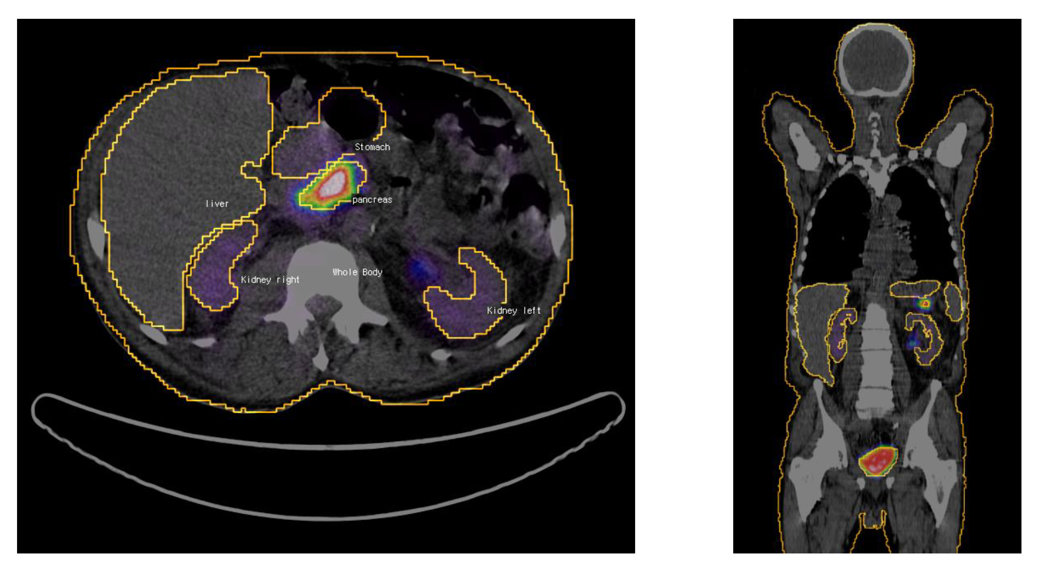
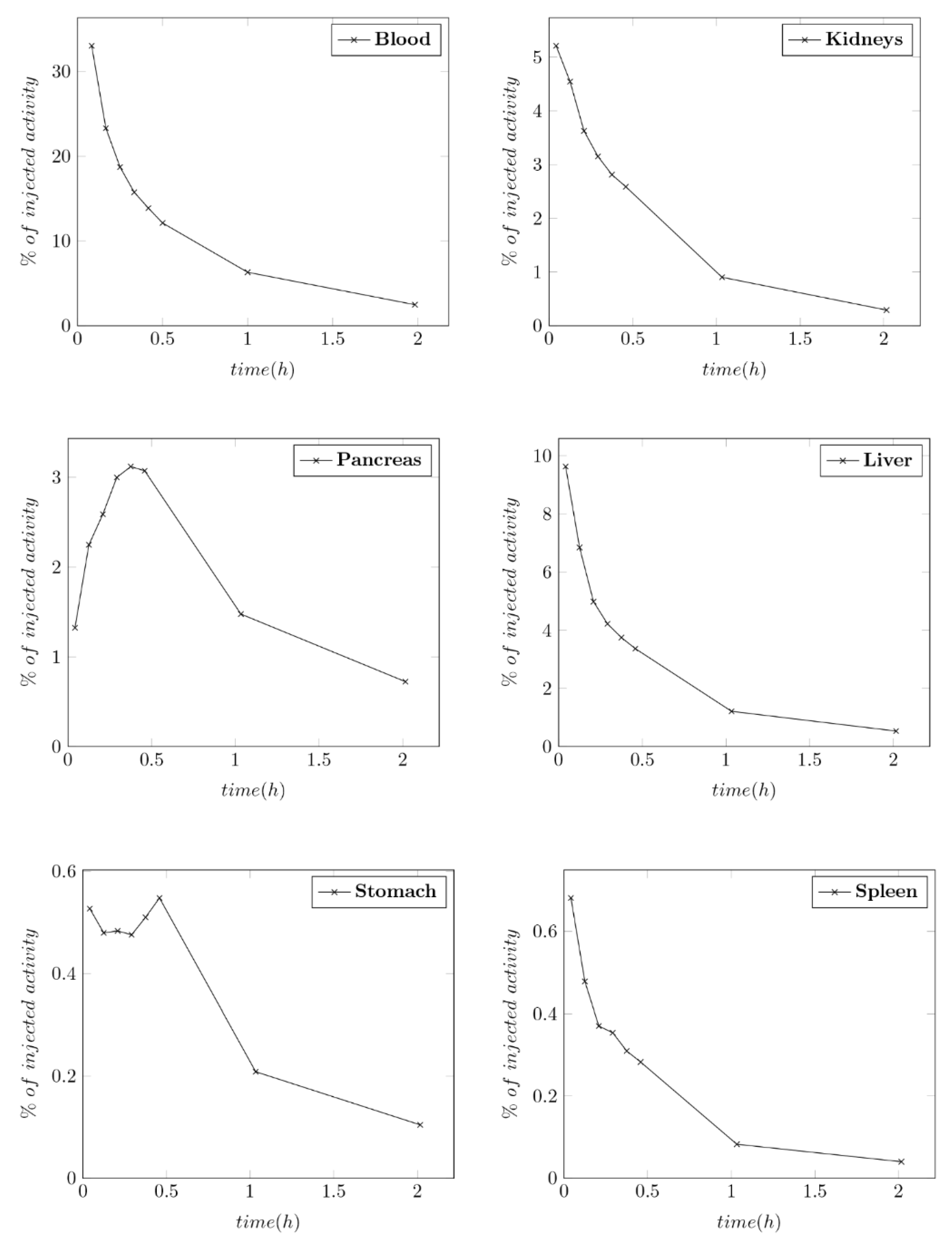
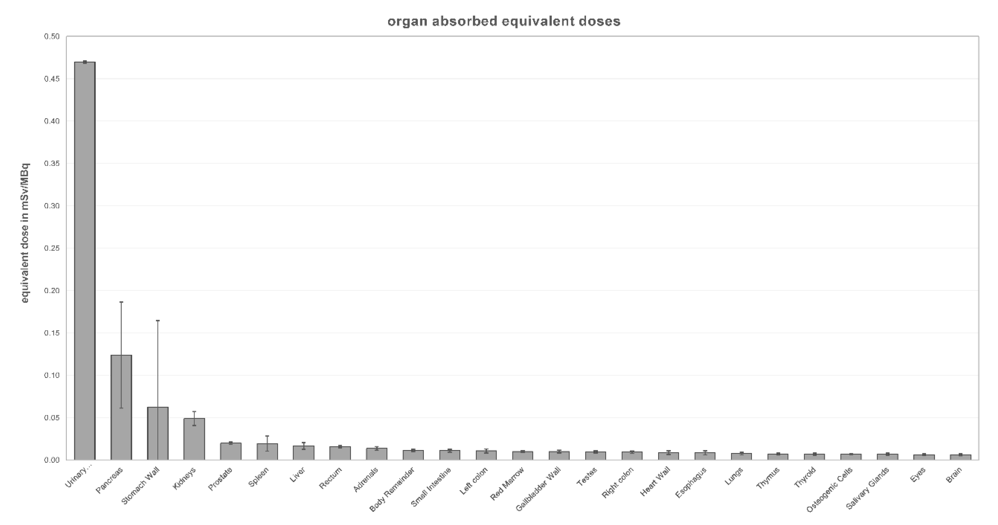
Eight venous blood samples were obtained at minute 5, 10, 15, 20, 25, 30, 60 and 120 from the arm opposite to the injection. 1 mL samples were analyzed in a gamma-counter (Wallac 1480 WIZARD 3″, Waltham, MA, USA) along with a known standard to determine activity (in MBq/mL).
Interview fusion software (MEDISO Medical Imaging Systems, Budapest, Hungary) was used to draw volumes of interest around source organs (pancreas, liver, stomach, spleen, kidneys, urinary bladder and the whole body) in the axial slices of the CT-images as shown in Figure 1. The total CT volume and the quantitative emission information (in kBq/mL) from co-registered PET images were used to determine total activity (in MBq) in source organs by multiplying the average activity concentration with the organ volume. The percentage of injected activity in source organs was determined for all time points by dividing the activity in the source organs by the total injected activity. Time–activity curves (TAC) were created and integrated using exponential curve fitting function in OLINDA/EXM version 2.0 software (Hermes Medical Solutions, Stockholm, Sweden) to determine residence times (MBq-h/MBq). Bi- or tri-exponential functions were fitted for all source organs as needed depending on the TAC indicating a monotonic decrease (blood, kidneys, liver, spleen, whole body) or a plateau phase followed by a monotonic decrease (pancreas, stomach). For the whole body TAC the total injected activity at the time of injection and the total activity from the whole body scans were used for curve fitting. Remainder of body residence time was determined by subtracting the residence times of all organs from the whole body residence time.
Since there is no report of RM2-uptake in blood components or bone marrow cells, the red-marrow TAC was calculated from the plasma TAC by multiplication with the red marrow-to-blood activity concentration ratio as described in the 2010 EANM Dosimetry Committee guidelines for bone marrow and whole-body dosimetry [22]. The urinary bladder residence time was determined using the voiding bladder model of OLINDA/EXM version 2.0 with the kidneys as the only excretory pathway and a bladder voiding interval of 2 h. Dosimetric analysis was performed with OLINDA/EXM version 2.0 using the IRCP-89 adult male phantom. Furthermore, a dosimetric analysis was performed with the female IRPC-P89 phantom to determine a sex-averaged effective dose valid for a reference person as described in the ICRP-P103 [23,24].

3. Results

The administration of [68Ga]Ga-RM2 was well tolerated by all patients without occurrence of side effects. Physiological uptake was seen in pancreas, stomach, spleen, liver, kidneys and urinary bladder. Rapid renal clearance of [68Ga]Ga-RM2 resulted in fast blood/background clearance and a high activity in the urinary bladder. In the first 30 min after injection, the pancreas and stomach showed an accumulation of activity followed by a washout phase. Other source organs showed monotonically decreasing activity (Figure 2).
As visible in Figure 3, three patients showed remarkable uptake in the cardiac part of the stomach, which was absent in other patients. Two patients showed notable uptake in the distal esophagus. Another patient had rather low pancreatic uptake due to atrophy of the pancreatic tail. No uptake was seen in the salivary glands.
No pathological uptake was seen. In four patients the imaging showed no reason for the rising PSA-levels in the form of local recurrence or distant metastases. In the patient with known osseous and liver metastases only a small metastasis next to the lower thoracic spine showed uptake (SUVmax 4.3) while the other tumors showed no uptake.
The tracer concentration was highest in the urinary bladder, pancreas and kidneys (maximal organ activity ± SD in kBq/mL: 103.31 ± 71.43; 38.23 ± 18.19; 19.28 ± 4.01). The mean residence time of [68Ga]Ga-RM2 was highest in the urinary bladder (0.400 MBq-h/MBq), followed by the stomach, spleen, red marrow, pancreas, liver, kidneys and remainder of the body (Table 2). The urinary bladder wall was the most irradiated organ with a mean organ absorbed dose of 0.470 mSv/MBq, followed by the pancreas (0.124 mSv/MBq), stomach wall (0.063 mSv/MBq), kidneys (0.049 mSv/MBq) and red marrow (0.010 mSv/MBq) (Table 3 and Figure 4). The mean effective dose was found to be 0.038 mSv/MBq, thus giving a total effective dose of 6.08 mSv from injected dose of 160 MBq. The mean effective dose is the sex-averaged effective dose as described in the ICRP-P103 [24]. A comparison with published data of other prostate-specific tracers can be found in Table 4.

4. Discussion

The wide use of radiolabeled peptides for imaging and treatment of neuroendocrine and prostate carcinoma has motivated the development of new peptides against other possible receptors [11]. [68Ga]Ga-RM2 is one such GRPR antagonist that has shown 88% sensitivity for primary PCa and 70% for lymph node metastasis [15]. It is also considered to be superior to PSMA in detection of low grade primary PCa or slow growing local recurrence in BCR [16]. To increase better detection of PCa, efforts are being done to create bispecific targeting agents or dual modality imaging with labeled GRPR antagonists [13,14]. This study presents the biodistribution and normal organ radiation absorbed dose analysis with [68Ga]Ga-RM2 in five prostatic carcinoma patients with intent of its compassionate use. Four of the five patients were post-prostatectomy and without obvious disease recurrence, one patient (patient 1, Table 1) had advanced widespread disease.
Biodistribution of [68Ga]Ga-RM2 revealed kidneys as sole route of excretion along with pancreas and urinary bladder as organs with highest uptake, consistent with the findings of its distribution in healthy males and other studies [16,21,26]. Low molecular weight characteristic of the peptide is responsible for its rapid washout from kidneys. This resulted in rapid blood clearance (Figure 2), low background and finally accumulation in the urinary bladder. Peak uptake in pancreas and stomach was seen less than half an hour after injection followed by rapid decline until 1 h after injection. This finding is probably due to non-specific binding of the tracer in these organs as in healthy pancreas and stomach GRPR-expression is low. Rapid excretion results in reduced background radiation facilitating interpretation of abdominal lesions. Incidental uptake in the cardiac end of stomach in three patients and lower esophagus in two patients was found potentially due to presence of GRPR receptors in inflamed tissue. Further, low uptake in the pancreas, secondary to fatty infiltration in one of the patients, shows individual variation in distribution.
In the current study, the mean residence time (MBq-h/MBq) was highest in the urinary bladder followed by other source organs consistent with previous studies [21,25]. However residence time of [68Ga]Ga-RM2 in urinary bladder (0.400 h) was lower than in healthy volunteers (0.53 h) in a study published recently [21]. Residence times in pancreas (0.0375 h) and kidneys (0.0316 h) were lower as described as well (0.11 h for pancreas and 0.050 h for kidneys). It was also found that mean residence time in stomach in our study was prolonged as compared to other studies. Uptake in cardiac end of stomach in three patients could be a reason for this inconsistent result.
The absorbed dose analysis in the normal organs revealed the urinary bladder as the most irradiated organ. The organ absorbed dose of the urinary bladder in this study was found to be lower, i.e., 0.470 mSv/MBq as compared to 0.61 mSv/MBq in healthy males [21] and [68Ga]Ga-RM26 (1.09 mSv/MBq) [26], but higher than for other [68Ga]Ga-labelled GRPR-antagonists (0.112 mSv/MBq; 0.307 mSv/MBq) [25,28] and [68Ga]Ga-PSMA (0.0671 mSv/MBq) [27]. The difference to the absorbed dose in healthy males might be due to a shorter bladder voiding interval in this study (2 h vs. 3.5 h). Proper hydration and frequent voiding are required to reduce the urinary bladder absorbed dose in these patients. Application of diuretic medication should be considered.
The organ absorbed dose to the pancreas was found to be 0.124 mSv/MBq, lower than for [68Ga]Ga-RM2 in healthy males (0.51 mSv/MBq) [21] as well as other GRPR antagonists (0.225–0.26 mSv/MBq) [25,26,28] but significantly higher than [68Ga]Ga-PSMA 0.0199 mSv/MBq [27]. The reason could be the higher uptake in healthy males of comparatively young age (mean age 52 years) as compared to old emaciated patients (mean age 77 years) in the current study, as well as fatty infiltration of the pancreas in one of our patients.
The kidney absorbed dose (0.049 mSv/MBq) was lower than recorded earlier (0.081 mSv/MBq) [21] but comparable with other antagonists [25,26,28] as well as significantly lower than for [68Ga]Ga-PSMA (0.413 mSv/MBq) [27]. Red marrow dose (0.010 mSv/MBq) was comparable to previous studies for GRPR antagonists and [68Ga]Ga-PSMA (0.00915–0.013 mSv/MBq) [21,25,26,27,28].
The radiation absorbed dose for the stomach wall varied among the patients. One patient had a much higher stomach wall dose compared to the other patients (0.245 mSv/MBq vs. 0.017 mSv/MBq mean dose of the other patients), probably due to above average proximity of pancreas and stomach and uptake in the cardiac end of stomach. Interindividual variations in organ doses were observed especially for the pancreas (0.0369–0.212 mSv/MBq) and stomach wall (0.0118–0.245 mSv/MBq). Therefore, it is advisable to perform an individual dosimetry before attempting [177Lu]Lu-RM2 treatment. Recently published data showed that for this treatment the pancreas was the critical organ, followed by the kidney and liver, although this study makes no statements about the absorbed dose to the urinary bladder [20]. The high urinary bladder dose may hamper application of bombesin tracers or therapeutics but may be reduced by proper hydration and frequent voiding.
As GRPR labeled agents are being sought for imaging as well as therapy, absence of salivary gland uptake, no risk of xerostomia, low kidney and bone marrow absorbed doses can be considered advantageous for its labeling with lutetium-177 for therapy as compared to [177Lu]Lu-PSMA.
With a value of 0.0380 ± 0.0138 mSv/MBq the effective dose of [68Ga]Ga-RM2 was found to be lower than in healthy males (0.051 mSv/MBq) [21] and [68Ga]Ga-RM26 [0.066 mSv/ MBq) [26], but slightly higher than other [68Ga]Ga-labelled GRPR-antagonists [25,28] as well as [68Ga]Ga-PSMA (0.0258 mSv/MBq) [27].
Compared to healthy males, deviations in residence times can be partly attributed to interindividual differences in GRPR-expression considering both studies included only five participants. Furthermore, differences in organ absorbed doses and the effective dose can be explained by differences in evaluation methodology and software. The study in healthy males used the OLINDA version 1 software utilizing the equation based first generation MIRD phantoms and the tissue weighting factors of the ICRP 60 publication, Olinda version 2 used in this study utilizes the second generation voxel-based phantoms published in the ICRP 89 and the tissue weighting factors from the ICRP 103 publication resulting in more up-to-date data.

5. Conclusions

We found that [68Ga]Ga-RM2 has a relatively high urinary bladder dose but comparable effective dose to [68Ga]Ga-PSMA and is therefore a safe diagnostic agent for prostatic carcinoma patients. Moreover, low kidney and bone marrow absorbed doses with proper hydration and frequent voiding of the lower urinary bladder absorbed dose may facilitate its application, especially for treatment, e.g., labeled with lutetium-177, although due to interindividual variations in organ absorbed doses an individual dosimetry is recommended before attempting such a treatment.

Author Contributions

M.H. and A.K. performed the measurements and data analysis. H.A. did patient preparation and protocol set-up, S.K. and M.M. provided the RM2 tracer; M.E., F.C.G. and R.A.B. planned the study and set up the protocol. All authors revised the manuscript, which was written in first-line by M.H. and A.K. All authors have read and agreed to the published version of the manuscript.

Funding

This research received no external funding.

Conflicts of Interest

The RM2 precursor was provided for free by Life Molecular Imaging GmbH Berlin, Germany based on a non-commercial research agreement with the University Medical Center Bonn. R.B. is a consultant for Bayer Healthcare (Leverkusen, Germany) and Eisai GmbH (Frankfurt, Germany). R.B. has a non-commercial research agreement and is on the speakers list with Mediso Medical Imaging (Budapest, Hungary). M.E. is a consultant for Bayer Healthcare (Leverkusen, Germany), Eisai GmbH (Frankfurt, Germany), IPSEN, and Novartis.

References

  1. Stewart, B.W.; Wild, C. World Cancer Report 2014; International Agency for Research on Cancer: Lyon, France; WHO Press World Health Organization: Geneva, Switzerland, 2014. [Google Scholar]
  2. Fox, J.; Schöder, H.; Larson, S.M. Molecular imaging of prostate cancer. Curr. Opin. Urol. 2012, 22, 320–327. [Google Scholar] [CrossRef] [Green Version]
  3. Pascale, M.; Azinwi, C.N.; Marongiu, B.; Pesce, G.; Stoffel, F.; Roggero, E. The outcome of prostate cancer patients treated with curative intent strongly depends on survival after metastatic progression. BMC Cancer 2017, 17, 651. [Google Scholar] [CrossRef] [PubMed] [Green Version]
  4. Freedland, S.J.; Humphreys, E.B.; Mangold, L.A.; Eisenberger, M.; Dorey, F.J.; Walsh, P.C.; Partin, A.W. Risk of Prostate Cancer–Specific Mortality Following Biochemical Recurrence After Radical Prostatectomy. JAMA 2005, 294, 433–439. [Google Scholar] [CrossRef] [Green Version]
  5. Minamimoto, R.; Sonni, I.; Hancock, S.; Vasanawala, S.S.; Loening, A.M.; Gambhir, S.S.; Iagaru, A. Prospective Evaluation of 68Ga-RM2 PET/MRI in Patients with Biochemical Recurrence of Prostate Cancer and Negative Findings on Conventional Imaging. J. Nucl. Med. 2017, 59, 803–808. [Google Scholar] [CrossRef] [PubMed] [Green Version]
  6. Mason, B.R.; Eastham, J.A.; Davis, B.J.; Lance, A.M.; Thomas, J.P.; Richard, J.L.; Joseph, E.I. Current Status of MRI and PET in the NCCN Guidelines for Prostate Cancer. J. Natl. Compr. Canc. Netw. 2019, 17, 506–513. [Google Scholar] [CrossRef] [PubMed] [Green Version]
  7. Kelloff, G.J.; Choyke, P.; Coffey, D.S. Challenges in Clinical Prostate Cancer: Role of Imaging. Am. J. Roentgenol. 2009, 192, 1455–1470. [Google Scholar] [CrossRef] [PubMed] [Green Version]
  8. Lütje, S.; Heskamp, S.; Cornelissen, A.S.; Poeppel, T.D.; Van den Broek, S.A.M.W.; Rosenbaum-Krumme, S.; Bockisch, A.; Gotthardt, M.; Rijpkema, M.; Boerman, O.C. PSMA Ligands for Radionuclide Imaging and Therapy of Prostate Cancer: Clinical Status. Theranostics 2015, 5, 1388–1401. [Google Scholar] [CrossRef] [Green Version]
  9. Eiber, M.; Fendler, W.P.; Rowe, S.P.; Calais, J.; Hofman, M.S.; Maurer, T.; Schwarzenboeck, S.M.; Kratowchil, C.; Herrmann, K.; Giesel, F.L. Prostate-Specific Membrane Antigen Ligands for Imaging and Therapy. J. Nucl. Med. 2017, 58, 67S–76S. [Google Scholar] [CrossRef] [Green Version]
  10. Kasperzyk, J.L.; Finn, S.P.; Flavin, R.; Fiorentino, M.; Lis, R.; Hendrickson, W.K.; Clinton, S.K.; Sesso, H.D.; Giovannucci, E.L.; Stampfer, M.J.; et al. Prostate-Specific Membrane Antigen Protein Expression in Tumor Tissue and Risk of Lethal Prostate Cancer. Cancer Epidemiol. Biomark. Prev. 2013, 22, 2354–2363. [Google Scholar] [CrossRef] [Green Version]
  11. Mansi, R.; Wang, X.; Forrer, F.; Waser, B.; Cescato, R.; Graham, K.; Borkowski, S.; Reubi, J.C.; Maecke, H.R. Development of a potent DOTA-conjugated bombesin antagonist for targeting GRPr-positive tumours. Eur. J. Nucl. Med. Mol. Imaging 2010, 38, 97–107. [Google Scholar] [CrossRef] [Green Version]
  12. Cornelio, D.; Roesler, R.; Schwartsmann, G. Gastrin-releasing peptide receptor as a molecular target in experimental anticancer therapy. Ann. Oncol. 2007, 18, 1457–1466. [Google Scholar] [CrossRef] [PubMed]
  13. Liolios, C.; Schäfer, M.; Haberkorn, U.; Eder, M.; Kopka, K. Novel Bispecific PSMA/GRPr Targeting Radioligands with Optimized Pharmacokinetics for Improved PET Imaging of Prostate Cancer. Bioconjug. Chem. 2016, 27, 737–751. [Google Scholar] [CrossRef] [PubMed]
  14. Zhang, H.; Desai, P.; Koike, Y.; Houghton, J.; Carlin, S.D.; Tandon, N.; Touijer, K.; Weber, W.A. Dual-Modality Imaging of Prostate Cancer with a Fluorescent and Radiogallium-Labeled Gastrin-Releasing Peptide Receptor Antagonist. J. Nucl. Med. 2017, 58, 29–35. [Google Scholar] [CrossRef] [PubMed] [Green Version]
  15. Kähkönen, E.; Jambor, I.; Kemppainen, J.; Lehtiö, K.; Grönroos, T.J.; Kuisma, A.; Luoto, P.; Sipilä, H.J.; Tolvanen, T.; Alanen, K.; et al. In Vivo Imaging of Prostate Cancer Using [68Ga]-Labeled Bombesin Analog BAY86-7548. Clin. Cancer Res. 2013, 19, 5434–5443. [Google Scholar] [CrossRef] [Green Version]
  16. Minamimoto, R.; Hancock, S.; Schneider, B. Pilot Comparison of ⁶⁸Ga-RM2 PET and ⁶⁸Ga-PSMA-11 PET in Patients with Biochemically Recurrent Prostate Cancer. J. Nucl. Med. 2016, 57, 557–562. [Google Scholar] [CrossRef] [Green Version]
  17. Wangerin, K.A.; Baratto, L.; Khalighi, M.M.; Hope, T.A.; Gulaka, P.K.; Deller, T.W.; Iagaru, A. Clinical Evaluation of 68Ga-PSMA-II and 68Ga-RM2 PET Images Reconstructed With an Improved Scatter Correction Algorithm. Am. J. Roentgenol. 2018, 211, 655–660. [Google Scholar] [CrossRef]
  18. Touijer, K.A.; Michaud, L.; Alvarez, H.A.V.; Gopalan, A.; Kossatz, S.; Gonen, M.; Beattie, B.; Sandler, I.; Lyaschenko, S.; Eastham, J.A.; et al. Prospective Study of the Radiolabeled GRPR Antagonist BAY86-7548 for Positron Emission Tomography/Computed Tomography Imaging of Newly Diagnosed Prostate Cancer. Eur. Urol. Oncol. 2019, 2, 166–173. [Google Scholar] [CrossRef]
  19. Wieser, G.; Popp, I.; Christian, R.H. Diagnosis of recurrent prostate cancer with PET/CT imaging using the gastrin-releasing peptide receptor antagonist 68Ga-RM2: Preliminary results in patients with negative or inconclusive 18FFluoroethylcholine-PET/CT. Eur. J. Nucl. Med. Mol. Imaging 2017, 44, 1463–1472. [Google Scholar] [CrossRef]
  20. Kurth, J.; Krause, B.J.; Schwarzenböck, S.M.; Carina, B.; Oliver, W.H.; Martin, H. First-in-human dosimetry of gastrin-releasing peptide receptor antagonist 177LuLu-RM2: A radiopharmaceutical for the treatment of metastatic castration-resistant prostate cancer. Eur. J. Nucl. Med. Mol. Imaging 2020, 47, 123–135. [Google Scholar] [CrossRef]
  21. Roivainen, A.; Luoto, P.; Borkowski, S.; Jambor, I.; Rantala, T.; Sipilä, H.; Sparks, R.; Suilamo, S.; Tolvanen, T.; Valencia, R.; et al. Plasma Pharmacokinetics, Whole-Body Distribution, Metabolism, and Radiation Dosimetry of 68Ga Bombesin Antagonist BAY 86-7548 in Healthy Men. J. Nucl. Med. 2013, 54, 867–872. [Google Scholar] [CrossRef] [Green Version]
  22. Hindorf, C.; Glatting, G.; Chiesa, C.; Lindén, O.; Flux, G. EANM Dosimetry Committee guidelines for bone marrow and whole-body dosimetry. Eur. J. Nucl. Med. Mol. Imaging 2010, 37, 1238–1250. [Google Scholar] [CrossRef] [PubMed]
  23. ICRP. P089 Basic Anatomical and Physiological Data for Use in Radiological Protection Reference Values; ICRP: Stockholm, Sweden, 2011. [Google Scholar]
  24. ICRP. P103 the 2007 Recommendations of the International Commission on Radiological Protection; ICRP: Stockholm, Sweden, 2007. [Google Scholar]
  25. Gnesin, S.; Cicone, F.; Mitsakis, P.; Van Der Gucht, A.; Baechler, S.; Miralbell, R.; Garibotto, V.; Zilli, T.; Prior, J.O. First in-human radiation dosimetry of the gastrin-releasing peptide (GRP) receptor antagonist 68Ga-NODAGA-MJ9. EJNMMI Res. 2018, 8, 108. [Google Scholar] [CrossRef] [PubMed]
  26. Zhang, J.; Niu, G.; Fan, X.; Lang, L.; Hou, G.; Chen, L.; Wu, H.; Zhu, Z.; Li, F.; Chen, X. PET Using a GRPR Antagonist68Ga-RM26 in Healthy Volunteers and Prostate Cancer Patients. J. Nucl. Med. 2018, 59, 922–928. [Google Scholar] [CrossRef] [PubMed] [Green Version]
  27. Green, M.A.; Eitel, J.A.; Fletcher, J.W.; Mathias, C.J.; Tann, M.A.; Gardner, T.; Koch, M.O.; Territo, W.; Polson, H.; Hutchins, G.D. Estimation of radiation dosimetry for 68Ga-HBED-CC (PSMA-11) in patients with suspected recurrence of prostate cancer. Nucl. Med. Biol. 2017, 46, 32–35. [Google Scholar] [CrossRef] [Green Version]
  28. Zhang, J.; Li, D.; Lang, L.; Zhu, Z.; Wang, L.; Wu, P.; Niu, G.; Li, F.; Chen, X. 68Ga-NOTA-Aca-BBN(7-14) PET/CT in Healthy Volunteers and Glioma Patients. J. Nucl. Med. 2016, 57, 9–14. [Google Scholar] [CrossRef] [Green Version]
Figure 1. Volume of interest drawn around source organs on axial (left) and coronal (right) slice.
Radiation 01 00004 g001
Figure 2. Time–activity curves (TAC) in source organs exemplary for one patient.
Radiation 01 00004 g002
Figure 3. Maximum intensity projection (MIP) of the first full body scan at one hour post injection for all patients.
Radiation 01 00004 g003
Figure 4. Mean organ absorbed equivalent doses (in mSv/MBq) with standard deviation. Urinary bladder was calculated with the OLINDA voiding bladder model and therefore has no relevant standard deviation.
Radiation 01 00004 g004
Table 1. Patient characteristics.
CharaccteristicsPt 1Pt 2Pt 3Pt 4Pt 5MeanSD
Age746672766971.43.97
Weight728972968081.810.59
Height178176176194182181.27.56
Injected activity165161164160164162.82.17
PSA-Level (ng/mL)2045 *1.827.510.12.2510.4112.01
Gleason score4 + 5 = 94 + 5 = 93 + 4 = 73 + 4 = 73 + 3 = 6--
Initial stageIV BII CII CIII AII C--
Initial grade32232--
Previous treatmentsHx, Bis, Ra, Lu, En, Arb, RTxRPE, Rtx, HxRPE, RTxRPE, Hx, RTxRPE, RTx--
* Not included in mean value. Abbreviations: RPE: radical prostatecomy, Hx: anti-hormonal treatment, Bis: bisphosphonates, Ra: Radium-223-dichlorid, Lu: Lu-177-PSMA, En: enzalutamide, Arb: abiraterone, RTx: radiation treatment.
Table 2. Residence times (MBq-h/MBq) of [68Ga]Ga-RM2.
Organ Pt 1Pt 2Pt 3Pt 4Pt 5MeanSD
Stomach0.2390.0120.0050.0060.0070.0540.104
Kidneys0.0260.0380.0250.0350.0330.0320.006
Liver0.0660.0530.0490.0330.0470.0490.012
Pancreas0.0620.0400.0110.0340.0400.0370.018
Red marrow0.0130.0120.0140.0230.0150.0150.004
Spleen0.0090.0040.0030.0060.0040.0050.002
Urinary bladder *0.4000.4000.4000.4000.4000.400-
Remainder of body0.960.880.780.611.050.810.15
* Urinary bladder residence time was calculated with OLINDAs bladder voiding model.
Table 3. Organ absorbed doses (mSv/MBq) and effective dose (mSv/MBq) for [68Ga]Ga-RM2.
OrganPt 1Pt 2Pt 3Pt 4Pt 5MeanSD
Adrenals0.0160.0140.0110.0120.0150.0140.002
Brain0.0060.0060.0050.0050.0080.0060.001
Esophagus0.0120.0080.0060.0070.0100.0080.002
Eyes0.0060.0060.0050.0050.0080.0060.001
Gallbladder wall0.0110.0100.0080.0080.0120.0100.002
Left colon0.0140.0110.0080.0090.0120.0110.002
Small intestine0.0120.0110.0090.0100.0130.0110.002
Stomach wall0.2450.0220.0120.0150.0190.0630.102
Right colon0.0090.0090.0080.0080.0110.0090.001
Rectum0.0160.0160.0150.0150.0180.0160.001
Heart wall0.0120.0080.0070.0070.0100.0090.002
Kidneys0.0420.0580.0390.0530.0520.0490.008
Liver0.0230.0170.0160.0120.0160.0170.004
Lungs0.0090.0070.0060.0060.0090.0080.002
Pancreas0.2120.1310.0370.1090.1300.1240.063
Prostate0.0200.0200.0190.0190.0220.0200.001
Salivary glands0.0060.0070.0060.0060.0090.0070.001
Red marrow0.0100.0090.0090.0100.0110.0100.001
Osteogenic cells0.0070.0060.0060.0070.0080.0070.001
Spleen0.0340.0170.0100.0210.0150.0190.009
Testes0.0090.0100.0090.0090.0120.0090.001
Thymus0.0080.0070.0060.0060.0090.0070.001
Thyroid0.0070.0070.0060.0060.0090.0070.001
Urinary bladder wall0.4690.4700.4690.4690.4720.4700.001
Body remainder0.0130.0110.0100.0100.0130.0110.001
Effective dose *0.0630.0330.0300.0310.0340.0380.014
* The effective dose is calculated as a sex-averaged value valid for a reference person.
Table 4. Comparison of organ absorbed doses (mSv/MBq) and effective doses (mSv/MBq) with published data.
Organs[68Ga]Ga-RM2 (Recent Study)[68Ga]Ga-Bombesin [21][68Ga]Ga-MJ9 [25][68Ga]Ga-RM26 [26][68Ga]Ga-PSMA [27]
MeanSDMeanSDMeanSDMeanSDMean
Adrenals0.0140.0020.0110.0010.0150.0020.0070.0030.017
Brain0.0060.0010.0060.0010.0020.0000.0010.0000.010
Esophagus0.0080.0020.0110.001
Eyes0.0060.0010.0080.000
Gallbladder wall0.0100.0020.0110.0010.0270.0060.0080.0030.016
Left colon0.0110.0020.0340.009
Small intestine0.0110.0020.0100.0000.0390.0100.0100.0030.014
Stomach wall0.0630.1020.0380.0090.0190.0020.0080.0030.014
Right colon0.0090.0010.0320.010
Rectum0.0160.0010.0330.010
Heart wall0.0090.0020.0280.0030.0210.0020.0060.0030.012
Kidneys0.0490.0080.0810.0110.0350.0030.0360.0020.413
Liver0.0170.0040.0230.0040.0140.0010.0160.0020.040
Lungs0.0080.0020.0070.0000.0130.0020.0060.0010.012
Pancreas0.1240.0630.5100.1600.2600.0610.2250.0380.020
Prostate0.0200.0010.0130.001
Salivary glands0.0070.0010.0220.0020.0090.000
Red marrow0.0100.0010.0130.0090.0090.0000.0090.0020.010
Osteogenic cells0.0070.0010.0130.0050.0070.0000.0100.0040.014
Spleen0.0190.0090.0230.0040.0140.0020.0190.0010.058
Testes0.0090.0010.0100.0000.0100.0010.0110.0010.011
Thymus0.0070.0010.0070.0010.0100.0000.0050.0030.011
Thyroid0.0070.0010.0270.0110.0090.0010.0050.0020.011
Urinary bladder Wall0.4700.0010.6100.0570.1120.0201.0900.2250.067
Body remainder0.0110.0010.0100.0000.0120.0010.0100.003
Effective dose *0.0380.0140.0510.0070.0190.0010.0660.0120.026
* The effective dose is calculated as a sex-averaged value valid for a reference person in this study.
Publisher’s Note: MDPI stays neutral with regard to jurisdictional claims in published maps and institutional affiliations.

