Systematic Review

The Role of 2-[18F]-FDG PET/CT in Detecting Richter Transformation in Chronic Lymphocytic Leukemia: A Systematic Review

1
Nuclear Medicine, ASST Spedali Civili Brescia, University of Brescia, 25123 Brescia, Italy
2
Nuclear Medicine Unit, IRCSS Regina Elena National Cancer Institute, 00144 Rome, Italy
3
Clinic of Nuclear Medicine, Imaging Institute of Southern Switzerland, Ente Ospedaliero Cantonale, 6500 Bellinzona, Switzerland
4
Faculty of Biomedical Sciences, Università della Svizzera Italiana (USI), 6900 Lugano, Switzerland
5
Clinic of Nuclear Medicine, University Hospital and University of Zürich, 8006 Zürich, Switzerland
6
Academic Education, Research and Innovation Area, Ente Ospedaliero Cantonale, 6500 Bellinzona, Switzerland
7
Department of Nuclear Medicine and Molecular Imaging, Lausanne University Hospital, University of Lausanne, 1011 Lausanne, Switzerland
*
Author to whom correspondence should be addressed.
Submission received: 25 January 2021 / Revised: 19 February 2021 / Accepted: 1 March 2021 / Published: 5 March 2021

Abstract

:

Simple Summary

Richter transformation (RT) is a phenomenon characterized by the transformation of B cell chronic lymphocytic leukemia (CLL) into a more aggressive lymphoma and it is considered a diagnostic challenge. In this review, we analyzed the published articles about the potential usefulness of 2-deoxy-2-[18F]-fluoro-D-glucose positron emission tomography/computed tomography (2-[18F]-FDG PET/CT) in detecting RT. Using evidence from 13 published studies, including 1336 CLL patients, we showed that 2-[18F]-FDG uptake expressed as maximum standardized uptake value (SUVmax) has a high negative predictive value. An SUVmax of 5 is the most frequent threshold used in the literature to detect RT.

Abstract

Richter transformation (RT) is a condition wherein B cell chronic lymphocytic leukemia (CLL) transforms into a more aggressive lymphoma variant. The incidence and the significance of RT detected by 2-[18F]-FDG PET/CT is a clinical challenge and it is not widely investigated in the literature. The aim of this systematic review was to analyze published data about the potential role of 2-[18F]-FDG PET/CT in detecting RT. A comprehensive computer literature search of the PubMed/MEDLINE, Embase and Cochrane library databases was conducted up to December 2020. Thirteen studies (1336 patients with CLL) were selected. The maximum standardized uptake value (SUVmax) was the most common metabolic parameter used to detect RT. An SUVmax of 5 had an average overall sensitivity of 87% (range: 71–96%), an average overall specificity of 49% (range: 4–80%), an average positive predictive value of 41% (range: 16–53%) and an average negative predictive value of 84% (range: 33–97%). Other metabolic variables were only marginally investigated, with promising results. 2-[18F]-FDG PET/CT imaging may play an important role in the detection of RT in CLL, based on the high metabolic activity of the nodal lesions that transformed into aggressive lymphomas. 2-[18F]-FDG PET/CT has high negative predictive value for evaluating RT.

1. Introduction

Chronic lymphocytic leukemia (CLL) is an indolent, low-grade B cell lymphoproliferative disease, characterized by the presence of high lymphocytosis, associated with nodal and splenic involvement, and irregular blood counts [1]. CLL derived from a clonal proliferation and accumulation of mature B cells (usually with a coexpression of CD5, CD19, CD20(dim) and CD23) in the bone marrow, blood, spleen and lymph nodes, and it is classified using to the Rai and Binet staging system [2]. In most cases, CLL is a lymphoproliferative disorder with good prognosis; however, in a small number of cases (about 2–10%), the patients developed a more aggressive condition called Richter transformation (RT) [3]. RT was first described by a pathologist, Maurice Richter, in 1928, and this definition was applied later to describe a subset of patients with CLL who developed large-cell lymphoma [4]. Nowadays, RT is defined when CLL or small lymphocytic lymphoma evolved into an aggressive lymphoma variant, more frequently diffuse large B-cell lymphoma (DLBCL) and less frequently Hodgkin lymphoma (HL) [5,6,7]. The prognosis of patients with RT is lower than CLL, with a median overall survival (OS) ranging from 2.5 to 10 months [5,6,7], also due to the absence of a shared gold standard efficient therapy for these kind of patients despite recent therapeutic improvements [8,9]. To confirm RT, histological diagnosis remains the gold standard and typically it is done by a core needle biopsy or excisional node biopsy, which are invasive procedures with potential side effects [9,10]. Several different clinical and pathological features, such as the LDH level, B symptoms, the asymmetric growth of bulky lymph nodes and some molecular factors, such as immunoglobulin heavy chain variable (IGHV) somatic hypermutation and TP53 interruption, have been studied, demonstrating significant roles in detecting RT; but, a strong debate is still present [10]. In this scenario, the potential usefulness of 2-deoxy-2-[18F]-fluoro-D-glucose positron emission tomography/computed tomography (2-[18F]-FDG PET/CT) in detecting RT is not completely understood, but with promising initial evidence [11,12]. For this reason, the aim of our systematic review was to investigate the published data about the role of 2-[18F]-FDG PET or PET/CT in detecting RT in patients affected by CLL.

2. Materials and Methods

2.1. Search Strategy

A comprehensive literature search of the PubMed/MEDLINE, Embase and Cochrane library databases was performed to find relevant published manuscripts about the role of 2-[18F]-FDG PET or PET/CT in detecting RT of CLL. We have created and used a search algorithm based on a combination of the terms (a) “chronic lymphocytic leukemia” OR “CLL” OR “Richter transformation” OR “Richter syndrome” AND (b) “positron emission tomography” OR “PET”. No beginning date limit was used; the search was updated until December 31, 2020. To expand our analysis, the references of the retrieved publications were also screened to search for additional articles related to the topic of interest. All the literature manuscripts collected were managed using EndNote®Basic (ClarivateTM).

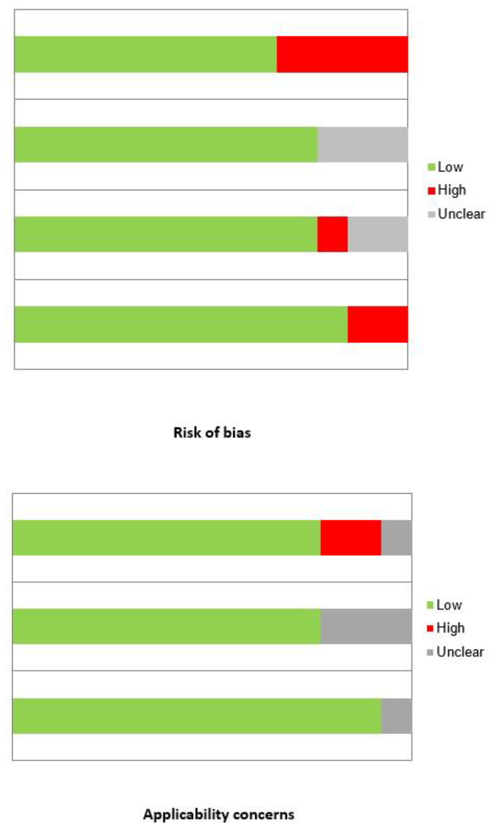
2.2. Study Selection and Quality Assessment

Studies or subsets in studies focused on detecting RT in patients with CLL by using 2-[18F]-FDG PET/CT PET or PET/CT were included. Exclusion criteria were (a) studies not in the field of interest of this systematic review; and (b) case reports or small case series (less than 5 patients with RT), review articles, meta-analyses, editorials, letters and conference proceedings. No language restriction was used. Two researchers (D.A. and F.B.) independently reviewed the titles and abstracts of the retrieved articles, applying the abovementioned inclusion and exclusion criteria. Articles were excluded in case of clear ineligibility. The same two researchers then independently reviewed the full-text version of the remaining publications to evaluate their suitability and performed the quality assessment of the included studies. Disagreements were discussed among the co-authors in a consensus meeting.
A quality assessment of the included articles was performed. This assessment included both the risk of bias assessment and applicability concerns by using the QUADAS-2 tool [13].

2.3. Data Extraction and Collection

For each included study, two researchers (DA and FB) collected data concerning the studies’ characteristics (author names, year of publication, country and study design), scanner-related features (type of scanner used, radiotracer injected activity, uptake time and image analysis), number of patients evaluated and number of cases with RT. Data about the sensitivity, specificity, positive predictive value (PPV) and negative predictive value (NPV) of 2-[18F]-FDG PET or PET/CT in detecting RT in patients with CLL were also extracted. The main findings of the articles included in this review are reported in the Results section.

3. Results

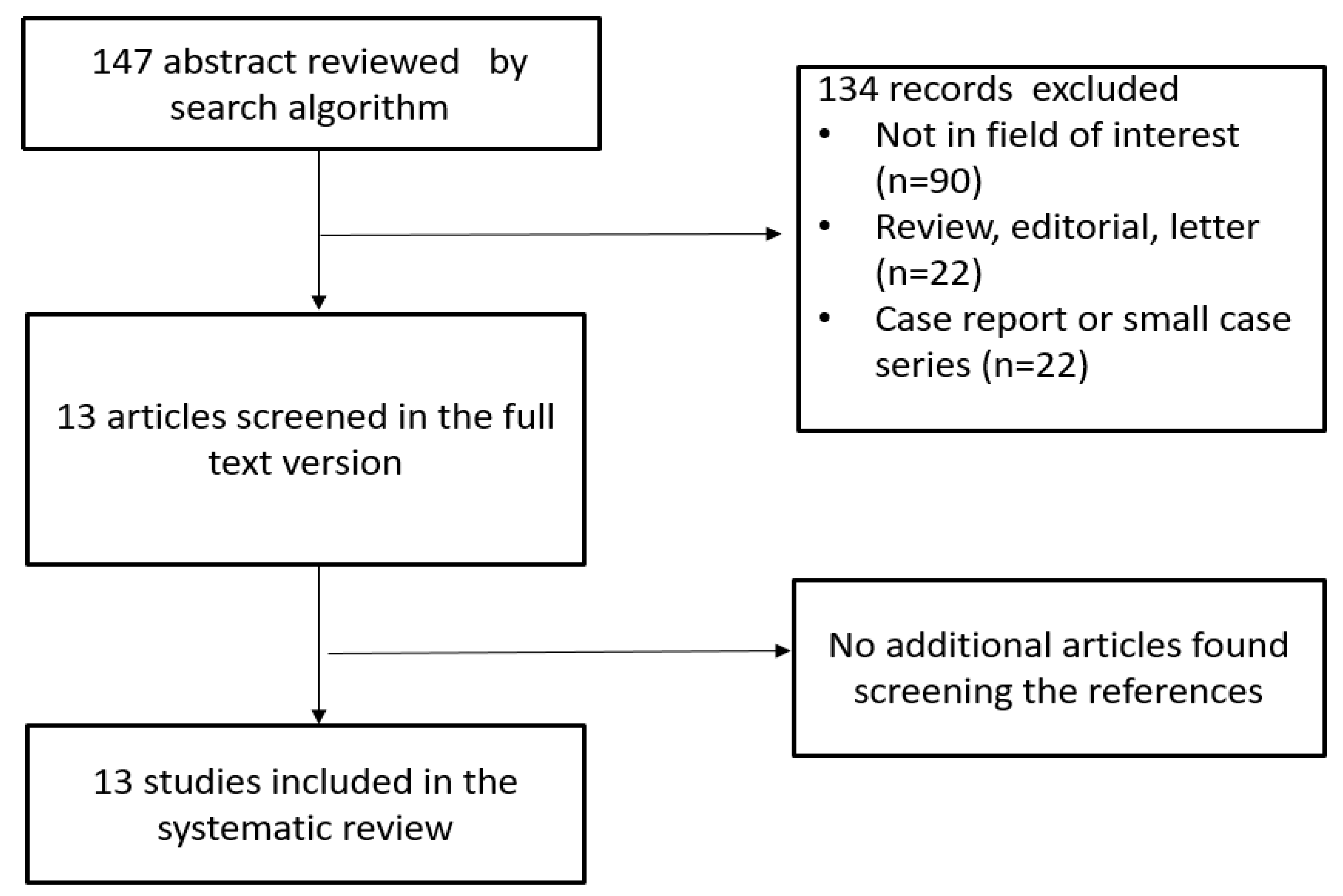
3.1. Literature Search

The comprehensive computer literature search from the selected databases revealed 147 manuscripts. After the revision of the titles and abstracts, 90 records were eliminated because they were not within the field of interest of the research of this review and 44 records were eliminated because they were editorials, comments, case reports, small case series, reviews or conference proceedings. Finally, 13 studies (for a total of 1336 patients affected by CLL who performed 2-[18F]-FDG PET or PET/CT) were screened in their full-text version and included in this systematic review [14,15,16,17,18,19,20,21,22,23,24,25,26] (Figure 1). No additional manuscripts were discovered while checking the references of the selected articles.

3.2. Qualitative Analysis

Table 1, Table 2 and Table 3 contain the main characteristics and results of the studies included. Of the 13 studies, 10 were of retrospective nature [14,15,16,19,20,21,23,24,25,26] and only 3 were prospective [17,18,22]. In two papers the scanner utilized was PET only [15,24], while in the other 10 cases the scanner used was hybrid PET/CT [14,16,17,18,20,21,22,23,25,26]; only in one manuscript both techniques were considered [19]. In all the included studies, PET or PET/CT images were analyzed visually and semi-quantitatively. Regarding the semi-quantitative analysis, the maximum standardized uptake value (SUVmax) was the most frequent metabolic parameter investigated. Metabolic tumor volume (MTV) and total lesion glycolysis (TLG) were analyzed only in two articles [23,26]. All 13 studies were evaluated qualitatively using the QUADAS-2 tool (Figure 2).
Among the 1336 patients included in these 13 studies, RT happened in 266 patients with a mean prevalence of 20% (ranging from 7% to 100%) (Table 1). The range is very wide, due to the high heterogeneity of the populations studied. In 2006, firstly Bruzzi et al. [14] analyzed the possible usefulness of 2-[18F]-FDG PET/CT in 37 patients who performed a total of 57 PET/CT scans and derived a threshold of an SUVmax of 5 with an overall good accuracy to detect RT (sensitivity 91%, specificity 80%, PPV 53% and NPV 97%). In three studies [15,16,17], only visual analysis of PET was used to detect RT and SUVmax was not applied. In the other 6 studies [18,19,20,22,24,25], an SUVmax of 5 was used to detect RT with an average overall sensitivity of 87% (range 71–96%), an average overall specificity of 49% (range 4–80%), an average overall PPV of 41% (range 16–53%) and an average overall NPV of 84% (range 33–97%). Albano et al. [26] and Michallet et al. [21] proposed a different SUVmax threshold as the best predictor for RT: an SUVmax of 9 and of 10, respectively, with a sensitivity, specificity, PPV and NPV of 67%, 90%, 67% and 90%, respectively, in the first case [26], and 91%, 95%, 29% and 99%, respectively, in the second one [21]. In two studies [22,24], different SUVmax cutoff values were directly compared in the same population. Notably, increasing the SUVmax threshold, the sensitivity of 2-[18F]-FDG PET/CT in detecting RT in patients with CLL usually decreased whereas the specificity increased (Table 3).
Other metabolic variables (such as the SUV corrected for lean body mass (SUVlbm), for body surface area (SUVbsa), lesion-to-blood-pool SUV ratio (L-BP SUV R), lesion-to-liver SUV ratio (L-L SUV R), total lesion glycolysis (TLG) and metabolic tumor volume (MTV)) were investigated only by Albano et al. [26], identifying as the best threshold 5.3 for SUVlbm (with a sensitivity and specificity of 72% and 83%, respectively), 1.7 for SUVbsa (with a sensitivity and specificity of 72% and 78%, respectively), 2 for L-L SUV R (with a sensitivity and specificity of 94% and 63%, respectively) and 4.8 for L-BP SUV (with a sensitivity and specificity of 72% and 92%, respectively). For these metabolic measures, there was a significant difference between RT patients and non RT patients applying the aforementioned thresholds. Instead, the cutoff values derived from MTV (14.5 cm3) and TLG (40) were not a significant predictor for RT [26].

4. Discussion

2-[18F]-FDG PET/CT is a non-invasive diagnostic tool useful for the study of different oncological diseases and with different purposes (e.g., staging, treatment response evaluation and restaging). The role of this imaging method for the study of lymphoma is well known with strong evidence, especially in those lymphoma histotypes considered 2-[18F]-FDG avid by definition, like Hodgkin lymphoma (HL), diffuse large B-cell lymphoma (DLBCL) and follicular lymphoma [27,28]. Recent publications have described a high 2-[18F]-FDG avidity also in other non-Hodgkin lymphoma variants (Burkitt lymphoma, mantle cell lymphoma) [29,30,31,32]. Instead, some lymphoma variants, such as mucosa-associated lymphoid tissue (MALT) and splenic marginal zone lymphomas (SMZL), present the usual low to moderate 2-[18F]-FDG uptake [33,34]. CLL also seems to be a lymphoproliferative disease with variable metabolic behavior and its 2-[18F]-FDG avidity is directly related to the aggressiveness and the risk of transformation into aggressive lymphoma [11,12]. The detection of patients that will develop RT seems to be fundamental because RT significantly influences the outcome and is associated with a lower efficacy of treatments [9]. Moreover, to date, the epidemiological, clinical and biological features considered as surely indicative of RT are not available [9].
2-[18F]-FDG PET/CT may help to define the disease status with the aim to detect the cases with a high risk of transformation in CLL patients and to choose the best personalized approach [11,12]. With this review, we underline that 2-[18F]-FDG PET/CT is helpful in evaluating CLL with the risk of transformation into aggressive lymphomas. Nevertheless, it is essential not to consider RT in all cases of hypermetabolic lesions at 2-[18F]-FDG PET/CT (for example, when SUVmax ≥ 10). The final confirmation of the RT remains to the prerogative of tissue biopsy and the PET/CT should only guide towards the choice of node to biopsy [14,15,16,17,18,19,20,21,22,23,24,25,26]. This is due to the low specificity and PPV of PET/CT in this field.
Our analysis shows a good NPV of PET/CT and its parameter in detecting RT, while PPV was shown to be less accurate despite the high heterogeneity of the results.
In this regard, another potential use of 2-[18F]-FDG PET/CT is to recognize the site with highest metabolic rate to identify sites for biopsy (to confirm the clinic suspicion of RT) as well as for CLL prognostication. The SUVmax at 2-[18F]-FDG PET may be considered a surrogate marker of metabolic activity, and a high value of SUVmax usually reflects aggressiveness and risk of transformation of CLL into more aggressive lymphomas [35]. First, Bruzzi et al. arbitrary proposed an SUVmax of 5 as a threshold to discriminate cases of RT, reporting good global accuracy [14]. This value was chosen on the basis of the institutional experience of that center and suggested as suspected for aggressive metabolic behavior. The cutoff of 5 was the most used in the literature, but other cutoffs are also suggested [21,22,24,26].
From a clinician’s point of view, the main aim is to obtain a high sensitivity by 2-[18F]-FDG PET/CT in detecting RT and in selecting sites for biopsy to confirm the suspicion of RT of CLL [11,12]. Based on the available data, when different SUVmax thresholds of 2-[18F]-FDG PET/CT are compared, the threshold value of 5 seems to guarantee the best sensitivity for detecting RT but with increased false positive results (lower specificity) compared to higher SUVmax thresholds [14,15,16,17,18,19,20,21,23,24,25,26]. Only one study was partially in contradiction to this evidence: Mato et al. [22] compared different threshold in a small population (n = 57), finding the best accuracy with an SUVmax value of 11, while applying 5 as the threshold NPV was very low (33%) and inferior to other SUVmax values.
The main source of false positive results is non-transformed CLL [14,15,16,17,18,19,20,21,22,23,24,25,26]. Conversely, increasing the SUVmax threshold for 2-[18F]-FDG PET/CT will result in a decrease in sensitivity with a higher risk of false negative results for RT [14,15,16,17,18,19,20,21,22,23,24,25,26].
The development of new PET/CT scanners and technical features could influence the choice of the best SUVmax threshold to detect RT and this topic needs to be better analyzed, especially in the future with the increased availability of digital PET/CT scanners [36].
Only one study [26] focused on the metabolic parameters different from the SUVmax corrected for body weight, which is the most famous and used metabolic parameter and very easy and rapid to calculate with modern workstations. However, SUV is a factor that includes intrinsically some weaknesses and it is affected by several elements, such as the blood glucose level, the uptake time, the risk of extravasation of the radiotracer, the radiotracer decay, the partial volume effect, the scanner features, the protocols of acquisition and elaboration and the potential residual activity in the syringe [37]. For these reasons, other SUV-related parameters were introduced that correct for a lean body mass and body surface area; but, except for body weight, all the other limitations persist. Subsequently, other PET parameters related to the metabolic volumes (MTV and TLG) were derived and were defined as parameters that represent the morphological and metabolic features in the same feature [37]. In the literature, other manuscripts investigated the potential role of MTV and TLG in lymphoproliferative diseases, especially in the prognostic field [38,39,40,41], but in CLL patients only two studies [23,26] analyzed these factors, reporting controversial results. For Pontoizeau et al. [23], baseline MTV was a predictor of overall survival in RT, while in the other paper [26], MTV and TLG were not related to survival.
New emerging radiotracers, such as [68Ga]Ga-Pentixafor, have been developed and tested in the study of CLL, showing promising results in the differential diagnosis with other carcinoma and lymphoma [42]; however, more studies are needed.
Overall, SUVmax remains the most commonly studied metabolic parameter at 2-[18F]-FDG PET/CT in CLL patients with suspicion of RT. An SUVmax ≥ 5 is the most used threshold in the literature, associated with a good NPV for RT in CLL patients, despite other SUVmax cut-off values with significant results that also have been suggested [14,15,16,17,18,19,20,21,22,23,24,25,26]. Further large and multicenter studies are needed to confirm or controvert these results. However, it seems reasonable to suggest to perform 2-[18F]-FDG PET/CT in patients with CLL and suspected RT, considering the good NPV. Instead, in case of a positive PET/CT (high SUVmax), it remains mandatory to perform biopsy to confirm the suspicious RT.

Limitations

Several limitations affect the quality of our systematic review on the role of 2-[18F]-FDG PET/CT in detecting RT in CLL patients, like the absence of multicenter studies and the heterogeneity among the included studies. In particular, this heterogeneity is due to different patient and study characteristics, different technical aspects about 2-[18F]-FDG PET/CT, different metabolic measures and related threshold values and different types of RT (Hodgkin and non-Hodgkin lymphomas). This heterogeneity prevented us from carrying out a meta-analysis with related pooled values of the diagnostic performance of 2-[18F]-FDG PET/CT in detecting RT in CLL patients; therefore, a systematic review only was preferred [43].
Another aspect that should be underlined is the possible selection bias in the included manuscripts. The prevalence of RT in the included articles is higher compared to that reported in the general population (2–10% of CLL patients) [1,2,3,4].

5. Conclusions

In spite of the heterogeneity of the studies (in most cases of retrospective nature) and the wide variability in the sample analyzed, with this systematic review we can conclude that 2-[18F]-FDG PET/CT is an important tool to detect RT in CLL patients with an overall high NPV. SUVmax may be considered the best metabolic parameter for detecting RT in CLL patients. An SUVmax equal to or greater than 5 remains the most frequent threshold suggested in the literature for this purpose. Larger prospective studies on this topic are warranted.

Author Contributions

Conceptualization of the study: D.A. and G.T.; methodology: G.T. and D.A.; literature search and systematic review of literature: R.G., S.A., F.D. and F.B.; writing—original draft preparation, all co-authors; writing—review and editing, G.T., L.C., L.G., R.G. and D.A. All authors have read and agreed to the published version of the manuscript.

Funding

This research received no external funding.

Conflicts of Interest

The authors declare no conflict of interest.

References

  1. Hallek, M. Chronic lymphocytic leukemia: 2020 update on diagnosis, risk stratification and treatment. Am. J. Hematol. 2019, 94, 1266–1287. [Google Scholar] [CrossRef] [Green Version]
  2. Rozman, C.; Montserrat, E. Chronic Lymphocytic Leukemia. N. Engl. J. Med. 1995, 333, 1052–1057. [Google Scholar] [CrossRef] [Green Version]
  3. Parikh, S.A.; Kay, N.E.; Shanafelt, T.D. How we treat Richter syndrome. Blood 2014, 123, 1647–1657. [Google Scholar] [CrossRef] [PubMed]
  4. Parikh, S.A.; Rabe, K.G.; Call, T.G.; Zent, C.S.; Habermann, T.M.; Ding, W.; Leis, J.F.; Schwager, S.M.; Hanson, C.A.; Macon, W.R.; et al. Diffuse large B-cell lymphoma (Richter syndrome) in patients with chronic lymphocytic leukaemia: A cohort study of newly diagnosed patients. Br. J. Haematol. 2013, 162, 774–782. [Google Scholar] [CrossRef]
  5. Rossi, D. Richter’s syndrome: Novel and promising therapeutic alternatives. Best Pract. Res. Clin. Haematol. 2016, 29, 30–39. [Google Scholar] [CrossRef]
  6. Mato, A.R.; Nabhan, C.; Barr, P.M.; Ujjani, C.S.; Hill, B.T.; Lamanna, N.; Skarbnik, A.P.; Howlett, C.; Pu, J.J.; Sehgal, A.R.; et al. Outcomes of CLL patients treated with sequential kinase inhibitor therapy: A real world experience. Blood 2016, 128, 2199–2205. [Google Scholar] [CrossRef] [PubMed] [Green Version]
  7. Ayers, E.C.; Mato, A.R. Richter’s Transformation in the Era of Kinase Inhibitor Therapy: A Review. Clin. Lymphoma Myeloma Leuk. 2017, 17, 1–6. [Google Scholar] [CrossRef]
  8. Khan, M.; Siddiqi, R.; Thompson, P.A. Approach to Richter transformation of chronic lymphocytic leukemia in the era of novel therapies. Ann. Hematol. 2018, 97, 1–15. [Google Scholar] [CrossRef]
  9. Condoluci, A.; Rossi, D. Treatment of Richter’s Syndrome. Curr. Treat. Options Oncol. 2017, 18, 75. [Google Scholar] [CrossRef]
  10. Eichhorst, B.; Robak, T.; Montserrat, E.; Ghia, P.; Niemann, C.; Kater, A.; Gregor, M.; Cymbalista, F.; Buske, C.; Hillmen, P.; et al. Chronic lymphocytic leukaemia: ESMO Clinical Practice Guidelines for diagnosis, treatment and follow-up. Ann. Oncol. 2021, 32, 23–33. [Google Scholar] [CrossRef]
  11. Rhodes, J.M.; Mato, A.R. PET/Computed Tomography in Chronic Lymphocytic Leukemia and Richter Transformation. PET Clin. 2019, 14, 405–410. [Google Scholar] [CrossRef] [PubMed]
  12. Shaikh, F.; Janjua, A.; Van Gestel, F.; Ahmad, A. Richter Transformation of Chronic Lymphocytic Leukemia: A Review of Fluorodeoxyglucose Positron Emission Tomography–Computed Tomography and Molecular Diagnostics. Cureus 2017, 9, e968. [Google Scholar] [CrossRef] [Green Version]
  13. Whiting, P.F.; Rutjes, A.W.; Westwood, M.E.; Mallett, S.; Deeks, J.J.; Reitsma, J.B.; Leeflang, M.M.; Sterne, J.A.; Bossuyt, P.M.M.; The QUADAS-2 Group. QUADAS-2: A Revised Tool for the Quality Assessment of Diagnostic Accuracy Studies. Ann. Intern. Med. 2011, 155, 529–536. [Google Scholar] [CrossRef]
  14. Bruzzi, J.F.; Macapinlac, H.; Tsimberidou, A.M.; Truong, M.T.; Keating, M.J.; Marom, E.M.; Munden, R.F. Detection of Richter’s transformation of chronic lymphocytic leukemia by PET/CT. J. Nucl. Med. 2006, 47, 1267–1273. [Google Scholar]
  15. Karam, M.; Novak, L.; Cyriac, J.; Ali, A.; Nazeer, T.; Nugent, F. Role of fluorine-18 fluoro-deoxyglucose positron emission tomography scan in the evaluation and follow-up of patients with low-grade lymphomas. Cancer 2006, 107, 175–183. [Google Scholar] [CrossRef] [PubMed]
  16. Taralli, S.; Calcagni, M.L.; Rufini, V.; Laurenti, L.; Sica, S.; Chiusolo, P.; Leone, G.; Giordano, A. Role of 18F-FDG PET-CT for evaluating the response to reduced-intensity conditioning allogeneic transplant in heavily pre-treated patients with chronic lymphocytic leukemia: Preliminary results in nine patients. Ann. Nucl. Med. 2012, 26, 764–768. [Google Scholar] [CrossRef] [PubMed]
  17. Papajik, T.; Myslivecek, M.; Urbanova, R.; Buriankova, E.; Kapitanova, Z.; Procházka, V.; Turcsanyi, P.; Formánek, R.; Henzlová, L.; Flodr, P.; et al. 2-[18F]fluoro-2-deoxy-d-glucose positron emission tomography/computed tomography examination in patients with chronic lymphocytic leukemia may reveal Richter transformation. Leuk. Lymphoma 2014, 55, 314–319. [Google Scholar] [CrossRef] [PubMed]
  18. Conte, M.J.; Bowen, D.A.; Wiseman, G.A.; Rabe, K.G.; Slager, S.L.; Schwager, S.M.; Call, T.G.; Viswanatha, D.S.; Zent, C.S. Use of positron emission tomography-computerized tomography (PET-CT) in the management of patients with chronic lymphocytic leukemia/small lymphocytic lymphoma (CLL). Leuk. Lymphoma 2014, 55, 2079–2084. [Google Scholar] [CrossRef] [Green Version]
  19. Falchi, L.; Keating, M.J.; Marom, E.M.; Truong, M.T.; Schlette, E.J.; Sargent, R.L.; Trinh, L.; Wang, X.; Smith, S.C.; Jain, N.; et al. Correlation between FDG/PET findings, histological features, clinical characteristics and survival in 332 patients with chronic lymphocytic leukemia. Blood 2014, 123, 2783–2790. [Google Scholar] [CrossRef] [Green Version]
  20. Mauro, F.R.; Chauvie, S.; Paoloni, F.; Biggi, A.; Cimino, G.; Rago, A.; Gentile, M.A.; Morabito, F.C.; Coscia, M.; Bello, M.J.; et al. Diagnostic and prognostic role of PET/CT in patients with chronic lymphocytic leukemia and progressive disease. Leukemia 2015, 29, 1360–1365. [Google Scholar] [CrossRef]
  21. Michallet, A.S.; Sesques, P.; Rabe, K.G.; Itti, E.; Tordot, J.; Tychyj-Pinel, C.; Baseggio, L.; Subtil, F.; Salles, G.; Dupuis, J.M.; et al. An 18F-FDG-PET maximum standardized uptake value > 10 represents a novel valid marker for discerning Richter’s Syndrome. Leuk. Lymphoma 2016, 57, 1474–1477. [Google Scholar] [CrossRef]
  22. Mato, A.R.; Wierda, W.G.; Davids, M.S.; Cheson, B.D.; Coutre, S.E.; Choi, M.; Furman, R.R.; Heffner, L.; Barr, P.M.; Eradat, H.; et al. Utility of PET-CT in patients with chronic lymphocytic leukemia following B-cell receptor pathway inhibitor therapy. Haematologica 2019, 104, 2258–2264. [Google Scholar] [CrossRef] [Green Version]
  23. Pontoizeau, C.; Girard, A.; Mesbah, H.; Haumont, L.-A.; Devillers, A.; Tempescul, A.; Salaün, P.-Y.; Lamy, T.; Le Jeune, F.; Palard-Novello, X. Prognostic Value of Baseline Total Metabolic Tumor Volume Measured on FDG PET in Patients With Richter Syndrome. Clin. Nucl. Med. 2020, 45, 118–122. [Google Scholar] [CrossRef]
  24. Wang, Y.; Rabe, K.G.; Bold, M.S.; Shi, M.; Hanson, C.A.; Schwager, S.M.; Call, T.G.; Kenderian, S.S.; Muchtar, E.; Hayman, S.R.; et al. The role of 18F-FDG-PET in detecting Richter’s transformation of chronic lymphocytic leukemia in patients receiving therapy with a B-cell receptor inhibitor. Haematologica 2020, 105, 2675–2678. [Google Scholar] [CrossRef] [PubMed]
  25. Porrazzo, M.; Nicolai, E.; Riminucci, M.; Vitale, C.; Coscia, M.; De Paoli, L.; Rago, A.; Buscicchio, G.; Maestrini, G.; Ligia, S.; et al. Prognostic Significance of PET/CT in Patients with Chronic Lymphocytic Leukemia (CLL) Treated with Frontline Chemoimmunotherapy. Cancers 2020, 12, 1773. [Google Scholar] [CrossRef] [PubMed]
  26. Albano, D.; Camoni, L.; Rodella, C.; Giubbini, R.; Bertagna, F. 2-[18F]-FDG PET/CT Role in Detecting Richter Transformation of Chronic Lymphocytic Leukemia and Predicting Overall Survival. Clin. Lymphoma Myeloma Leuk. 2021, 21, e277–e283. [Google Scholar] [CrossRef] [PubMed]
  27. Cheson, B.D.; Fisher, R.I.; Barrington, S.F.; Cavalli, F.; Schwartz, L.H.; Zucca, E.; Lister, T.A. Recommendations for Initial Evaluation, Staging, and Response Assessment of Hodgkin and Non-Hodgkin Lymphoma: The Lugano Classification. J. Clin. Oncol. 2014, 32, 3059–3067. [Google Scholar] [CrossRef]
  28. Barrington, S.F.; Mikhaeel, N.G.; Kostakoglu, L.; Meignan, M.; Hutchings, M.; Müeller, S.P.; Schwartz, L.H.; Zucca, E.; Fisher, R.I.; Trotman, J.; et al. Role of Imaging in the Staging and Response Assessment of Lymphoma: Consensus of the International Conference on Malignant Lymphomas Imaging Working Group. J. Clin. Oncol. 2014, 32, 3048–3058. [Google Scholar] [CrossRef]
  29. Albano, D.; Bosio, G.; Re, A.; Pagani, C.; Giubbini, R.; Bertagna, F. Metabolic behavior and prognostic value of early and end of treatment 18F-FDG PET/CT in adult Burkitt’s lymphoma: The role of Deauville and IHP criteria. Leuk. Lymphoma 2019, 60, 326–333. [Google Scholar] [CrossRef]
  30. Albano, D.; Bertagna, F.; Giubbini, R. 18F-FD PET/CT role in Burkitt lymphoma. Clin. Transl. Imaging 2020, 8, 39–45. [Google Scholar] [CrossRef]
  31. Albano, D.; Treglia, G.; Gazzilli, M.; Cerudelli, E.; Giubbini, R.; Bertagna, F. 18F-FDG PET or PET/CT in Mantle Cell Lymphoma. Clin. Lymphoma Myeloma Leuk. 2020, 20, 422–430. [Google Scholar] [CrossRef] [PubMed]
  32. Albano, D.; Ferro, P.; Bosio, G.; Fallanca, F.; Re, A.; Tucci, A.; Ferreri, A.J.M.; Angelillo, P.; Gianolli, L.; Giubbini, R.; et al. Diagnostic and Clinical Impact of Staging 18F-FDG PET/CT in Mantle-Cell Lymphoma: A Two-Center Experience. Clin. Lymphoma Myeloma Leuk. 2019, 19, e457–e464. [Google Scholar] [CrossRef] [PubMed]
  33. Albano, D.; Durmo, R.; Treglia, G.; Giubbini, R.; Bertagna, F. 18F-FDG PET/CT or PET Role in MALT Lymphoma: An Open Issue not Yet Solved—A Critical Review. Clin. Lymphoma Myeloma Leuk. 2020, 20, 137–146. [Google Scholar] [CrossRef] [PubMed]
  34. Albano, D.; Giubbini, R.; Bertagna, F. 18F-FDG PET/CT in splenic marginal lymphoma. Abdom. Radiol. 2018, 43, 2721–2727. [Google Scholar] [CrossRef] [PubMed]
  35. Alessandrino, F.; DiPiro, P.J.; Jagannathan, J.P.; Babina, G.; Krajewski, K.M.; Ramaiya, N.H.; Giardino, A.A. Multimodality imaging of indolent B cell lymphoma from diagnosis to transformation: What every radiologist should know. Insights Imaging 2019, 10, 25. [Google Scholar] [CrossRef] [Green Version]
  36. Constantino, C.S.; Oliveira, F.P.M.; Silva, M.; Oliveira, C.; Castanheira, J.C.; Silva, Â.; Vaz, S.C.; Vieira, P.; Costa, D.C. Are lesion features reproducible between 18F-FDG PET/CT images when acquired on analog or digital PET/CT scanners? Eur. Radiol. 2020, 1–9. [Google Scholar] [CrossRef]
  37. Kostakoglu, L.; Chauvie, S. Metabolic Tumor Volume Metrics in Lymphoma. Semin. Nucl. Med. 2018, 48, 50–66. [Google Scholar] [CrossRef] [PubMed]
  38. Albano, D.; Bosio, G.; Bianchetti, N.; Pagani, C.; Re, A.; Tucci, A.; Giubbini, R.; Bertagna, F. Prognostic role of baseline 18F-FDG PET/CT metabolic parameters in mantle cell lymphoma. Ann. Nucl. Med. 2019, 33, 449–458. [Google Scholar] [CrossRef] [PubMed]
  39. Albano, D.; Bosio, G.; Pagani, C.; Re, A.; Tucci, A.; Giubbini, R.; Bertagna, F. Prognostic role of baseline 18F-FDG PET/CT metabolic parameters in Burkitt lymphoma. Eur. J. Nucl. Med. Mol. Imaging 2019, 46, 87–96. [Google Scholar] [CrossRef]
  40. Albano, D.; Bertoli, M.; Battistotti, M.; Rodella, C.; Statuto, M.; Giubbini, R.; Bertagna, F. Prognostic role of pretreatment 18F-FDG PET/CT in primary brain lymphoma. Ann. Nucl. Med. 2018, 32, 532–541. [Google Scholar] [CrossRef] [PubMed]
  41. Albano, D.; Bosio, G.; Camoni, L.; Farina, M.; Re, A.; Tucci, A.; Giubbini, R.; Bertagna, F. Prognostic role of baseline 18F-FDG PET/CT parameters in MALT lymphoma. Hematol. Oncol. 2019, 37, 39–46. [Google Scholar] [CrossRef] [PubMed] [Green Version]
  42. Mayerhoefer, M.E.; Jaeger, U.; Staber, P.; Raderer, M.; Wadsak, W.; Pfaff, S.; Kornauth, C.; Senn, D.; Weber, M.; Wester, H.-J.; et al. [68Ga]Ga-Pentixafor PET/MRI for CXCR4 Imaging of Chronic Lymphocytic Leukemia: Preliminary Results. Investig. Radiol. 2018, 53, 403–408. [Google Scholar] [CrossRef] [PubMed]
  43. Sadeghi, R.; Treglia, G. Systematic reviews and meta-analyses of diagnostic studies: A practical guideline. Clin. Transl. Imaging 2017, 5, 83–87. [Google Scholar] [CrossRef]
Figure 1. Literature search flowchart.
Radiation 01 00006 g001
Figure 2. QUADAS 2 score of all included studies.
Radiation 01 00006 g002
Table 1. Basic study and patient characteristics of the articles included in this systematic review.
First AuthorYearCountryStudy DesignCLL PatientsM:F RatioMean Age (Range)Patients with RT
Bruzzi J.F. [14]2006USARetrospective3726:1161 (40–82)11 (30%)
Karam M. [15]2006USARetrospective15nrnr1 (7%)
Taralli S. [16]2012ItalyRetrospective98:157.7 (49–70)1 (11%)
Papajik T. [17]2014Czech RepublicProspective44nrnr8 (18%)
Conte M.J. [18]2014USAProspective272197:7561.5 * (21–91)25 (9%)
Falchi L. [19]2014USARetrospective332218:11468 * (31–85)95 (29%)
Mauro F.R. [20]2015ItalyRetrospective9065:2561.2 * (31–81)17 (19%)
Michallet A.S. [21]2016FranceRetrospective24094:14662 (21–91)24 (10%)
Mato A.R. [22]2019USAProspective57nr67 * (28–85)8 (14%)
Pontoizeau C. [23]2020FranceRetrospective2822:671 * (36–89)28 (100%)
Wang Y. [24]2020USARetrospective9269:2368 * (43–89)25 (27%)
Porrazzo M. [25]2020ItalyRetrospective4031:962 * (35–92)5 (13%)
Albano D. [26]2020ItalyRetrospective8058:2261 (27–83)18 (22.5%)
Legend: M = male; F = female; CLL = chronic lymphocytic leukemia; RT = Richter’s transformation; nr: not reported; * = median.
Table 2. Main technical features of the articles included in this systematic review.
First AuthorDeviceMean Radiopharmaceutical Injected ActivityUptake Time (min)Image AnalysisPET Semi-Quantitative Parameters
Bruzzi J.F. [14]PET/CT555 MBq60Visual and semiquantitativeSUVmax
Karam M. [15]PET592–700 MBq45Visual and semiquantitativeSUVmax
Taralli S. [16]PET/CT166–318 MBq60Visual and semiquantitativeSUVmax
Papajik T. [17]PET/CT400 MBq60 ± 3Visual and semiquantitativeSUVmax
Conte M.J. [18]PET/CTnrnrVisual and semiquantitativeSUVmax
Falchi L. [19]PET & PET/CTnrnrVisual and semiquantitativeSUVmax
Mauro F.R. [20]PET/CTnrnrVisual and semiquantitativeSUVmax
Michallet A.S. [21]PET/CTnrnrVisual and semiquantitativeSUVmax
Mato A.R. [22]PET/CTnrnrVisual and semiquantitativeSUVmax
Pontoizeau C. [23]PET/CTnrnrVisual and semiquantitativeSUVmax, MTV, TLG
Wang Y. [24]PETnrnrVisual and semiquantitativeSUVmax
Porrazzo M. [25]PET/CT4 MBq/Kg60 ± 10Visual and semiquantitativeSUVmax
Albano D. [26]PET/CT3.5–4.5 MBq/Kg60Visual and semiquantitativeSUVbw, SUVlbm, SUVbsa, L-L SUV R, L-BP SUV R, MTV, TLG
Legend: PET = positron emission tomography; CT = computed tomography; MBq = MegaBecquerel; SUV = standardized uptake value; max = maximum; bw = body weight; lbm = lean body mass; bsa = body surface area; L-L SUV R = lesion to liver SUV ratio, L-BP SUV R = lesion to blood pool SUV ratio; MTV = metabolic tumor volume; TLG = total lesion glycolysis; nr = not reported.
Table 3. Diagnostic accuracy of 2-[18F]-FDG PET or PET/CT in detecting Richter transformation.
First AuthorCLL PatientsRT PatientsSUVmax Cut-Off UsedSensitivitySpecificityPPVNPV
Bruzzi J.F. [14]3711 (30%)591%80%53%97%
Karam M. [15]151 (7%)nanananana
Taralli S. [16]91 (11%)nanananana
Papajik T. [17]448 (18%)nanananana
Conte M.J. [18]27225 (9%)5nananana
Falchi L. [19]33295 (29%)588%47%38%92%
Mauro F.R. [20]9017 (19%)587%71%51%94%
Michallet A.S. [21]24024 (10%)1091%95%29%99%
Mato A.R. [22]578 (14%)571%4%16%33%
1071%50%26%88%
1171%61%31%89%
1257%68%31%86%
Pontoizeau C. [23]2828 (100%)nanananana
Wang Y. [24]9225 (27%)596%21%51%86%
692%28%52%80%
784%45%57%76%
876%62%63%75%
972%72%69%75%
1056%76%67%67%
1152%83%72%67%
1244%86%73%64%
1340%93%83%64%
1428%93%78%63%
1528%93%78%60%
Porrazzo M. [25]405 (13%)580%74%31%96%
Albano D. [26]8018 (22.5%)967%90%67%90%
Legend: na = not available; CLL = chronic lymphocytic leukemia; RT = Richter’s transformation; PPV = positive predictive value; NPV = negative predictive value.
Publisher‘s Note: MDPI stays neutral with regard to jurisdictional claims in published maps and institutional affiliations.

