Article

Doxorubicin Enhances Procoagulant Activity of Endothelial Cells after Exposure to Tumour Microparticles on Microfluidic Devices

Department of Biomedical Science, The University of Hull, Hull HU6 7RX, UK
*
Author to whom correspondence should be addressed.
Submission received: 11 June 2020 / Revised: 17 July 2020 / Accepted: 21 July 2020 / Published: 23 July 2020

Abstract

:
The majority of cancer patients undergoing chemotherapy have a significantly increased risk of venous thromboembolism via a mechanism not yet fully elucidated but which most probably involves tumour microparticles (MP) combined with damaged/activated endothelium. Tumour cell lines (ES-2 and U87) were cultured as 3D spheroids and transferred to biochips connected through to a second chip precultured with an endothelial cell layer (human umbilical vein endothelial cells [HUVECs]). Media were introduced with and without doxorubicin (DOX) to the spheroids in parallel chips under constant flow conditions. Media samples collected pre- and post-flow through the biochip were analysed for tissue factor microparticles (TFMP) and procoagulant activity (PCA). HUVECs were also harvested and tested for PCA at a constant cell number. TFMP levels in media decreased after passing over HUVECs in both conditions over time and this was accompanied by a reduction in PCA (indicated by a slower coagulation time) of the media. The relationship between PCA and TFMP was correlated (r = −0.85) and consistent across experiments. Harvested HUVECs displayed increased PCA when exposed to tumour spheroid media containing TFMP, which was increased further after the addition of DOX, suggesting that the TFMP in the media had bound to HUVEC cell surfaces. The enhanced PCA of HUVECs associated with the DOX treatment was attributed to a loss of viability of these cells rather than additional MP binding. The data suggest that tumour MP interact with HUVECs through ligand-receptor binding. The model described is a robust and reproducible method to investigate cytotoxic agents on tumour spheroids and subsequent downstream interaction with endothelial cells.

1. Introduction

Cancer is considered a prothrombotic or hypercoagulable state commonly attributed to the ability of malignancy to activate the hemostatic system and interfere with blood clotting [1]. The haematological balance between pro- and anti-coagulation factors is tipped in favour of a more procoagulant phenotype, particularly in patients with pancreatic or ovarian tumours. This haematological procoagulant state is thought to be driven, or compounded, by tumour-derived microparticles (MP) that are released into the blood; surgical removal of the tumour has been shown to significantly reduce circulating MP levels in pancreatic cancer patients [2]. Chemotherapy is an independent risk factor for venous thromboembolism (VTE) in patients and a current hypothesis is that the increased risk of VTE is due to an increased release of tumour MP into the blood via tumour apoptosis.
Initially, MP were believed to be little more than inert cellular debris or dust [3,4,5], sometimes called “platelet dust” [6]. However, the reality is that MP play many roles depending on their parental cells and the antigens the MP retain [7,8]. Extracellular vehicles (EVs) are either released by cells through multivesicular bodies, broken off from the cell surface, or a combination of both [9]. EV in the blood circulation can be subdivided by size, i.e., MP are larger (100–1000 nm) whereas exosomes are typically < 100 nm. Human blood contains circulating tissue factor which is mainly in the form of microparticles (TFMP) [10], and we have previously shown that the procoagulant activity of in vitro-released tumour MP is dependent upon the concentration of the TFMP [11].
Date et al. (2013) reported that precise mechanisms of cancer-associated VTE are largely uncharacterised, though several have been postulated [12]. One mechanism is that chemotherapy induces the release of cytokines and procoagulant molecules, such as TF, during treatment-induced cell damage and associated tumour lysis [12,13,14]. This process causes damage to the vascular endothelium, prompting the coagulation cascade. Chemotherapy is also capable of causing reductions in the production of endogenous anticoagulant proteins such as protein C and protein S [15,16]. Without these critical anticoagulant proteins, the cellular environment becomes more procoagulant which further promotes the development of thrombi. It is well established that patients treated with chemotherapy have a two- to six fold increased risk for VTE [17,18]. An earlier study reported that cisplatin-based chemotherapy is associated with a 9% risk of thromboembolic events [19]. Moreover, hypertension induced by cisplatin might result in acute cardiovascular complications [20]. Finally, it has also been shown that the procoagulant activity of endothelial cells and macrophages is influenced by doxorubicin (DOX), which changes the fluidity of the membrane and promotes TF activation [21,22]. Further studies have shown that DOX-induced apoptosis in endothelial cells results in a hypercoagulable state [23,24].
Microfluidic technology offers new opportunities to investigate the tumour microenvironment, having been recently reviewed in a number of articles [25,26]. To further investigate the interaction between tumour MP, chemotherapy and the endothelium, an in vitro microfluidic flow model system was developed wherein tumour spheroids maintained on one device could be treated with chemotherapy agents, and the effluent media directly flowed over a second chip precoated with viable endothelial cells. The procoagulant activity (PCA) was assessed both pre- and post-treatment for both the spheroid and endothelial cells.

2. Materials and Methods

2.1. Cell Lines and Culture

The ovarian carcinoma cell line ES2 (ATCC, Scotland, UK) and glioblastoma U87 cell line (ATCC, UK were seeded at 1 × 10⁶/mL cells into 25 cm2 tissue culture flask (Sarstedt, Leicester, UK) and left to adhere overnight at 37 °C in a 5% CO2 incubator and maintained in McCoy’s 5A media or DMEM media respectively, supplemented with 10% (v/v) foetal bovine serum (FBS) and 100 units of both penicillin and streptomycin (all Lonza, Slough, UK). Spheroids were formed using ultra-low adherence 96-well plates (ThermoFisher, Loughborough, UK) seeded with 2 × 10⁵ cells and cultivated over 5–7 days prior to use. Primary human umbilical vein endothelial cells (HUVECs; PromoCell, Heidelberg, Germany) were cultured in complete endothelial cell growth media (ECGM) (PromoCell). HUVECs were seeded at 1 × 10⁶/mL cells into 25 cm2 cell tissue culture flasks (Sarstedt) and cultured at 37 °C in a 5% CO2 incubator. HUVECs were purchased as a growing culture (passage 1) and utilised for experiments when enough cells had grown, between passages 3–6.

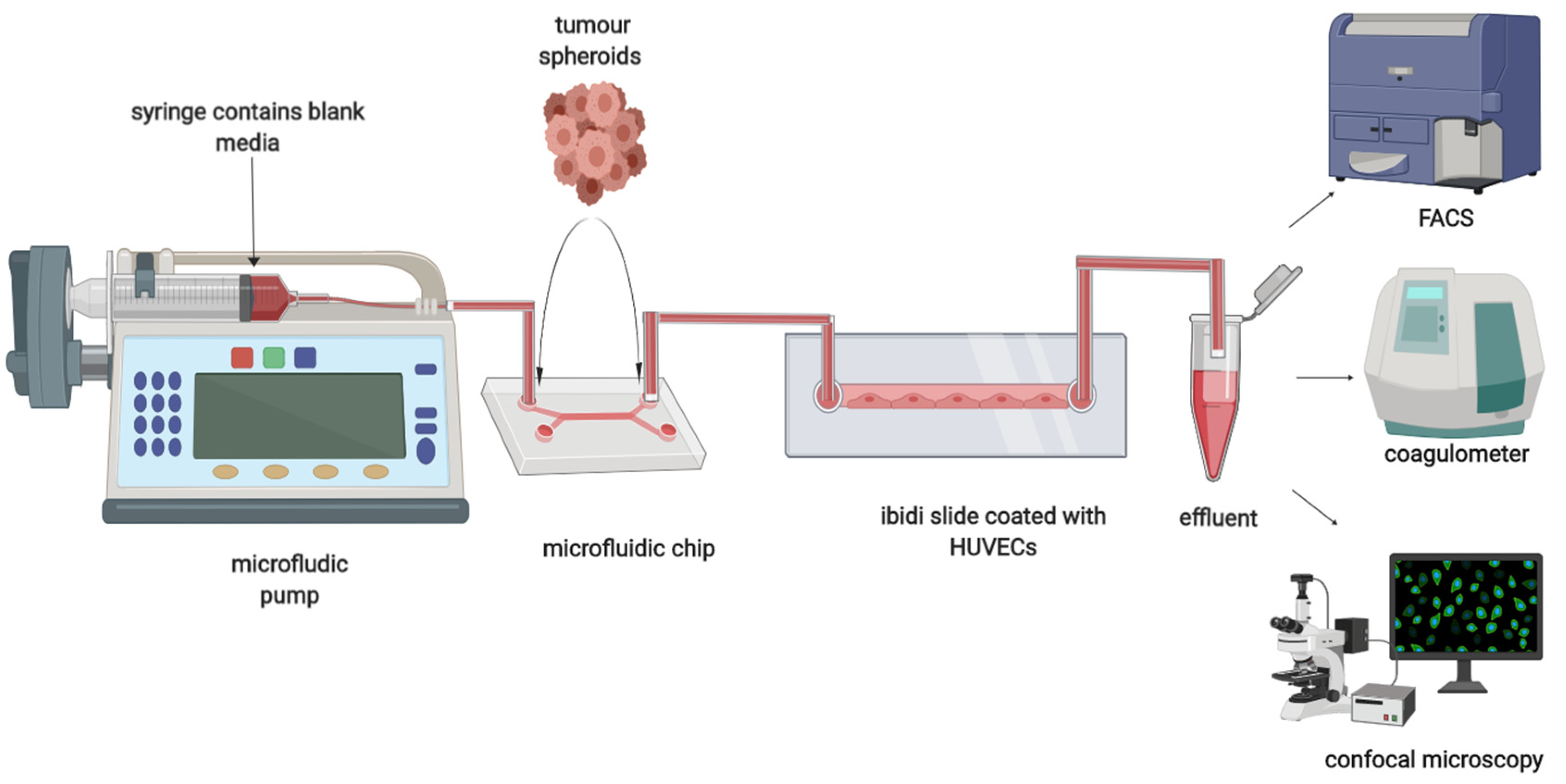
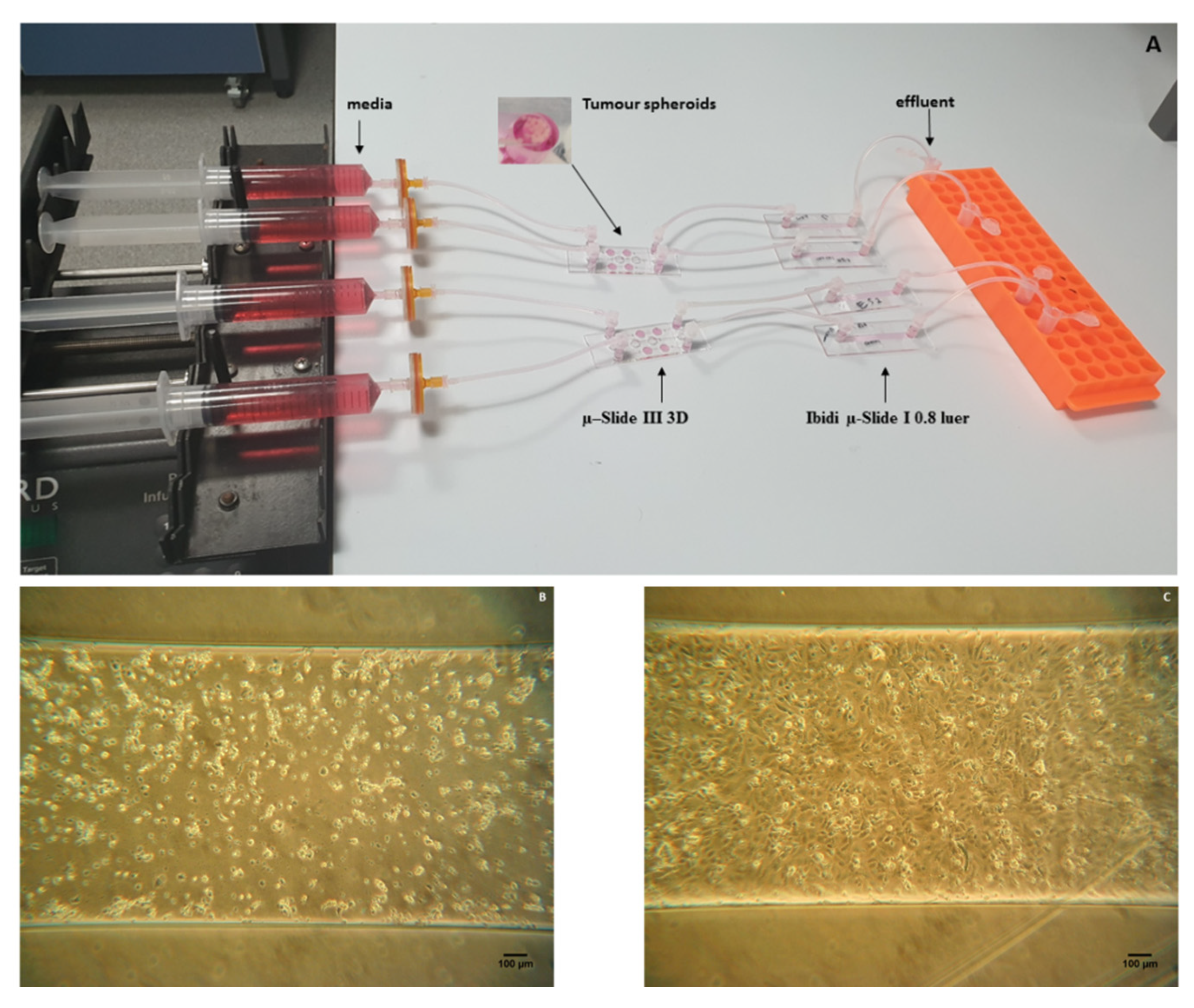
2.2. Dual Chip Microfluidic Setup

HUVECs were cultured on a μ-Slide I Luer (Ibidi, Gräfelfing, Germany). Slides were treated with UV irradiation for 20 min and coated by dispensing approximately 12 µL of type B 2% v/v gelatin (Sigma Aldrich, Gillingham, UK) into the channel. Then, the biochips were incubated for 24 h at 4 °C. Cultured HUVECs (2000 cells) were added into each channel and the reservoirs filled with 60 µL of media. The biochips were incubated in the CO2 incubator for 24 h at 37 °C. A µ-slide III 3D perfusion was used to hold the spheroids, then flow was applied via a Harvard infusion PHD22/200 syringe pump (4 μL/min). The µ-Slide I Luer was attached to the output of the µ-slide III 3D chip as shown in Figure 1 and Figure 2A–C. Samples were then collected via the output of the µ-Slide I Luer into sterile 1.5 mL polypropylene tubes.

2.3. Procoagulant Activity
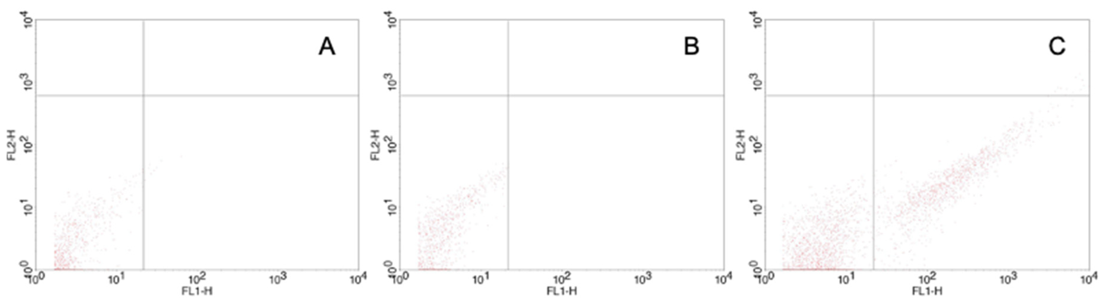
The procoagulant potential of cell-free supernatant or cells (300,000 per assay) was measured using the semi-automated Thrombotrack SOLO coagulometer. Samples (100 µL) were placed into a cuvette containing a steel ball and 25 mM CaCl2 (100 µL) was added; finally, 100 µL of control plasma (NormTrol, Helena Biosciences, Gateshead, UK) was added and the time taken for clot formation (prothrombin time) was automatically determined.

2.4. Flow Cytometry

TF labelled MP released from ES2 and U87 tumour cells were quantified by flow cytometry before and after, being passed through the dual chips. MP were analysed immediately, directly from samples of effluent media which was centrifuged to remove any larger cell debris (1000× g, 5 min) prior to addition of antibodies. Samples of supernatant (50 µL) were incubated with 5 μL of anti-TF: FITC (Bio-Rad, clone CLB/TF-5) for 30 min then analysed by flow cytometry by adding an equal volume of Accucheck beads (Invitrogen, UK) and 150 µL of 0.2 µm-filtered sterile PBS [27,28]. A flow cytometer (BD FACSCalibur) was set up with Megamix SSC beads (Biocytex, France) that were used to define an MP gate from 200–500 nm according to side-scatter characteristics of the beads following the International Society of Thrombosis and Haemostasis current recommendation for standardisation of MP measurement by flow cytometry [29]. An FITC-conjugated isotype negative control antibody (murine IgG1, MCA928F, Bio-Rad) was used to establish parameters (Figure 3).

2.5. Doxorubicin Treatment

Spheroids, in 180 μL of cell culture media, were transferred to 96-well sterile tissue culture plates. Various concentrations of doxorubicin were added in a volume of 20 μL into the cells and incubated for 24 and 48 h in an incubator (37 °C, 5% CO2). Cell proliferation, relative to the controls (no drug), was determined by a cytotoxicity assay according to the manufacturers’ instructions (CellTitre Aqueous One, Promega, Southampton, UK). MTS reagent (3-[4,5-dimethylthiazol-2-yl]-5-[3-carboxymethoxyphenyl]-2-[4-sulfophenyl]-2H-tetrazolium) uses a colorimetric method for the sensitive quantification of viable cells. After incubation, plates were centrifuged at 400× g for 5 min and the supernatant was removed. Fresh medium (180 μL) was then added followed by MTS reagent (20 μL) and the plates incubated (at 37 °C, 5% CO2) for 4 h. The absorbance was determined at 490 nm using a microplate reader (BioTek synergy HT, BioTeK, Swindon, UK). For on-chip experiments, DOX was added to media at a final concentration of 0.75 μM prior to perfusion by syringe pump (Figure 2A).

2.6. Statistical Analysis

Pearson correlation coefficients were calculated and analyses of variance were used to compare conditions. p values less than 0.05 were considered significant.

3. Results

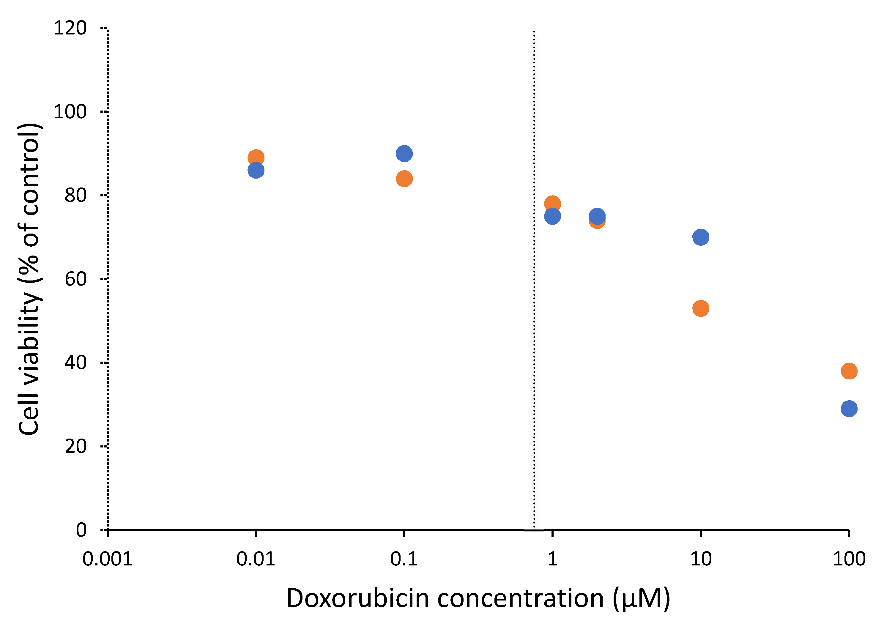
3.1. Cytotoxicity of Doxorubicin on Cells

To investigate the effects of a cytotoxic agent on the interaction between tumour MP and HUVECs, doxorubicin (DOX) was used. Initially the minimum concentration of DOX that would have an effect on cell viability was determined using a cell proliferation assay (MTS); various concentrations of DOX were used on U87 and ES-2 tumour cells (Figure 4) and a concentration of 0.75 μM DOX was selected for further study. At this concentration, HUVEC viability also decreased to 71% as compared with control.

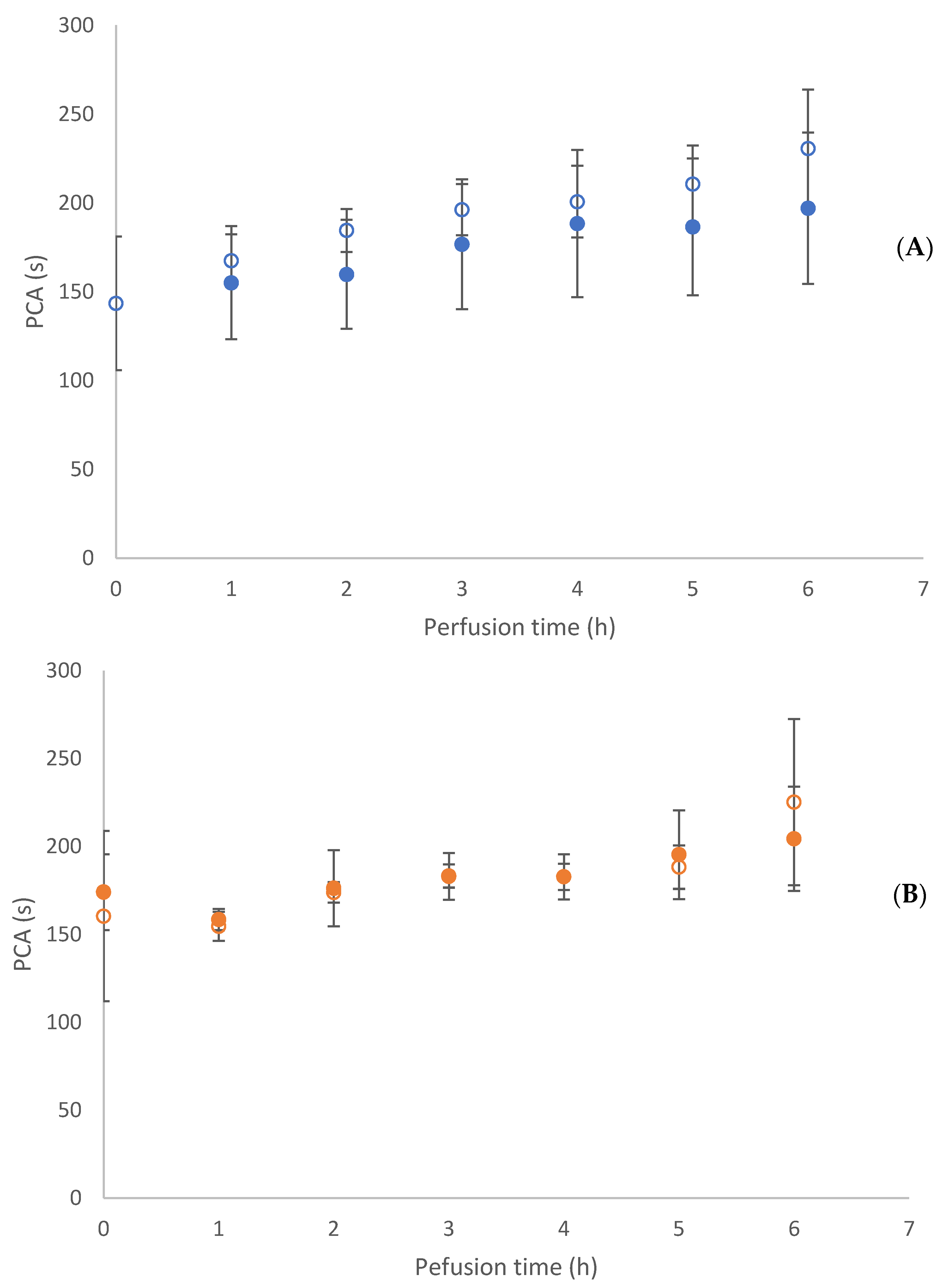
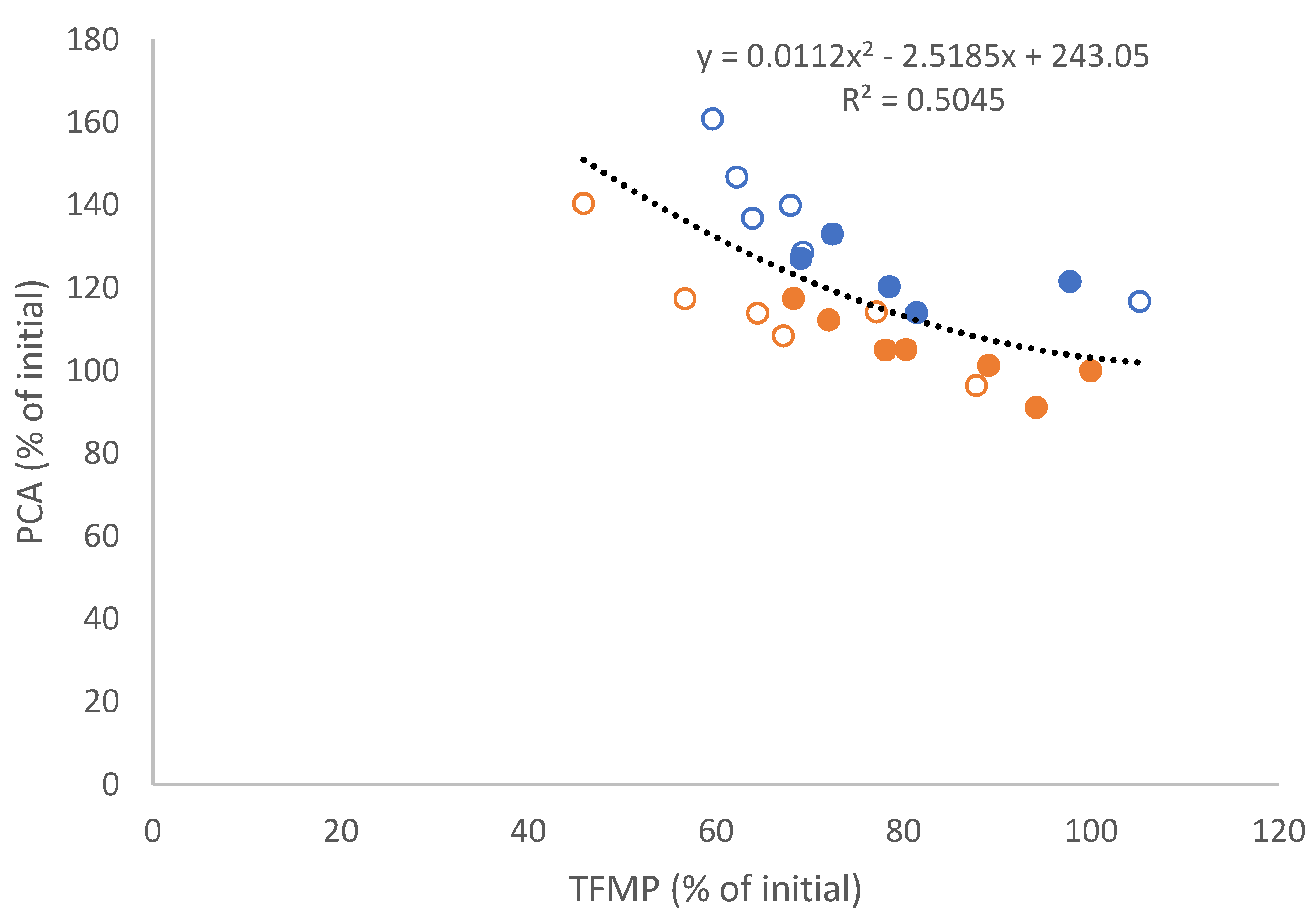
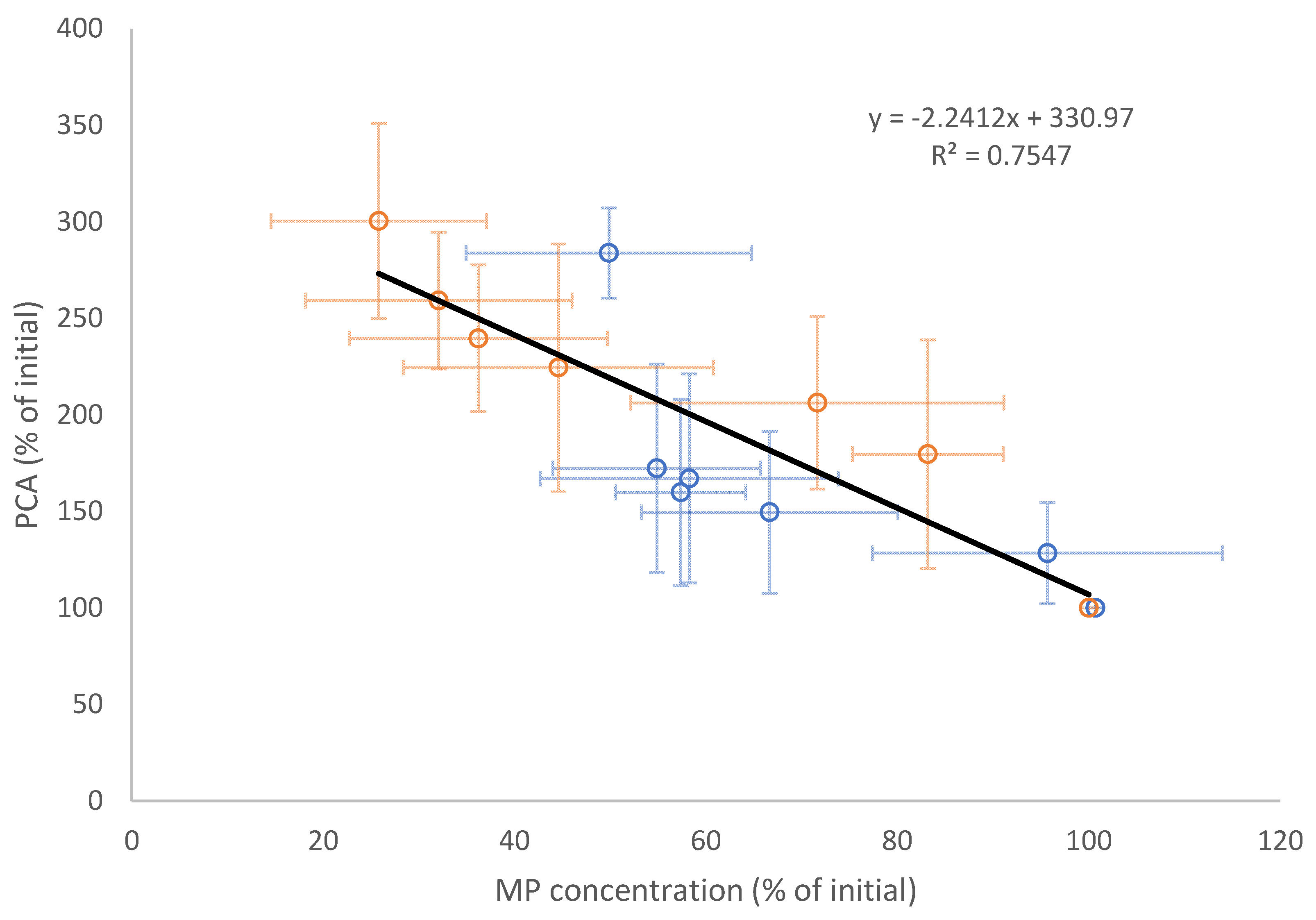
3.2. Tumour Spheroid Flow Experiments

Further experiments were carried as follows: U87 and ES-2 tumour spheroids were formed and then transferred into 3D Ibidi chips and media with or without the addition of DOX (0.75 μM), which was then flowed across the spheroids, collected and analysed for procoagulant activity (PCA) and TFMP prior to the commencement of the experiment and then every hour for 6 h. A continual loss of PCA (reflected as a slower coagulation time) was observed in the collected media over the 6 h window for both U87 and ES-2 (Figure 5A,B). The relationship between PCA and concentration of TFMP observed in the collected media showed a correlation which was unaltered by the presence of DOX. Overall a consistent negative correlation was observed between TFMP and PCA with an average Pearson’s rank correlation of −0.85 ± 0.05. To further confirm this relationship, the loss of PCA and loss of TFMP was calculated as a percentage of the initial values prior to the start of the experiments. Again, a negative correlation was observed (Figure 6, r = −0.70) for all data, independent of time.

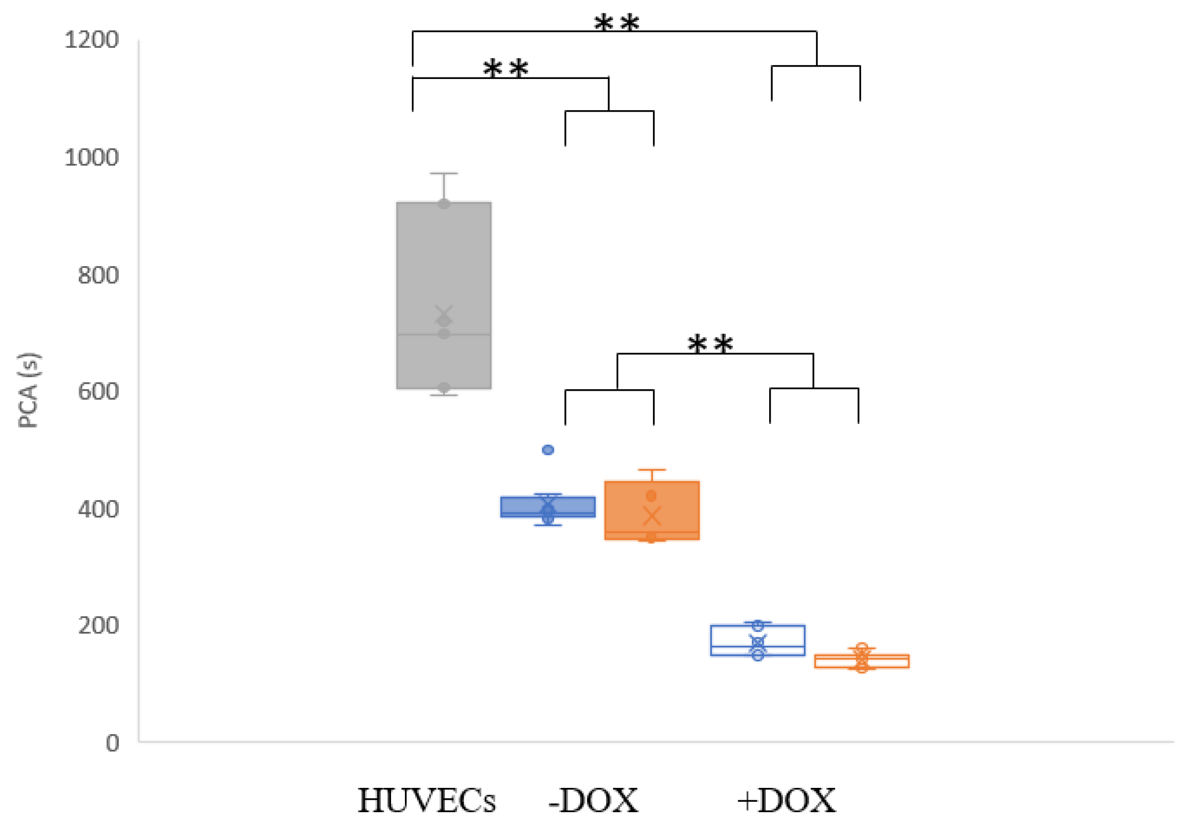
3.3. Flow Experiments Analysing HUVECs’ Response

Having observed no differential loss of PCA or TFMP from media flowed over HUVECs in the absence or presence of DOX (Figure 5A,B and Figure 6), a further series of experiments were undertaken in which the HUVECs themselves were subsequently harvested from the chips at the completion of each experiment for cell analysis. Firstly, TFMP and PCA in the media were again assessed, and the gradual loss of TFMP and PCA were observed across this series of independent experiments, confirming the previous experiments. The largest reduction in TFMP concentration was generally observed within the first 3 h, when there was an approximate loss of 50%; following this a plateau was seen from 3 (ES-2) or 4 (U87) to 6 h where less TFMP per hour were lost (Figure 7). This was most pronounced for ES-2, where there was practically no further loss of MP from 3–6 h, whereas the U87 TFMP continued to slowly decline.
A decrease in PCA was associated with the loss of TFMP, and the relationship between these factors was again consistent with the TFMP in the media most likely being responsible for PCA (r = −0.87, Figure 8).
Finally, HUVECs from these experiments were subsequently harvested from the chips and analysed for PCA at a constant cell number, after 24 h. Control chips containing HUVECs were subjected to flow without the presence of tumour spheroids in the upstream 3D Ibidi chip. DOX controls are described as “no DOX” within the dual chip setup and finally the same experiments in the presence of DOX were all run in parallel on five separate occasions. The PCA of HUVEC cells was shown to be significantly increased (faster clotting time) when tumour media from the spheroids was flowed through for 24 h (p < 0.001, Figure 9), compared with HUVECs with media alone. Furthermore, in the presence of DOX the HUVECs were shown to have a significantly increased PCA over both the control HUVECs and the no DOX (but tumour-spent media) conditions. No significant differences were observed in PCA of HUVECs in response to exposure to media from the two different cell lines, most likely due to ES-2 and U87 possessing similar PCA properties.
The viability of HUVECs after 24 h of direct flow with tumour spheroid-derived MP with or without DOX was assessed by trypan blue exclusion. In the absence of DOX, HUVEC viability was 88.5 ± 3.4%, whereas in the presence of the drug viability decreased to 69.3 ± 4.0%. In static (no flow) conditions, the viability of HUVECs in the presence of DOX was found to be 77.6 ± 6.0%. This increased loss of viability under flow conditions could be due to the fact the DOX is perfused over the HUVECs at a constant concentration rather than being added at a single time and thus is continually replenished. Another factor to consider is the concentration of DOX that comes into contact with the HUVECs is unknown as the tumour spheroids are exposed first in the dual chip flow-through model.

4. Discussion

Chemotherapy is an independent risk factor for VTE in cancer patients. Here we showed that TFMP, released from 3D tumour spheroids under flow, decreased in concentration after perfusion through biochips coated with HUVECs and this effect is associated with a concurrent, proportional increase of procoagulant activity (PCA) of the endothelial cells (reflected as a faster coagulation time). The increased PCA of the HUVECs was further increased in the presence of doxorubicin treatment of the tumour spheroids. We have previously shown that tumour MP are able to bind to endothelial cells, transfer and increase the PCA of endothelial cells in static and flow conditions [27,28], and here we demonstrate that the incubation of a cytotoxic agent with the tumour spheroids further increased the PCA of endothelial cells when exposed to tumour MP in flow conditions. The average loss of MP over the HUVEC layer was similar but slightly enhanced in the presence of DOX compared to media alone. As the DOX concentration used was the lowest that caused an effect on viability of the tumour spheroids and HUVECs, the increased PCA of HUVECs harvested after 24 h perfusion can most likely be attributed to a loss of viability (increased procoagulant phospholipid exposure) rather than any significant increase in MP binding.
Perhaps the most interesting finding observed was the cessation of loss of TFMP from media perfused over HUVECs after 3 h (Figure 7, indicated by blue arrow). This may well suggest that MP are binding to HUVECs through a specific receptor-mediated mechanism which becomes saturated or sterically inaccessible due to the number of MP attached to the cell surface. MP are known to interact with target cells via multiple mechanisms; direct ligand-receptor interaction, membrane fusion leading to internalisation and acquisition of MP cargo by the target cell or by transferring surface receptors [30,31]. The data herein suggest that the interaction between tumour MP and endothelial cells is the former. Additionally, if this was indeed the case then it may be expected that a proportional response of the recipient endothelial cells may be observed and we have recently shown a positive correlation between endothelial cell MP release in response to exposure of increasing concentrations of tumour MP [32].
It has previously been proposed that ligand-receptor interaction between MP and target cells accounts for subsequent biological effects showing the role of MP in cell–cell communication [33]. This has been demonstrated in endothelial cells where MP carrying Sonic Hedgehog were shown to induce nitric oxide production in endothelial cells and improve endothelial function after ischaemia/reperfusion [34] and also modulate neovascularisation [35]. Platelet MP have also been shown to activate endothelial cells [36] and are able to transfer the adhesion molecule CD41. The data presented add to this body of work suggesting that endothelial cells are receptive to MP binding via specific mechanisms.
Here we describe a dual chip microfluidic flow model for study of cytotoxic agents on tumour spheroids and endothelial cells. Doxorubicin treatment of tumour spheroids resulted in an enhanced PCA of an endothelial cell layer under flow. This increased PCA was not MP-associated but due to the presence of doxorubicin within the media which reduced HUVECs’ viability by 20% and could have relevance to the mechanism by which cancer patients undergoing chemotherapy have an increased risk of VTE. Endothelial cell damage by chemotherapy agents may contribute to the hypercoagulable state.

5. Conclusions

Doxorubicin treatment of tumour spheroids resulted in enhanced PCA of an endothelial cell layer under flow. This increased PCA may have relevance to the mechanism by which cancer patients undergoing chemotherapy have an increased risk of VTE.

Author Contributions

Conceptualisation, L.A.M.; methodology, L.A.M., A.A.; formal analysis, A.A., L.A.M.; investigation, A.A..; writing—original draft preparation, L.A.M.,. A.A.; writing—review and editing, L.A.M.,. A.A., J.G.; supervision, L.A.M., J.G.; project administration, L.A.M.; All authors have read and agreed to the published version of the manuscript.

Funding

This research was funded through a PhD scholarship from the Northern Border University (Saudi Arabia) to A.A.

Conflicts of Interest

The authors declare no conflict of interest.

References

  1. Seaman, S.; Nelson, A.; Noble, I. Cancer-associated thrombosis, low-molecular-weight heparin, and the patient experience: A qualitative study. Patient Prefer. Adherence 2014, 8, 453–461. [Google Scholar]
  2. Echrish, H.; Xiao, Y.; Madden, L.; Allgar, V.; Cooke, J.; Wedgwood, K.; Dasgupta, D.; Greenman, J.; Maraveyas, A. Effect of resection of localized pancreaticobiliary adenocarcinoma on angiogenic markers and tissue factor related pro-thrombotic and pro-angiogenic activity. Thromb. Res. 2014, 134, 479–487. [Google Scholar] [CrossRef]
  3. Aleman, M.M.; Gardiner, C.; Harrison, P.; Wolberg, A.S. Differential contributions of monocyte- and platelet-derived microparticles towards thrombin generation and fibrin formation and stability. J. Thromb. Haemost. 2011, 9, 2251–2261. [Google Scholar] [CrossRef] [Green Version]
  4. Shai, E.; Varon, D. Development, cell differentiation, angiogenesis—Microparticles and their roles in angiogenesis. Arterioscler. Thromb. Vasc. Biol. 2011, 31, 10–14. [Google Scholar] [CrossRef]
  5. Campello, E.; Spiezia, L.; Radu, C.M.; Bulato, C.; Castelli, M.; Gavasso, S.; Simioni, P. Endothelial, platelet, and tissue factor-bearing microparticles in cancer patients with and without venous thromboembolism. Thromb. Res. 2011, 127, 473–477. [Google Scholar] [CrossRef]
  6. Brodsky, S.V.; Zhang, F.; Nasjletti, A.; Goligorsky, M.S. Endothelium-derived microparticles impair endothelial function in vitro. Am. J. Physiol. -Heart Circ. Physiol. 2004, 286, H1910–H1915. [Google Scholar] [CrossRef] [Green Version]
  7. Barteneva, N.S.; Fasler-Kan, E.; Bernimoulin, M.; Stern, J.N.H.; Ponomarev, E.D.; Duckett, L.; Vorobjev, I.A. Circulating microparticles: Square the circle. BMC Cell Biol. 2013, 14, 23. [Google Scholar] [CrossRef] [Green Version]
  8. Nomura, S.; Shimizu, M. Clinical significance of procoagulant microparticles. J. Intensive Care 2015, 3, 2. [Google Scholar] [CrossRef] [Green Version]
  9. Gold, B.; Cankovic, M.; Furtado, L.V.; Meier, F.; Gocke, C.D. Do Circulating Tumor Cells, Exosomes, and Circulating Tumor Nucleic Acids Have Clinical Utility? J. Mol. Diagn. 2015, 17, 209–224. [Google Scholar] [CrossRef] [Green Version]
  10. Manly, D.A.; Boles, J.; Mackman, N. Role of tissue factor in venous thrombosis. Annu. Rev. Physiol. 2011, 73, 515–525. [Google Scholar] [CrossRef] [Green Version]
  11. Yates, K.R.; Welsh, J.; Echrish, H.H.; Greenman, J.; Maraveyas, A.; Madden, L.A. Pancreatic cancer cell and microparticle procoagulant surface characterization: Involvement of membrane-expressed tissue factor, phosphatidylserine and phosphatidylethanolamine. Blood Coagul. Fibrinolysis 2011, 22, 680–687. [Google Scholar] [CrossRef] [PubMed]
  12. Date, K.; Hall, J.; Greenman, J.; Maraveyas, A.; Madden, L.A. Tumour and microparticle tissue factor expression and cancer thrombosis. Thromb. Res. 2013, 131, 109–115. [Google Scholar] [CrossRef] [PubMed]
  13. Haddad, T.C.; Greeno, E.W. Chemotherapy-induced thrombosis. Thromb. Res. 2006, 118, 555–568. [Google Scholar] [CrossRef] [PubMed]
  14. Swystun, L.L.; Shin, L.Y.Y.; Beaudin, S.; Liaw, P.C. Chemotherapeutic agents doxorubicin and epirubicin induce a procoagulant phenotype on endothelial cells and blood monocytes. J. Thromb. Haemost. 2009, 7, 619–626. [Google Scholar] [CrossRef]
  15. Furie, B.C.; Furie, B. Cancer-associated thrombosis. Blood Cellsmolecules Dis. 2006, 36, 177–181. [Google Scholar] [CrossRef]
  16. Falanga, A.; Russo, L. Epidemiology, risk and outcomes of venous thromboembolism in cancer. Hamostaseologie 2012, 32, 115–125. [Google Scholar]
  17. Barni, S.; Labianca, R.; Agnelli, G.; Bonizzoni, E.; Verso, M.; Mandalà, M.; Brighenti, M.; Petrelli, F.; Bianchini, C.; Perrone, T. Chemotherapy-associated thromboembolic risk in cancer outpatients and effect of nadroparin thromboprophylaxis: Results of a retrospective analysis of the PROTECHT study. J. Transl. Med. 2011, 9, 179. [Google Scholar] [CrossRef] [Green Version]
  18. Moore, R.A.; Adel, N.; Riedel, E.; Bhutani, M.; Feldman, D.R.; Tabbara, N.E.; Soff, G.; Parameswaran, R.; Hassoun, H. High incidence of thromboembolic events in patients treated with cisplatin-based chemotherapy: A large retrospective analysis. J. Clin. Oncol. 2011, 29, 3466–3473. [Google Scholar] [CrossRef]
  19. Nuver, J.; Smit, A.; Sleijfer, D.T.; Van Gessel, A.; Van Roon, A.; Van Der Meer, J.; van den Berg, M.; Burgerhof, J.; Hoekstra, H.; Sluiter, W. Microalbuminuria, decreased fibrinolysis, and inflammation as early signs of atherosclerosis in long-term survivors of disseminated testicular cancer. Eur. J. Cancer 2004, 40, 701–706. [Google Scholar] [CrossRef]
  20. Soultati, A.; Mountzios, G.; Avgerinou, C.; Papaxoinis, G.; Pectasides, D.; Dimopoulos, M.-A.; Papadimitriou, C. Endothelial vascular toxicity from chemotherapeutic agents: Preclinical evidence and clinical implications. Cancer Treat. Rev. 2012, 38, 473–483. [Google Scholar] [CrossRef]
  21. Hoshi, A.; Matsumoto, A.; Chung, J.; Isozumi, Y.; Koyama, T. Activation of coagulation by a thalidomide-based regimen. Blood Coagul. Fibrinolysis 2011, 22, 532–540. [Google Scholar] [CrossRef] [PubMed]
  22. Walsh, J.; Wheeler, H.R.; Geczy, C.L. Modulation of tissue factor on human monocytes by cisplatin and adriamycin. Br. J. Haematol. 1992, 81, 480–488. [Google Scholar] [CrossRef] [PubMed]
  23. Kim, E.J.; Lim, K.M.; Kim, K.Y.; Bae, O.N.; Noh, J.Y.; Chung, S.M.; Shin, S.; Yun, Y.P.; Chung, J.H. Doxorubicin-induced platelet cytotoxicity: A new contributory factor for doxorubicin-mediated thrombocytopenia. J. Thromb. Haemost. 2009, 7, 1172–1183. [Google Scholar] [CrossRef]
  24. Ben Aharon, I.; Bar Joseph, H.; Tzabari, M.; Shenkman, B.; Farzam, N.; Levi, M.; Shalgi, R.; Stemmer, S.M.; Savion, N. Doxorubicin-Induced Vascular Toxicity—Targeting Potential Pathways May Reduce Procoagulant Activity. PLoS ONE 2013, 8, e75157. [Google Scholar] [CrossRef]
  25. Sontheimer-Phelps, A.; Hassell, B.A.; Ingber, D.E. Modelling cancer in microfluidic human organs-on-chips. Nat. Rev. Cancer 2019, 19, 65–81. [Google Scholar] [CrossRef]
  26. Garcia-Cordero, J.L.; Maerkl, S.J. Microfluidic systems for cancer diagnostics. Curr. Opin. Biotechnol. 2020, 65, 37–44. [Google Scholar] [CrossRef] [Green Version]
  27. Adesanya, M.A.; Maraveyas, A.; Madden, L.A. Cancer microvesicles induce tissue factor-related procoagulant activity in endothelial cells in vitro. Blood Coagul. Fibrinolysis 2017, 28, 365–372. [Google Scholar] [CrossRef]
  28. Algarni, A.; Greenman, J.; Madden, L.A. Procoagulant tumor microvesicles attach to endothelial cells on biochips under microfluidic flow. Biomicrofluidics 2019, 13, 064124. [Google Scholar] [CrossRef]
  29. Cointe, S.; Judicone, C.; Robert, S.; Mooberry, M.; Poncelet, P.; Wauben, M.; Nieuwland, R.; Key, N.; Dignat-George, F.; Lacroix, R. Standardization of microparticle enumeration across different flow cytometry platforms: Results of a multicenter collaborative workshop. J. Thromb. Haemost. 2017, 15, 187–193. [Google Scholar] [CrossRef]
  30. Benameur, T.; Osman, A.; Parray, A.; Ait Hssain, A.; Munusamy, S.; Agouni, A. Molecular mechanisms underpinning microparticle-mediated cellular injury in cardiovascular complications associated with diabetes. Oxidative Med. Cell. Longev. 2019, 6475187. [Google Scholar] [CrossRef] [Green Version]
  31. Camussi, G.; Deregibus, M.C.; Bruno, S.; Cantaluppi, V.; Biancone, L. Exosomes/microvesicles as a mechanism of cell-to-cell communication. Kidney Int. 2010, 78, 838–848. [Google Scholar] [CrossRef] [PubMed] [Green Version]
  32. Faulkner, L.G.; Alqarni, S.; Maraveyas, A.; Madden, L.A. Isolated tumour microparticles induce endothelial microparticle release in vitro. Blood Coagul. Fibrinolysis 2020, 31, 35–42. [Google Scholar] [CrossRef] [PubMed]
  33. Maas, S.L.; Breakefield, X.O.; Weaver, A.M. Extracellular vesicles: Unique intercellular delivery vehicles. Trends Cell Biol. 2017, 27, 172–188. [Google Scholar] [CrossRef] [PubMed] [Green Version]
  34. AgouTii, A.; Ahmed Mostefai, H.; Porro, C.; Carusio, N.; Favre, J.; Richard, V.; Henrion, D.; Carmen Martinez, M.; Andriantsitohaina, R. Sonic hedgehog carried by microparticles corrects endothelial injury through nitric oxide release. FASEB J. 2007, 21, 2735–2741. [Google Scholar] [CrossRef] [Green Version]
  35. Benameur, T.; Soleti, R.; Porro, C.; Andriantsitohaina, R.; Martínez, M.C. Microparticles carrying Sonic hedgehog favor neovascularization through the activation of nitric oxide pathway in mice. PLoS ONE 2010, 5, e12688. [Google Scholar] [CrossRef] [Green Version]
  36. Barry, O.P.; Praticò, D.; Lawson, J.A.; FitzGerald, G.A. Transcellular activation of platelets and endothelial cells by bioactive lipids in platelet microparticles. J. Clin. Investig. 1997, 99, 2118–2127. [Google Scholar] [CrossRef] [Green Version]
Figure 1. Schematic showing the dual microfluidic setup. Media with or without doxorubicin was perfused though a chip containing established spheroids of ES-2 or U87 tumour cells and subsequently a second chip coated with a human umbilical vein endothelial cell (HUVEC) monolayer. Samples were then collected and HUVECs harvested after 24 h for analysis.
Hemato 01 00006 g001
Figure 2. Experimental dual chip setup (A) and HUVECs monolayers before (B) and after (C) the application of flow.
Hemato 01 00006 g002
Figure 3. Representative fluorescence scatter plots for positive microparticle (MP) events showing (A) tumour-spent media alone, (B) tumour-spent media with isotype-matched negative control and (C) tissue factor (TF)-labelled MP within tumour-spent media. The lower right quadrant in (C) was established and used for quantification of positive events.
Hemato 01 00006 g003
Figure 4. Cell viability (n = 2) in various static cancer cell cultures when applying between 0 and 100 μM of doxorubicin (DOX) to 3D tumour spheroids of ES-2 (●) or U87 (●). Dashed line represents a concentration 0.75 μM of DOX.
Hemato 01 00006 g004
Figure 5. Procoagulant activity (PCA) of media samples after perfusion through the dual chip setup of (A) ES-2 and (B) U87 spheroids with (unfilled) and without (filled) the presence of DOX (n = 6 independent observations per point, error bars represent SD).
Hemato 01 00006 g005
Figure 6. Data represented as a percentage of initial values, independent of time. Conditions were ES-2 (blue) no DOX (filled), ES-2 DOX (unfilled), U87 (orange) no DOX (filled) and U87 DOX (unfilled).
Hemato 01 00006 g006
Figure 7. Loss of TFMP (expressed as % of initial concentration and averaged) from ES-2 or U87 spheroid media perfused over HUVECs for up to 6 h in the presence of DOX (0.75 μM, n = 5 independent experiments, error bars represent SD). Arrow indicates possible point of saturation of HUVECs with MP at 3 h perfusion with ES2 spheroid media.
Hemato 01 00006 g007
Figure 8. The relationship between loss of PCA and loss of TFMP from media collected after perfusion with HUVECs from tumour spheroid 3D culture of ES-2 or U87 cells in the presence of DOX (0.75 μM); r = −0.87.
Hemato 01 00006 g008
Figure 9. PCA of HUVECs harvested after 24 h of flow from (grey) blank, coated 3D chips, ES-2 or U87 spheroid 3D chip without DOX (filled) and ES-2 or U87 in the presence of DOX (0.75 μM, unfilled). ** p < 0.01.
Hemato 01 00006 g009

