Article

Nucleoredoxin Downregulation Reduces β-Catenin Levels and Shifts Hematopoietic Differentiation towards Myeloid Lineage In Vitro

by
Alejandro Pérez-Fernández
1,2,
Guillermo López-Ruano
1,2,
Rodrigo Prieto-Bermejo
1,2,
Carmen Sánchez-Bernal
1,2,
Jesús Sánchez-Yagüe
1,2 and
Ángel Hernández-Hernández
1,2,*
1
Department of Biochemistry and Molecular Biology, University of Salamanca, 37007 Salamanca, Spain
2
Institute for Biomedical Research of Salamanca (IBSAL), 37007 Salamanca, Spain
*
Author to whom correspondence should be addressed.
Submission received: 23 February 2021 / Revised: 26 March 2021 / Accepted: 27 March 2021 / Published: 4 April 2021

Abstract

:
The importance of dissecting signaling pathways governing cell differentiation is based on their relevance not only for understanding basic biological phenomena but also for better comprehending the underlying mechanisms of pathologic alterations such as cancer. A paradigm of cell differentiation processes is hematopoiesis, where a single stem cell gives rise to multiple, fully differentiated, cell lineages. Nucleoredoxin (Nrx), a member of the thioredoxin family, is an important redox-sensitive modulator of Wnt/β-catenin signaling, a key pathway for the control of hematopoiesis. In this work, the relevance of Nrx for the differentiation of mouse hematopoietic progenitor cells has been analyzed in vitro. Nrx silencing leads to a dramatic reduction in the size of the Lin and LSK progenitor populations. Moreover, there is also a remarkable decrease in CD3+ cells and an enhancement in the percentage of CD11b+Gr1 myeloid cells. This myeloid bias would agree with the inhibition of the Wnt/β-catenin pathway. Interestingly, a reduction in β-catenin at the protein level was observed upon Nrx silencing. Our results strongly support the importance of Nrx for hematopoietic differentiation, which could be mediated by the regulation of the Wnt/β-catenin pathway.

1. Introduction

Blood is a rather heterogeneous tissue in terms of cellular components, a feature that allows it to perform many different functions. However, all hematopoietic lineages emerge from a single cell type: the hematopoietic stem cell (HSC) [1]. Consequently, hematopoiesis must be a fine-tuned process in which the balance between the repopulation ability of HSCs and the generation of fully differentiated cells (lacking that property) needs to meet the organism’s demands.
This equilibrium is maintained at the level of signaling pathways governing differentiation and self-renewal. The Wnt/β-catenin pathway is a pivotal regulator of those cellular phenomena at different developmental stages. For instance, an activation of this signaling cascade is required at a particular dose and moment for the emergence of HSCs from the aorta-gonad-mesonephros [2]. Once they have settled in the bone marrow, the activation intensity of the pathway will dictate their outcome in terms of self-renewal ability and lineage commitment [3]. More precisely, the central effector of the pathway, β-catenin, influences the expression of several adhesion genes, which in turn has an effect on the quiescence of HSCs [4]. Of note, the stability and cellular localization of β-catenin are regulated by a myriad of extra- and intracellular cues beyond the interaction of Wnt ligands with frizzled (Fzd) receptors and the subsequent downstream events. Therefore, it is worth studying the possible crosstalk between well-documented players in cell differentiation and in β-catenin levels and localization in the context of hematopoiesis.
Among the wide variety of cell-extrinsic and -intrinsic factors modulating the differentiation of hematopoietic cells, reactive oxygen species (ROS) have gained attention over the last two decades. ROS production is increased upon growth factor stimulation, and their intracellular levels dictate lineage decisions (reviewed in [5]). Indeed, hematopoietic cells subject to pro-differentiative stimuli undergo ROS generation in a regulated manner through the activation of NADPH oxidases (NOXs), an event required to fully trigger the process of megakaryocytic differentiation [6]. Mechanistically, ROS interfere with proteins involved in cell signaling, thus altering their enzymatic activity or their interactions. Well-known examples of this are the protein tyrosine phosphatases (PTPs), whose catalytic activity can be transiently switched off through mild oxidation of a key cysteine residue located at the active center [7]. We have previously described the close relationship between different PTPs and β-catenin in this context. Protein tyrosine phosphatase non-receptor type 13 (PTPN13) is a negative regulator of megakaryocytic differentiation, and its levels are linked to β-catenin stability [8]. In addition, the transient oxidation and inactivation of the SRC homology domain containing protein tyrosine phosphatases 1 (SHP1) and 2 (SHP2) is required to trigger the same differentiation processes. Interestingly, downregulation of these proteins results in a decrease in β-catenin levels [9]. This work supports the influence of redox signaling on the Wnt/β-catenin pathway and encourages the study of redox-sensitive proteins that may link these two cellular processes.
In this regard, nucleoredoxin (Nrx) must be mentioned as a protein subject to redox regulation [10,11]. It belongs to the thioredoxin family and displays oxidoreductase activity undertaken by two oxidation-sensitive cysteine residues located within the active center motif Trp-Cys-Pro-Pro-Cys [12]. The mouse and human homologs share 99% identity, and its active center is conserved across different species [13]. It is worth noting that its redox status affects its interaction with the protein disheveled (Dvl), a positive regulator of Wnt/β-catenin signaling that sequesters the destruction complex. Interestingly, the oxidation of Nrx prompts different outcomes in β-catenin levels in a context-dependent manner. In the absence of kelch-like 12 (KLHL12), Nrx oxidation destabilizes its interaction with Dvl and releases the latter for further sequestration of the destruction complex, thus impairing Wnt/β-catenin signaling [10]. On the other hand, KLHL12 targets Dvl for proteasomal degradation, which has a negative impact on the canonical Wnt pathway. Under these circumstances, reduced Nrx is able to disrupt the KLHL12–Dvl interaction, thus maintaining a pool of Dvl that sustains Wnt/β-catenin signaling [14]. Interestingly, it has been reported that stimulation of cells with the canonical ligand Wnt3a triggers Nox1-derived ROS production, which in turn oxidize Nrx and prevent the interaction with Dvl, thereby contributing to β-catenin stabilization [11]. Moreover, Nrx expression augments during adipocyte differentiation in mouse cell lines, with a negative impact on nuclear accumulation of β-catenin and expression of its downstream target cyclin D1 [15].
Against this background, the hypothesis that Nrx might take part in the regulation of hematopoietic differentiation was tested in this work. A decrease in β-catenin levels was observed in a human hematopoietic cell line subjected to NRX knockdown. In addition, the downregulation of the Nrx gene caused a severe reduction in immunophenotypic HSCs after in vitro culture of murine primary hematopoietic stem and progenitor cells (HSPCs). The results presented here support the influence of this protein on the differentiation of hematopoietic cells.

2. Materials and Methods

2.1. Knockdown of Nucleoredoxin

The expression of nucleoredoxin was reduced by lentiviral transduction with constructs generating small hairpin RNAs (shRNAs) as previously detailed [6]. The specific sequences targeting nucleoredoxin mRNA are detailed in Table 1.

2.2. Cell Culture

HEL cells (ACC-11) were purchased from DSMZ (Braunschweig, Germany) and grown in RPMI medium supplemented with 10% FBS, 100 U/mL penicillin, 100 U/mL streptomycin and 2 mM L-glutamine. NIH/3T3 cells (kindly provided by Dr. C. Guerrero, CIC, Salamanca, Spain) were cultured in DMEM supplemented with 10% NCS, 100 U/mL penicillin, 100 U/mL streptomycin and 2 mM L-glutamine.
Lineage-negative (Lin) cells from C57BL/6 mice were purified and transduced as described before [4], and seeded in 96-well plates at 105 cells/well in IMDM supplemented with 20% FBS, 100 U/mL penicillin, 100 U/mL streptomycin, 2 mM L-glutamine and 20 μg/mL ciprofloxacin. To prevent lineage biases, the next cytokines from Miltenyi Biotec (Madrid, Spain) were added to a final concentration of 10 ng/mL: mouse recombinant interleukin-3 (mrIL-3), stem cell factor (mrSCF), thrombopoietin (mrTPO) and FLT3 ligand (mrFLT3L). Cells were cultured for 5 days in this medium prior to flow cytometry analysis. All experimental protocols involving animals were approved by the Bioethics Committee of the University of Salamanca.

2.3. Assessment of Cell Proliferation

HEL cells were seeded at the indicated densities, and basal metabolic activity was determined by MTT assays, as described before [16]. Replicate plates were cultured for 48 h, and metabolic activity was compared with basal level after that period.

2.4. Study of Cell Adhesion

HEL cells were seeded at a density of 1.5, 2.0 and 2.5 × 10⁵ cells/mL in 200 μL of culture medium in duplicate on 100 μg/mL collagen- or 20 μg/mL fibronectin-coated plates and incubated for 3 h at 37 °C. Then, non-attached cells were removed from one of the replicates by pipetting the supernatant and gently washing the wells three times with PBS. Finally, metabolic activity was measured by MTT assays, and percentage of adhesion was calculated by dividing metabolic activity of wells with only attached cells by that of wells with all cells.

2.5. Flow Cytometry

Megakaryocytic differentiation of HEL cells was assessed by measuring the surface antigens CD41 and CD61, as described previously [6,8]. Briefly, 5 × 10⁴ cells were seeded per well in a 96-well plate, and cells were treated with 20 nM phorbol 12-myristate-13-acetate (PMA) for 48 h. Relative mean fluorescence intensity (Relative MFI in Figures) was calculated as the ratio of MFI for every surface marker in a given condition against the MFI for the same surface marker in the control PMA-untreated cells. For this last condition, this ratio was considered to be 100. Differentiation status of Lin- cells was studied through staining with the following antibodies from Miltenyi Biotec (Madrid, Spain): Lineage cell detection kit, Sca-1, c-kit, CD3, B220, CD41, Ter-119, CD11b and Gr-1. Gating strategy for surface marker analysis can be found in Figures S1–S3.

2.6. Western Blot

Protein extracts were obtained from HEL cells, and expression of β-catenin and GAPDH was measured as previously [6,8]. GAPDH was used as loading control.

2.7. Quantitative PCR (qPCR)

RNA extraction and gene expression analysis was performed as before [4], using oligonucleotides against human and mouse beta actin coding genes. Expression of human and mouse nucleoredoxin coding genes was measured by using the oligonucleotides detailed in Table 2.

2.8. Statistical Analysis

Differences between control and nucleoredoxin knocked down cells were investigated by two-tailed unpaired Student’s t-test. Significant differences were declared when p < 0.05 (*), p < 0.01 (**) or p < 0.001 (***). Error bars represent the standard deviation (SD).

3. Results

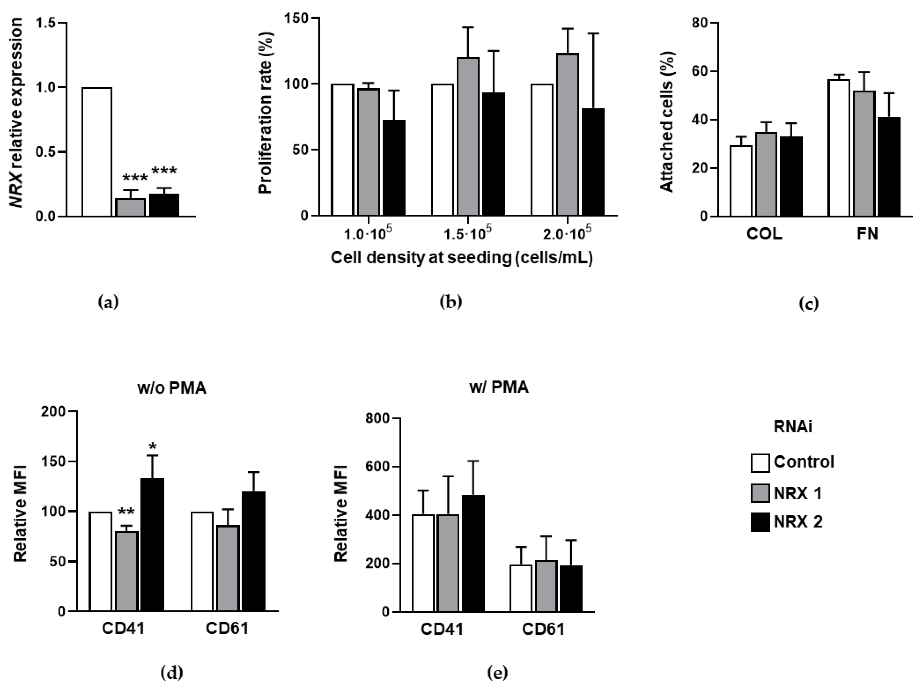
3.1. Effect of NRX Downregulation on Megakaryocytic Differentiation of HEL Cells

HEL cells were subject to effective downregulation of NRX with two different sequences (Figure 1a), and effects on different cellular parameters were evaluated. Given that proliferation capacity is usually lost concomitantly with cell differentiation, it was evaluated in NRX-silenced cells. No relevant changes were observed in this regard, irrespective of the cell density at the time of seeding (Figure 1b). Adhesion is another feature related to differentiation in this system, with cells expressing integrins throughout the process. This was the rationale for testing the adhesiveness of NRX knocked down cells. Two different proteins, collagen (COL) and fibronectin (FN), were used as adhesive substrates to cell surface integrins. In line with cell proliferation, no significant changes in the percentage of attached cells could be observed (Figure 1c). Finally, the expression of the surface markers CD41 and CD61, a common measurement of megakaryocytic differentiation [6,8], was assessed. The observed changes were mild and inconsistent without stimulation of cells with phorbol 12-myristate-13-acetate (PMA) (Figure 1d) and absent when cells were exposed to this compound (Figure 1e). All in all, these results did not allow us to attribute a role for NRX in the differentiation of HEL cells to the megakaryocytic lineage.

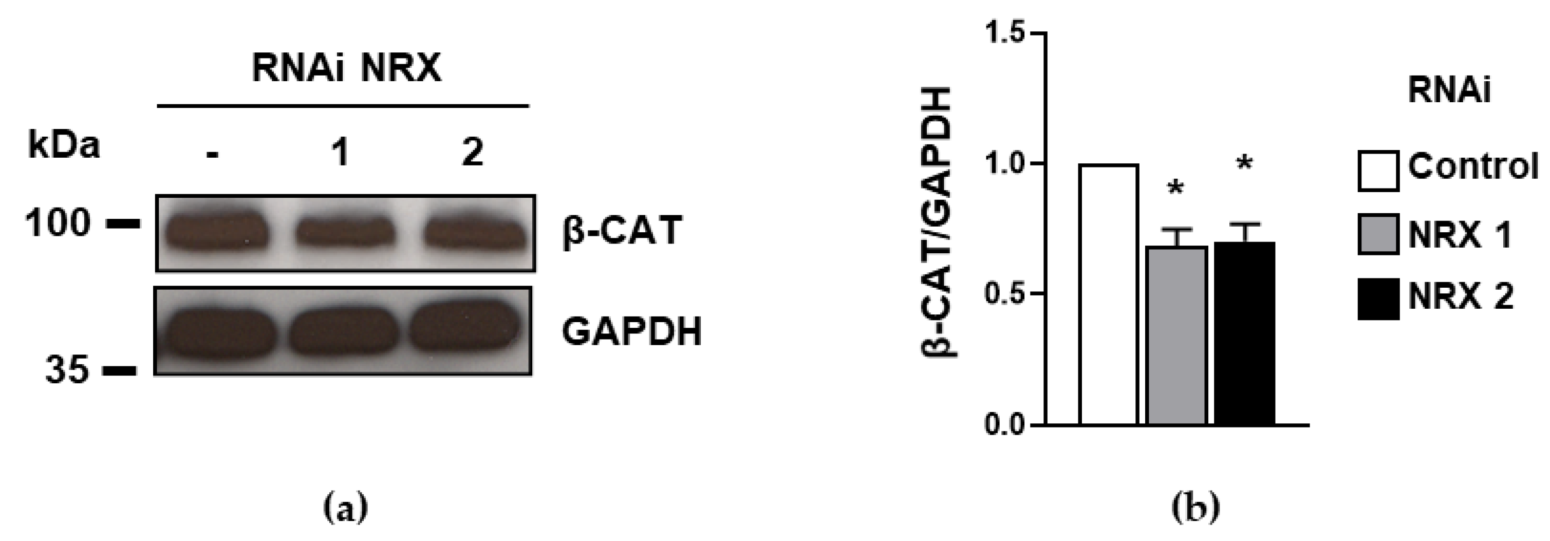
3.2. The Downregulation of NRX Prompts a Decrease in β-Catenin Levels in HEL Cells

To test whether NRX might have a role on Wnt/β-catenin signaling in our system, the levels of the central effector β-catenin were studied in NRX-downregulated HEL cells. A consistent decrease in this protein could be observed with the two sequences employed (Figure 2), which supports the idea that NRX might have a positive role in the canonical Wnt pathway in this system.

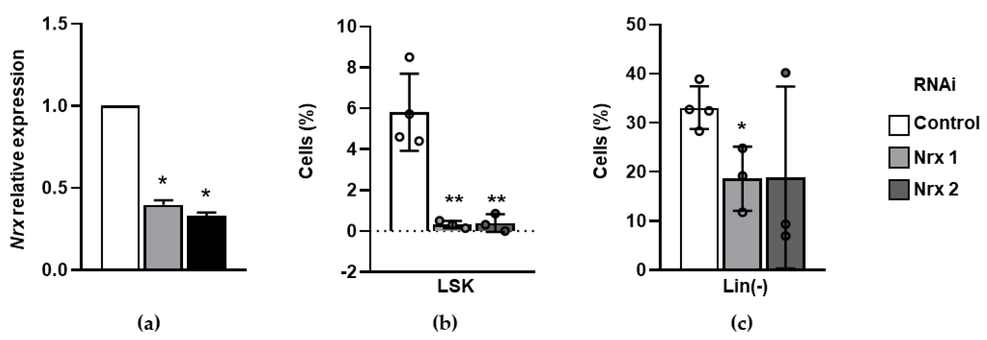
3.3. Nrx Downregulation Dramatically Decreases Murine Hematopoietic Progenitors In Vitro

To address the possible contribution of Nrx to hematopoietic differentiation in a wider range of blood lineages, mouse Lin- HSPCs were subject to lentiviral transduction with shRNA constructs targeting the mRNA of that protein. Prior to this, shRNA efficiency was validated in the mouse fibroblastic cell line NIH/3T3 (Figure 3a), thus overcoming the limitation of biological material characteristic of Lin cells.
After 5 days of in vitro culture, cell surface markers were studied to gain insights into possible lineage biases triggered by Nrx knockdown. The most striking effect observed was a dramatic reduction in Lin Sca-1+ c-Kit+ (LSK) cells, an immunophenotype-defined population enriched in HSCs in the in vivo setting (Figure 3b). This result was consistent with a less pronounced but significant decrease in Lin- cells, which contains the LSK population, together with more committed progenitors (Figure 3c). Overall, these data suggest that Nrx is crucial to maintain the HSC characteristic undifferentiated phenotype in vitro.

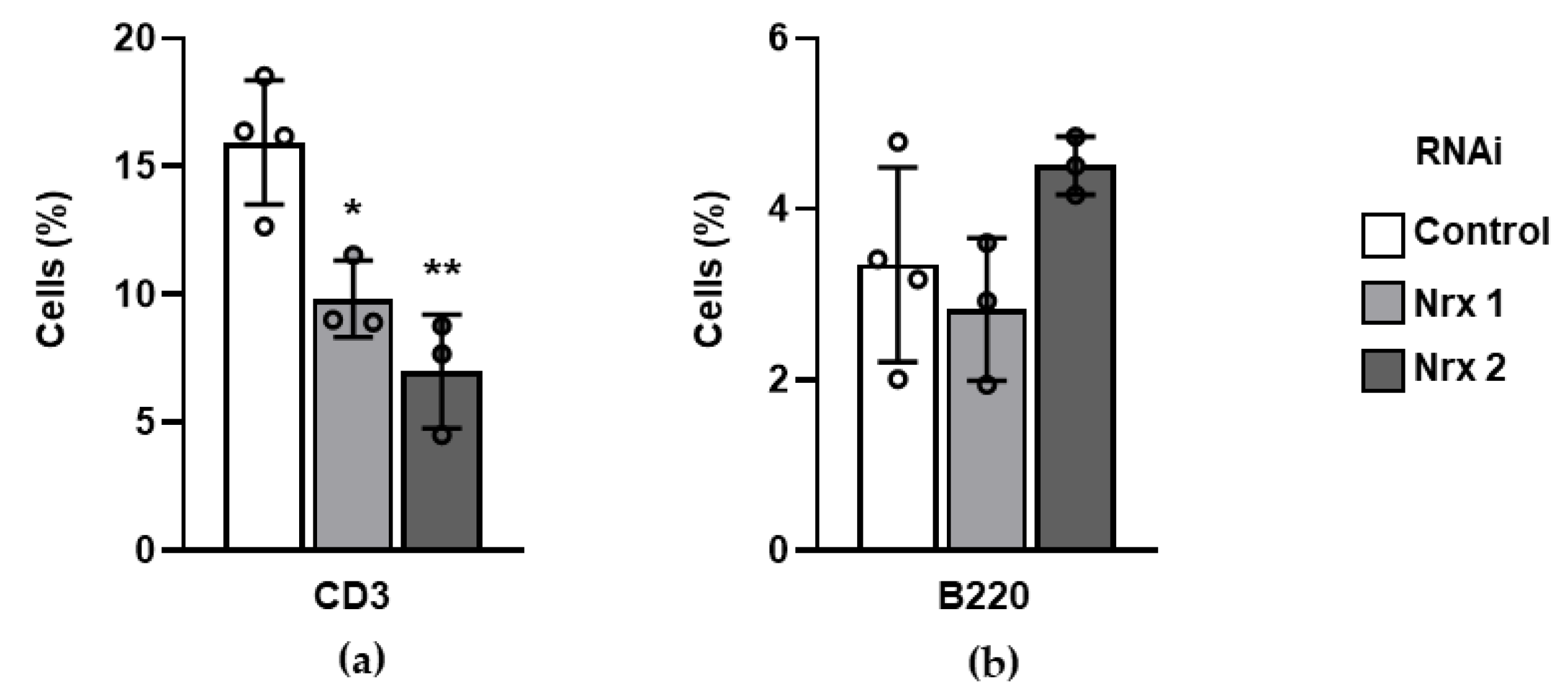
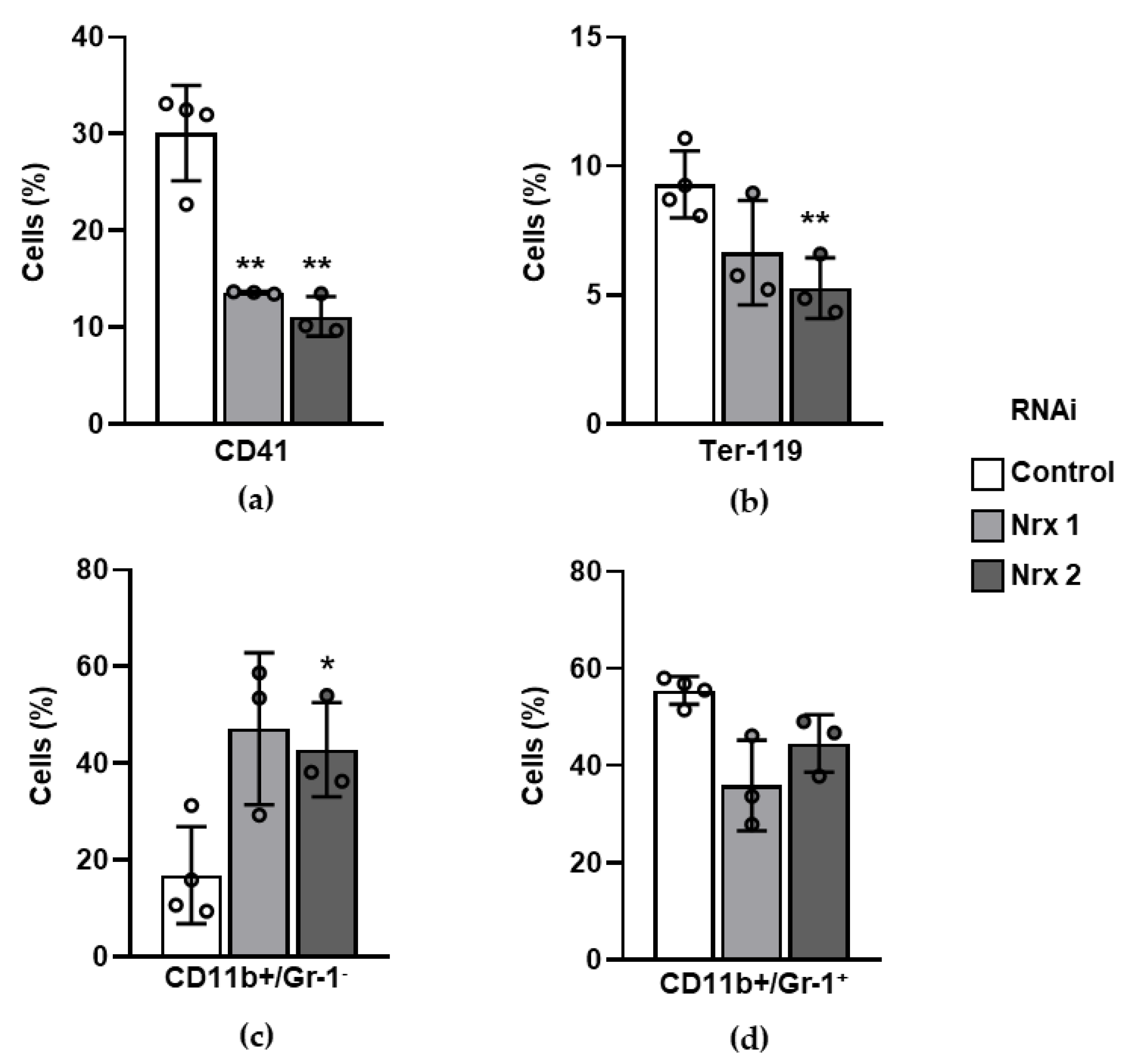
3.4. Surface Markers of Mature Blood Lineages Are Altered in Nrx-Silenced Mouse Hematopoietic Progenitors In Vitro

The previously described results supported an altered differentiation process of mouse HSPCs with reduced expression of Nrx. For this reason, characteristic surface markers of different blood cell types were used to assess possible lineage biases caused by the downregulation of Nrx. Lymphoid differentiation was studied by using antibodies against CD3, a specific marker of T lymphocytes, and B220, which distinguishes B lymphocytes. Whereas a notorious and consistent decrease in the expression of CD3 was observed (Figure 4a), no changes were detected for B220 (Figure 4b), thus pointing to an impairment in cell differentiation towards T cells, while sparing the B cell branch.
Regarding the myeloid lineage markers, there was a general decrease in the erythro-megakaryocytic branch, since Nrx-downregulated progenitors displayed diminished expression of both CD41 (Figure 5a) and Ter-119 (Figure 5b). When granulo-monocytic specific markers were studied, a clear increasing trend could be observed for CD11b+Gr-1 cells, a surface immunophenotype characteristic of monocytes, with RNAi 1, and a significant increase was detected in this population with RNAi 2 (Figure 5c). On the other hand, no consistent changes were found for CD11b and Gr-1 double-positive cells, a combination which allows the identification of granulocytes in blood compartments in vivo.
All these things considered, it could be hypothesized that the reduction in Nrx expression impairs T cell differentiation, without a consistent effect on the generation of B cells. Regarding the myeloid lineage, the previously described data point to a general bias towards monocytic differentiation, with a concomitant reduction in the surface markers distinctive of the rest of the main myeloid branches.

4. Discussion

Redox signaling has been unveiled over the last two decades as a regulator of hematopoietic differentiation [17], and its dysregulation often leads to hematologic malignancies [5], further supporting its relevance in the whole process. A seminal contribution by Jang and Sharkis revealed the lineage decisions made by hematopoietic progenitors depending on their intracellular levels of ROS [18]. Knowledge of how these ROS act to modulate intracellular signaling would allow us to better comprehend the whole process of hematopoiesis, as well as to identify altered pathways underlying pathologic conditions.
In this respect, thiol-based proteins are well-known examples of intracellular targets of ROS. They are involved in both redox homeostasis and signaling pathways [19]. PTPs are probably the best studied representatives of the second group, with reversible oxidation of their active center as an important mechanism to modulate their catalytic activity [7]. Indeed, they have a direct role in hematopoietic differentiation [8,9]. On the other hand, thioredoxin family proteins have been mainly studied in the context of redox homeostasis rather than cellular signaling. Therefore, the main goal of the present study was to assess the possible role of nucleoredoxin, a thioredoxin family member, in the differentiation of hematopoietic cells. This attempt was supported by the direct implication of this protein in Wnt/β-catenin signaling [10,14,15], a pathway with great relevance in hematopoiesis [2,3,8,20].
First, the downregulation of NRX has shown a concomitant decrease in β-catenin levels in the human hematopoietic cell line HEL. NRX reportedly has a dual role in the regulation of this central effector of canonical Wnt signaling: on one hand, it sequesters DVL and releases the destruction complex to target β-catenin for degradation [10]. Conversely, it is required for the displacement of KLHL12 from its interaction with DVL, which in turn prevents the degradation of the latter and promotes canonical Wnt signaling [14]. The results presented here would be consistent with the second scenario, given the correlation between NRX expression and β-catenin levels. Further studies will be required to assess whether KLHL12 is operating in the hematopoietic system and its direct involvement in cell differentiation. This would help to clarify the link between NRX and β-catenin levels and the redox control of Wnt signaling from a broader perspective.
Nucleoredoxin has been shown to be required for embryonic development of the cardiovascular system [14]. It is worth noting that both the cardiovascular and hematopoietic systems share a mesodermal origin [1], and therefore, it might be also needed for the emergence and maintenance of hematopoietic progenitors. This hypothesis would agree with the results described here, showing a pronounced decrease in both Lin- and LSK populations.
Regarding the surface markers of mature blood cells, a remarkable decrease in CD3+ was observed upon Nrx downregulation. This would be consistent with a decrease in β-catenin in murine progenitors, as found in HEL cells, since this protein is required for the activation of transcription factor 7, T cell specific (Tcf7), a protein required for T cell development [21]. Further studies using Nrx-downregulated thymocytes would help to address this issue. Under this same premise, the augmented percentage of CD11b+Gr1 cells would be expected, according to a report showing that ex vivo treatment of murine LT-HSCs with the non-canonical ligand Wnt5a, a reported inhibitor of canonical Wnt pathway, increases this surface immunophenotype [22].
On the contrary, our own reports have shown a positive effect of β-catenin downregulation on the percentage of LSKs in vivo [4], as well as on megakaryocytic differentiation of murine progenitors ex vivo [8]. In line with this, Paluru et al. have reported that Wnt signaling prevents both megakaryocytic and erythrocytic differentiation in vitro in mouse embryonic stem cells [23]. Given the dose-dependent effects of Wnt signaling in hematopoiesis [24], none of the hypotheses should be discarded.
As a thioredoxin family protein, the effects seen on overall cell differentiation might be explained by an alteration of the redox status of hematopoietic progenitors. Indeed, under the premise that Nrx could also present ROS scavenger functions within the cell, its downregulation might increase intracellular ROS levels. This, in turn, would reduce the most primitive hematopoietic cells and induce a lineage bias towards myeloid differentiation, which would agree well with previous work by Jang and Sharkis [18].
All in all, the results shown here strongly support the involvement of Nrx in hematopoietic differentiation. Given the reported role of this protein in the Wnt/β-catenin pathway and its relevance for the whole process, we hypothesize that this effect might be mediated by this signaling cascade. Importantly, our work supports the strong implication of redox targets in signaling pathways controlling hematopoietic differentiation, and sheds light on a particular protein which participates in the process. The molecular mechanism underlying these phenomena warrants further investigation in the field of redox signaling.

Supplementary Materials

The following are available online at https://0-www-mdpi-com.brum.beds.ac.uk/article/10.3390/biochem1010003/s1, “Supplementary Figures” file contains gating strategies for Lin and LSK cells (Figure S1); CD3+, B220+ and CD61+ cells (Figure S2); and Ter-119+, CD11b+Gr1 and CD11b+Gr1+ cells (Figure S3).

Author Contributions

Conceptualization, Á.H.-H.; formal analysis, A.P.-F., G.L.-R. and R.P.-B.; investigation, A.P.-F., G.L.-R. and R.P.-B.; resources, Á.H.-H.; writing—original draft preparation, A.P.-F. and Á.H.-H.; writing—review and editing, A.P.-F., R.P.-B., J.S.-Y., C.S.-B. and Á.H.-H.; visualization, A.P.-F.; supervision, Á.H.-H.; project administration, Á.H.-H.; funding acquisition, J.S.-Y., C.S.-B. and Á.H.-H. All authors have read and agreed to the published version of the manuscript.

Funding

This research was funded by the Spanish Ministry of Economy and Competitiveness, grant number BFU2014-56490-R. The salaries of A.P.-F., G.L.-R. and R.P.-B. were co-funded by the Junta de Castilla y León and European Social Fund (Ayudas destinadas a la contratación predoctoral de personal investigador).

Acknowledgments

We thank C. Guerrero for kindly providing the NIH/3T3 cell line.

Conflicts of Interest

The authors declare no conflict of interest.

References

  1. Orkin, S.H.; Zon, L.I. Hematopoiesis: An Evolving Paradigm for Stem Cell Biology. Cell 2008, 132, 631–644. [Google Scholar] [CrossRef] [Green Version]
  2. Ruiz-Herguido, C.; Guiu, J.; D’Altri, T.; Ingles-Esteve, J.; Dzierzak, E.; Espinosa, L.; Bigas, A. Hematopoietic Stem Cell Development Requires Transient Wnt/β-Catenin Activity. J. Exp. Med. 2012, 209, 1457–1468. [Google Scholar] [CrossRef] [PubMed] [Green Version]
  3. Luis, T.C.; Naber, B.A.E.; Roozen, P.P.C.; Brugman, M.H.; De Haas, E.F.E.; Ghazvini, M.; Fibbe, W.E.; Van Dongen, J.J.M.; Fodde, R.; Staal, F.J.T. Canonical Wnt Signaling Regulates Hematopoiesis in a Dosage-Dependent Fashion. Cell Stem Cell 2011, 9, 345–356. [Google Scholar] [CrossRef] [Green Version]
  4. López-Ruano, G.; Prieto-Bermejo, R.; Ramos, T.L.; San-Segundo, L.; Sánchez-Abarca, L.I.; Sánchez-Guijo, F.; Pérez-Simón, J.A.; Sánchez-Yagüe, J.; Llanillo, M.; Hernández-Hernández, Á.; et al. PTPN13 and β-Catenin Regulate the Quiescence of Hematopoietic Stem Cells and Their Interaction with the Bone Marrow Niche. Stem Cell Rep. 2015, 5, 516–531. [Google Scholar] [CrossRef] [Green Version]
  5. Prieto-Bermejo, R.; Romo-González, M.; Pérez-Fernández, A.; Ijurko, C.; Hernández-Hernández, Á. Reactive Oxygen Species in Haematopoiesis: Leukaemic Cells Take a Walk on the Wild Side. J. Exp. Clin. Cancer Res. 2018, 37. [Google Scholar] [CrossRef] [PubMed] [Green Version]
  6. Sardina, J.L.; López-Ruano, G.; Sánchez-Abarca, L.I.; Pérez-Simón, J.A.; Gaztelumendi, A.; Trigueros, C.; Llanillo, M.; Sánchez-Yagüe, J.; Hernández-Hernández, A. P22phox-Dependent NADPH Oxidase Activity Is Required for Megakaryocytic Differentiation. Cell Death Differ. 2010, 17, 1842–1854. [Google Scholar] [CrossRef] [Green Version]
  7. Yu, Z.-H.; Zhang, Z.-Y. Regulatory Mechanisms and Novel Therapeutic Targeting Strategies for Protein Tyrosine Phosphatases. Chem. Rev. 2018, 118, 1069–1091. [Google Scholar] [CrossRef] [Green Version]
  8. Sardina, J.L.; López-Ruano, G.; Prieto-Bermejo, R.; Sánchez-Sánchez, B.; Pérez-Fernández, A.; Sánchez-Abarca, L.I.; Pérez-Simón, J.A.; Quintales, L.; Sánchez-Yagüe, J.; Llanillo, M.; et al. PTPN13 Regulates Cellular Signalling and β-Catenin Function during Megakaryocytic Differentiation. Biochim. Biophys. Acta - Mol. Cell Res. 2014, 1843, 2886–2899. [Google Scholar] [CrossRef] [PubMed]
  9. Pérez-Fernández, A.; López-Ruano, G.; Prieto-Bermejo, R.; Ijurko, C.; Díez-Campelo, M.; Sánchez-Guijo, F.; Hernández-Hernández, Á. SHP1 and SHP2 Inhibition Enhances the Pro-Differentiative Effect of Phorbol Esters: An Alternative Approach against Acute Myeloid Leukemia. J. Exp. Clin. Cancer Res. 2019, 38, 80. [Google Scholar] [CrossRef] [PubMed]
  10. Funato, Y.; Michiue, T.; Asashima, M.; Miki, H. The Thioredoxin-Related Redox-Regulating Protein Nucleoredoxin Inhibits Wnt–β-Catenin Signalling through Dishevelled. Nat. Cell Biol. 2006, 8, 501–508. [Google Scholar] [CrossRef]
  11. Kajla, S.; Mondol, A.S.; Nagasawa, A.; Zhang, Y.; Kato, M.; Matsuno, K.; Yabe-Nishimura, C.; Kamata, T. A Crucial Role for Nox 1 in Redox-Dependent Regulation of Wnt-β-Catenin Signaling. FASEB J. 2012, 26, 2049–2059. [Google Scholar] [CrossRef] [PubMed]
  12. Kurooka, H.; Kato, K.; Minoguchi, S.; Takahashi, Y.; Ikeda, J.; Habu, S.; Osawa, N.; Buchberg, A.M.; Moriwaki, K.; Shisa, H.; et al. Cloning and Characterization of the Nucleoredoxin Gene That Encodes a Novel Nuclear Protein Related to Thioredoxin. Genomics 1997, 39, 331–339. [Google Scholar] [CrossRef] [PubMed]
  13. Funato, Y.; Miki, H. Nucleoredoxin, a Novel Thioredoxin Family Member Involved in Cell Growth and Differentiation. Antioxid. Redox Signal. 2007, 9, 1035–1057. [Google Scholar] [CrossRef] [PubMed]
  14. Funato, Y.; Terabayashi, T.; Sakamoto, R.; Okuzaki, D.; Ichise, H.; Nojima, H.; Yoshida, N.; Miki, H. Nucleoredoxin Sustains Wnt/β-Catenin Signaling by Retaining a Pool of Inactive Dishevelled Protein. Curr. Biol. 2010, 20, 1945–1952. [Google Scholar] [CrossRef] [Green Version]
  15. Bahn, Y.J.; Lee, K.-P.; Lee, S.-M.; Choi, J.Y.; Seo, Y.-S.; Kwon, K.-S. Nucleoredoxin Promotes Adipogenic Differentiation through Regulation of Wnt/β-Catenin Signaling. J. Lipid Res. 2015, 56, 294–303. [Google Scholar] [CrossRef] [Green Version]
  16. Sanchez-Sanchez, B.; Gutierrez-Herrero, S.; Lopez-Ruano, G.; Prieto-Bermejo, R.; Romo-Gonzalez, M.; Llanillo, M.; Pandiella, A.; Guerrero, C.; Miguel, J.F.S.S.; Sanchez-Guijo, F.; et al. NADPH Oxidases as Therapeutic Targets in Chronic Myelogenous Leukemia. Clin. Cancer Res. 2014, 20, 4014–4025. [Google Scholar] [CrossRef] [Green Version]
  17. Sardina, J.L.; López-Ruano, G.; Sánchez-Sánchez, B.; Llanillo, M.; Hernández-Hernández, A. Reactive Oxygen Species: Are They Important for Haematopoiesis? Crit. Rev. Oncol. Hematol. 2012, 81, 257–274. [Google Scholar] [CrossRef]
  18. Jang, Y.Y.; Sharkis, S.J. A Low Level of Reactive Oxygen Species Selects for Primitive Hematopoietic Stem Cells That May Reside in the Low-Oxygenic Niche. Blood 2007, 110, 3056–3063. [Google Scholar] [CrossRef] [Green Version]
  19. Groitl, B.; Jakob, U. Thiol-Based Redox Switches. Biochim. Biophys. Acta 2014, 1844, 1335–1343. [Google Scholar] [CrossRef] [Green Version]
  20. Cain, C.J.; Manilay, J.O. Hematopoietic Stem Cell Fate Decisions Are Regulated by Wnt Antagonists: Comparisons and Current Controversies. Exp. Hematol. 2013, 41, 3–16. [Google Scholar] [CrossRef]
  21. Verbeek, S.; Izon, D.; Hofhuis, F.; Robanus-Maandag, E.; te Riele, H.; van de Wetering, M.; Oosterwegel, M.; Wilson, A.; MacDonald, H.R.; Clevers, H. An HMG-Box-Containing T-Cell Factor Required for Thymocyte Differentiation. Nature 1995, 374, 70–74. [Google Scholar] [CrossRef] [PubMed]
  22. Florian, M.C.; Nattamai, K.J.; Dörr, K.; Marka, G.; Uberle, B.; Vas, V.; Eckl, C.; Andrä, I.; Schiemann, M.; Oostendorp, R.A.J.J.; et al. A Canonical to Non-Canonical Wnt Signalling Switch in Haematopoietic Stem-Cell Ageing. Nature 2013, 503, 392–396. [Google Scholar] [CrossRef] [PubMed] [Green Version]
  23. Paluru, P.; Hudock, K.M.; Cheng, X.; Mills, J.A.; Ying, L.; Galvão, A.M.; Lu, L.; Tiyaboonchai, A.; Sim, X.; Sullivan, S.K.; et al. The Negative Impact of Wnt Signaling on Megakaryocyte and Primitive Erythroid Progenitors Derived from Human Embryonic Stem Cells. Stem Cell Res. 2013, 12, 441–451. [Google Scholar] [CrossRef] [PubMed] [Green Version]
  24. Luis, T.C.; Ichii, M.; Brugman, M.H.; Kincade, P.; Staal, F.J.T. Wnt Signaling Strength Regulates Normal Hematopoiesis and Its Deregulation Is Involved in Leukemia Development. Leukemia 2012, 26, 414–421. [Google Scholar] [CrossRef] [PubMed] [Green Version]
Figure 1. Effect of nucleoredoxin (NRX) downregulation on megakaryocytic differentiation-related features in HEL cells. (a) Expression levels of NRX in HEL cells transduced with shRNA constructs targeting that gene (n = 3); (b) Cell proliferation in NRX-downregulated HEL cells seeded at different densities (n = 3); (c) Adhesiveness of NRX-downregulated HEL cells to different protein substrates (n = 4); (d) Basal expression levels of megakaryocytic markers in NRX-downregulated HEL cells (n = 4); (e) Expression levels of surface megakaryocytic markers in NRX-downregulated HEL cells after 48 h stimulation with 20 nM phorbol 12-myristate-13-acetate (PMA) (n = 4).
Biochem 01 00003 g001
Figure 2. Knockdown of NRX reduces β-catenin levels in HEL cells. (a) Representative image of a Western blot showing the levels of β-catenin in NRX-silenced HEL cells; (b) Densitometric quantification of the chemiluminescence signals from the protein bands recorded on X-ray films (n = 3).
Biochem 01 00003 g002
Figure 3. NRX downregulation drastically decreases the percentage of mouse hematopoietic stem and progenitor cells (HSPCs) in vitro. (a) Expression levels of Nrx in NIH/3T3 cells transduced with shRNA constructs targeting that gene (n = 2); (b) Percentage of LSK cells after 5 days of in vitro culture of Lin- cells expressing shRNA constructs against Nrx; (c) Percentage of Lin- cells after Nrx downregulation and 5 days of in vitro culture.
Biochem 01 00003 g003
Figure 4. The downregulation of Nrx affects lymphoid cell differentiation in vitro. (a) Percentage of CD3+ cells after a 5-day culture of Nrx-downregulated Lin cells; (b) Percentage of B220+ cells after a 5-day culture of Nrx-downregulated Lin cells.
Biochem 01 00003 g004
Figure 5. The downregulation of Nrx originates a myeloid differentiation bias towards monocytes in vitro. (a) Percentage of CD41+ cells after a 5-day culture of Nrx-downregulated Lin- cells; (b) Percentage of Ter-119+ cells after a 5-day culture of Nrx-downregulated Lin cells; (c) Percentage of CD11b+Gr-1 cells after a 5-day culture of Nrx-downregulated Lin cells; (d) Percentage of CD11b+Gr-1+ cells after a 5-day culture of Nrx-downregulated Lin cells.
Biochem 01 00003 g005
Table 1. Specific sequences for small hairpin RNA (shRNA) targeting of nucleoredoxin.
TargetIDSequence (5′-3′)
Human NRXNRX 1
NRX 2
AAACAGTACTTCAGTGAGA
AGAAAATCATTGCCAAGTA
Mouse NrxNrx 1
Nrx 2
AGATCATTGCCAAGTACAA
ATACCGAGTCTCCAACATT
Table 2. Oligonucleotides used for quantitative PCR (qPCR) reactions.
Target GeneTarget StrandSequence (5′-3′)
Human NRXForward
Reverse
ACCCAGAAGGTCTGGAGTTC
CCAATGTGCGGAGAAATAGA
Mouse NrxForward
Reverse
TCGTTAGTGCAGACAGGTCA
TGCCTTGGATTCCATACAGT
Human ACTBForward
Reverse
CACCACACCTTCTACAATGA
ACATGATCTGGGTCATCTTC
Mouse ActbForward
Reverse
CAGCCTTCCTTCTTGGGTAT
TGGCATAGAGGTCTTTACGG
Publisher’s Note: MDPI stays neutral with regard to jurisdictional claims in published maps and institutional affiliations.

