Article

Glucocorticoid-Induced Osteocytic Cell Death in a Hypoxic Environment Is Associated with Necroptosis

1
Department of Orthopaedic Surgery, Kanazawa Medical University, Daigaku 1-1, Uchinada-machi, Kahoku-gun, Ishikawa 920-0293, Japan
2
Department of Pathology 2, Kanazawa Medical University, Daigaku 1-1, Uchinada-machi, Kahoku-gun, Ishikawa 920-0293, Japan
*
Author to whom correspondence should be addressed.
These authors contributed equally to this work.
Submission received: 14 April 2021 / Revised: 5 July 2021 / Accepted: 9 August 2021 / Published: 30 August 2021

Abstract

:
Neither the underlying pathophysiology of nor prophylactic strategies for glucocorticoid-associated femoral head osteonecrosis have yet been established. In neurovascular and cardiac ischemic disorders, necroptosis has been reported as a new concept of cell death. Here we investigated the involvement of necroptosis in glucocorticoid-induced osteonecrosis in vitro, the putative cause of which is ischemia. Murine osteocytic cells (MLO-Y4) to which 1 µM dexamethasone (Dex) was added and were cultured in 1% O2 (hypoxia) are thought to resemble the in vivo environment in which glucocorticoid-induced osteonecrosis occurs (H-D stress environment). Using such cells cultured for 24 h (Dex(+)/hypoxia(+) group), immunofluorescent staining and Western blotting were performed with receptor-interacting protein (RIP) 1 and RIP3, which are necroptosis expression factors. In addition, the necroptosis inhibitor necrostatin-1 (Nec-1) was added to Dex(+)/hypoxia(+) and cultured for 12 h and 24 h. Then using an Apoptotic/Necrotic Cells Detection Kit the numbers of apoptotic and necrotic cells were counted and compared. In Dex(+)/hypoxia(+) group, expression of both RIP1 and RIP3 was found. Additionally, in Western blotting, the addition of Nec-1 attenuated their expression. A decrease in the number of cell deaths was also found following Nec-1 administration. Necroptosis has been implicated as a cause of death in osteocytic cell necrosis. Use of the necroptosis inhibitor, Nec-1, suggests a possible approach to preventing osteocytic cell necrosis even in an H-D stress environment when given within 12 h.

1. Introduction

Although numerous studies have focused on issues, such as the underlying pathophysiology and prevention of glucocorticoid-associated femoral head osteonecrosis, its causes and preventative strategies remain to be established. Glucocorticoid-associated femoral head osteonecrosis has been generally attributed to an ischemic-hypoxia event, but more recently factors such as oxidative injury and mitochondrial injury have also been implicated [1,2,3].
Apoptosis, which is a kind of programmed cell death, is considered to be an essential function in vivo, but an increase in the incidence of apoptosis alone does not lead to osteonecrosis, with the apoptotic cells being only metabolized and absorbed. However, here our attention was focused on necrosis occurring when trauma and/or severe stress were added to such a state. Necrosis is defined as rapid, accidental or uncontrolled cell death characterized by cell swelling and membrane rupture induced by an inflammatory reaction [4,5]. In conditions such as cerebral ischemia the importance of necrosis has been recognized, but because of its unregulated property, analysis of its molecular mechanisms has not progressed much. However, recent research has clarified that necrosis, like apoptosis, is affected by certain molecular mechanisms, and programmed necrosis has been reported. Usually, apoptosis occurs due to caspase dysfunction, with the resultant necrosis documented in a number of publications, and referred to as necroptosis. Moreover, while necroptosis is a kind of necrosis, it is considered to be a programmed necrosis [6,7]. For necroptosis to occur, activity of both receptor-interacting protein (RIP) 1 and RIP3 is believed to be necessary, and is thought to be regulated by these two proteins [8,9,10,11]. Hitherto, the morphology of apoptosis and necrosis has been considered separately, while, at present, various types of other cell death, such as necroptosis, ferroptosis and autophagy, are also being reported [12].
Recently, necroptosis has been implicated in ischemic injury occurring in conditions such as cerebral and myocardial infarction [13,14], and moreover in glucocorticoid-associated femoral head osteonecrosis, which has generally been attributed to ischemia as well. The possibility that necroptosis plays a role in glucocorticoid-associated femoral head osteonecrosis in vivo has also been reported [15]. However, because diverse environmental factors have been found to influence this condition in animal models, it is difficult to evaluate any one factor in isolation. This makes in vitro studies that make investigations possible at the cellular level especially valuable. Moreover, if it could also be documented in vitro that necroptosis, which is a programmed necrosis, is involved in the development of difficult-to-prevent glucocorticoid-induced osteonecrosis, this new information could help to promote the development of novel prophylactic strategies, as well as achieve a better understanding of the mechanisms underlying the osteonecrosis process. Additionally, when considering a preventative role against necroptosis, it will be necessary to determine the timing of necrostatin-1 (Nec-1) administration that best inhibits it.
It has been reported that the rate of osteocytic cell necrosis increases in vitro in a hypoxia environment with added dexamethasone (Dex) (H-D stress environment), which is equivalent to the in vivo environment in which glucocorticoid-induced osteonecrosis is likely to occur [16,17,18]. Here using MLO-Y4, which are cultured osteocytic cells, we investigated the role of necroptosis in an H-D stress environment prone to the development of osteocytic cell necrosis. In addition, since in cultured osteocytic cells, osteocytic cell necrosis has been successfully inhibited by avoiding an H-D stress environment for at least 12 h [19], we added Nec-1 to cells when an H-D stress environment was established and after 12 h and compared the rates of osteocytic cell necrosis so as to determine the best timing for Nec-1 administration.

2. Materials and Methods

2.1. Cell Culture

MLO-Y4 murine cultured osteocytic cells, which were used previously, were cultured [20]. The cells were plated on type I collagen-coated dishes (BD Biosciences, Bedford, MA, USA) and cultured in α-minimal essential medium (α-MEM) supplemented with 2.5% (v/v) FBS, 2.5% (v/v) FCS, streptomycin (100 μg/mL) and penicillin (100 units/mL). Then, for the hypoxia experiments, the cells were incubated for 24 h in a CO2/tri-gas incubator (Astec, Fukuoka, Japan) set to a mixture of 5% (v/v) CO2 and 1% (v/v) O2 balanced with N2 (hypoxia).

2.2. Cell Viability Assay

MLO-Y4 cells seeded in type I collagen-coated 4-chamber culture slides (BD Biosciences, Bedford, MA, USA) were cultured overnight. A total of 1 µM Dex (MSD, Tokyo, Japan) was added to MLO-Y4 in a hypoxia environment (H-D stress environment) [3], and then, after the addition of 50 µM necrostatitin-1 (Nec-1, abcam, Cambridge, UK), cultured for 24 h (Dex(+)/hypoxia(+)/Nec-1 group). In addition, MLO-Y4 was cultured for 12 h in an H-D stress environment, followed by the addition of Nec-1. A further group cultured for 12 h (total 24 h) was also prepared (HDN group). As a control group osteocytic cells cultured for 24 h in 20% O2 without addition of Dex were prepared as the Dex(−)/normoxia group. Additionally, osteocytic cells to which Dex was added, and cultured in hypoxia for 24 h were prepared as the Dex(+)/hypoxia(+) group.
Viability assays were then performed using an Apoptotic/Necrotic Cells Detection Kit (PromoKine, Heidelberg, Germany) according to the manufacturer’s instructions, and the percentages of apoptotic/necrotic cells relative to the total cell number were determined. In the viability assays, apoptotic cells can be detected by staining with fluorescein-labeled annexin V (green fluorescence) and necrotic cells by staining with Ethidium homodimer III, a highly positively charged nucleic acid probe, which is impermeant to live cells and early apoptotic cells, but stains necrotic cells and late apoptotic cells (entering into secondary necrosis) with red fluorescence. Fluorescence-positive cells were evaluated by phase contrast and fluorescence (470 nm and 530 nm LED modules) microscopy using Axiovert.A1 FL-LED (Carl Zeiss, Jena, Germany).

2.3. Immunocytochemistry

The presence/absence of RIP1 and RIP3 expression in Dex(+)/hypoxia(+) was investigated to confirm whether the conditions required for the development of necroptosis in osteocytic cells in an H-D stress environment were satisfied. Cultured cells were fixed in 4% paraformaldehyde, washed in phosphate buffered saline (PBS), and permeabilized with 0.3% Triton X-100 in PBS. Nonspecific binding was blocked by incubating sections with 10% bovine serum albumin (Dako Cytomation, Santa Clara, CA, USA) in PBS for 15 min. They were incubated with anti-RIP1 (Bioss, MA, USA) and anti-RIP3 (abcam, Cambridge, UK) antibody for 2 h at concentrations of 20.0 or 5.0 µg/mL, followed by a fluorescent-labeled secondary antibody (Alexa 594, or 488, Thermo Fisher Scientific, Waltham, MA, USA) and by DAPI for 30 min. After washing, a prolong diamond antifade mountant (Thermo Fisher Scientific) was added, and cover slips were mounted. Images were acquired using a Zeiss-LSM710.

2.4. Western Blotting

To quantitatively determine whether necroptosis was inhibited by Nec-1 Western blotting was performed. Cells were washed briefly with ice-cold PBS, and cell pellets were lysed with Laemmli Sample buffer containing 5% β-mercaptoethanol and heated at 95 °C for 5 min. Approximately 50 μg of cell lysate was electrophoresed on a 10% polyacrylamide gel and transferred on to a PVDF membrane (Merck Millipore, Darmstadt, Germany) using Trans-Blot SD Cell (Bio-Rad Laboratories, Hercules, CA, USA). After blocking with 5% skim milk in 0.05% TBS-Tween, the membranes were incubated with anti-RIP1 antibody (proteintech, Rosemont, IL, USA) or anti-RIP3 antibody (abcam) or anti-β-actin antibody (Sigma-Aldrich, Saint Louis, MO, USA), and then incubated with anti-rabbit IgG-HRP (Dako Cytomation, Tokyo, Japan). The bands were visualized with enhanced chemiluminescence (ECL) reagent (GE Healthcare Life Sciences, Little Chalfont, UK), and the images were captured using an LAS-4000 imaging system (Fujifilm, Tokyo, Japan). Protein expression was quantified via densitometry analysis using ImageJ [21,22]. The quantitative densitometric values for RIP1 and RIP3 protein were normalized to β-actin.

2.5. Statistical Analysis

All quantified results were expressed as the mean ± SD. Statistical significance in the comparison of apoptosis or necrosis, and RIP1/β-actin or RIP3/β-actin between the control and each of the experimental groups was analyzed with Dunnett’s multiple comparison test. p-values less than 0.05 were accepted as statistically significant. The statistical analysis was performed using StatView J-5.0 software (SAS Institute, Cary, NC, USA).

3. Results

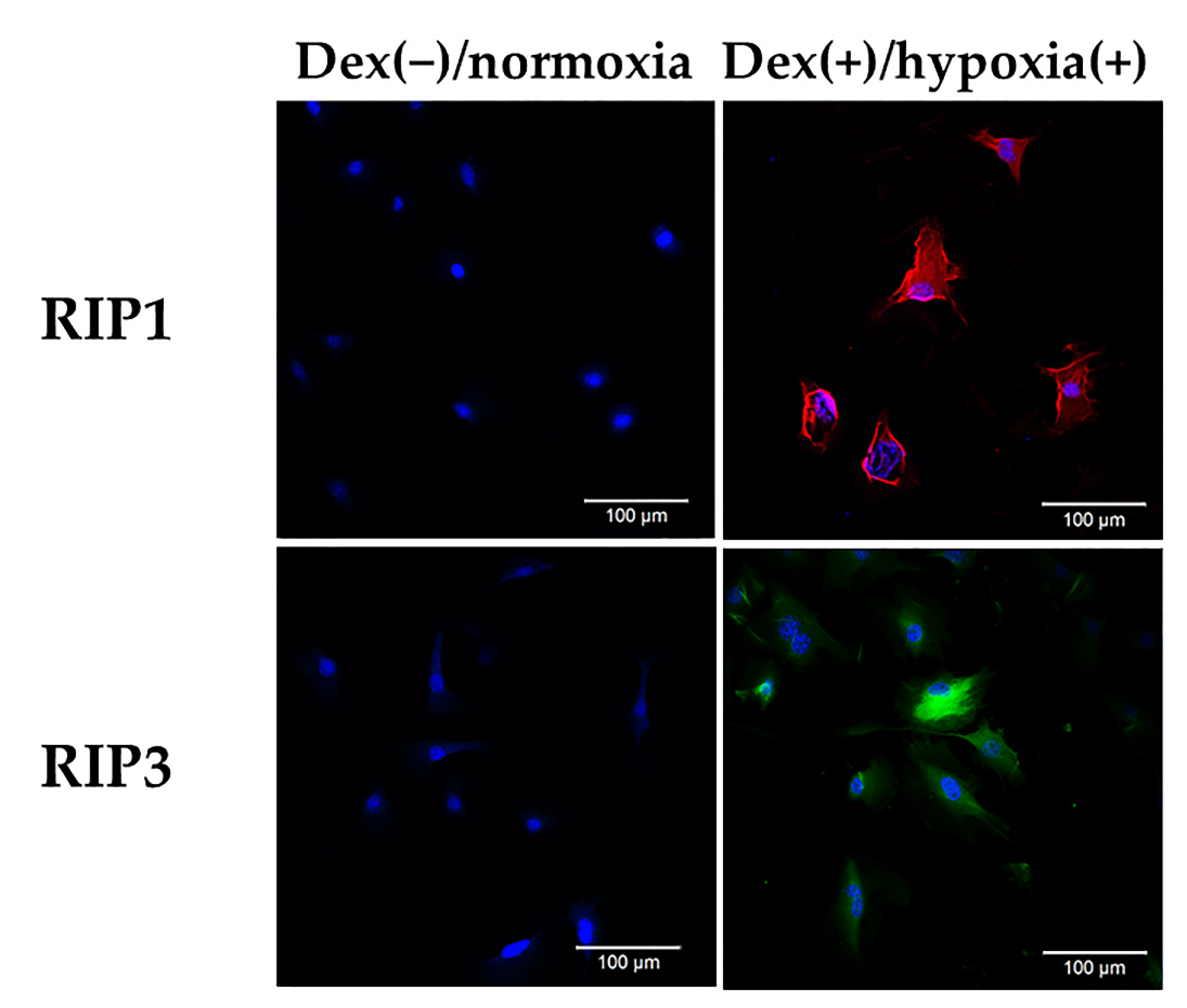
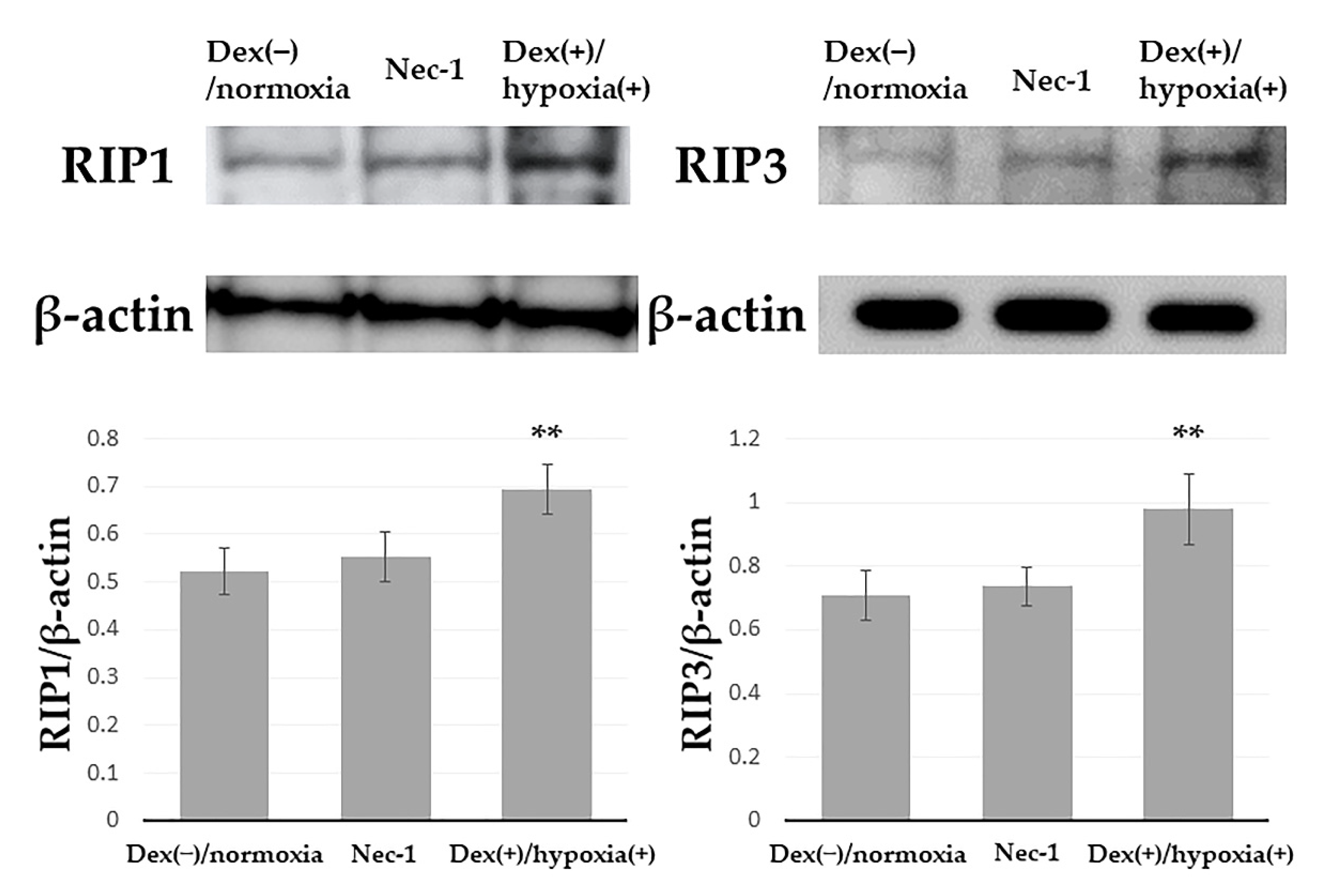
3.1. Expression of RIP1 and RIP3 in Osteocytic Cells under an H-D Stress Environment

In Dex(−)/normoxia group expression of both RIP1, RIP3 was found. In Dex(+)/hypoxia(+) group as compared with Dex(−)/normoxia group significant expression of both RIP1 and RIP3 was found (Figure 1). This demonstrates that in an H-D stress environment, expression of both RIP1 and RIP3 is present. This finding, that both RIP1 and RIP3 were expressed, confirmed that necroptosis develops in osteocytes placed in an H-D stress environment.

3.2. Inhibition of Expression of RIP1, RIP3 by Necrostation-1

While by Western blotting, in Dex(+)/hypoxia(+) group, enhanced expression of both RIP1, RIP3 was found, in Dex(+)/hypoxia(+)/Nec-1 group as compared with Dex(+)/hypoxia(+) group expression was significantly inhibited (** = p < 0.05). In Dex(−)/normoxia group no significant difference noted as compared with Dex(+)/hypoxia(+)/Nec-1 group (Figure 2). In an H-D stress environment an inhibitory effect on RIP1, RIP3 was exerted by Nec-1. Quantification using Western blotting confirmed the development of necroptosis in an H-D stress environment and its significant inhibition by Nec-1.

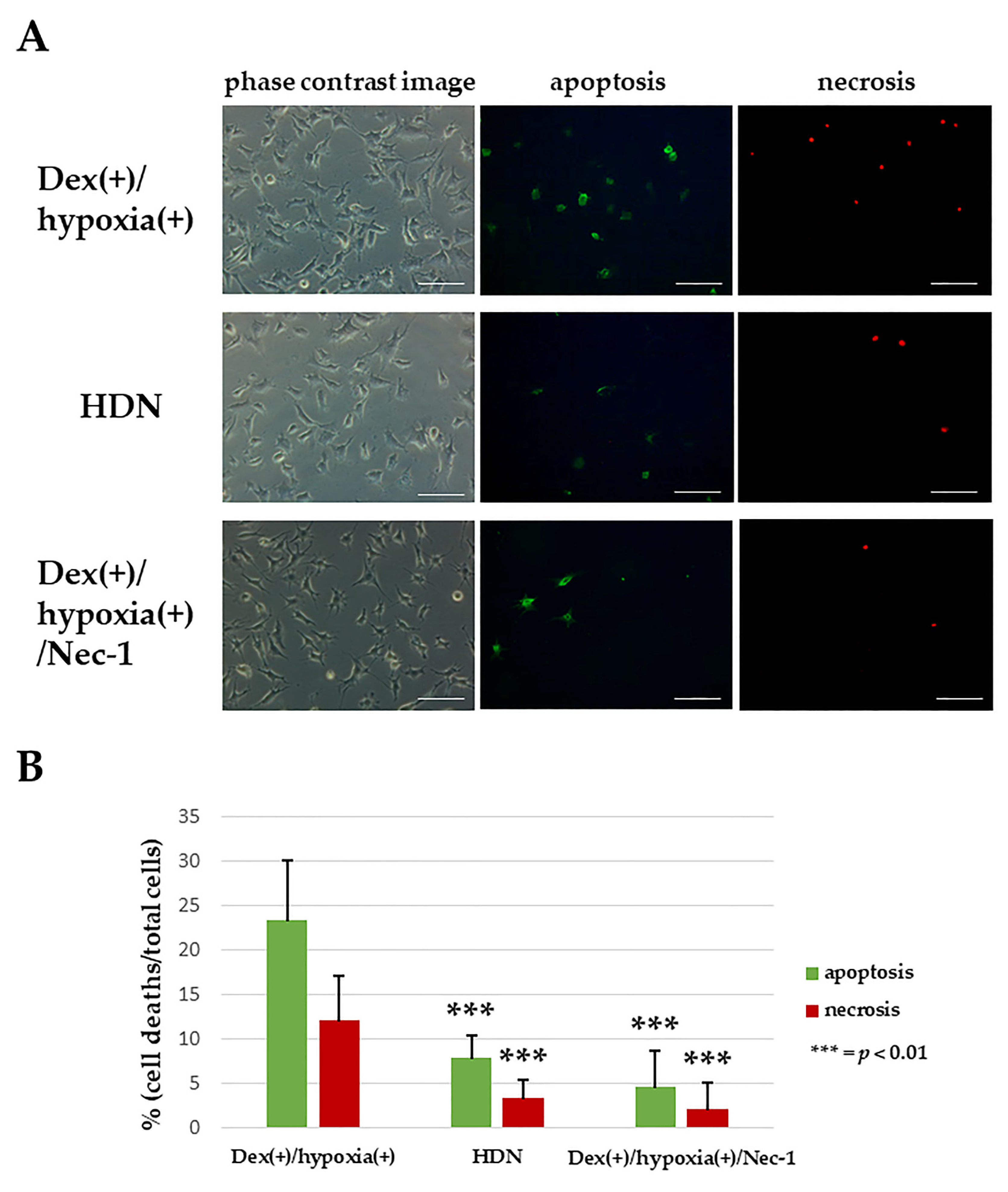
3.3. Inhibition of Osteocytic Cell Death in an H-D Stress Environment by Necrostatin-1

In Dex(+)/hypoxia(+) group the rate of apoptosis was 23.4 ± 6.8%, and that of necrosis 12.2 ± 4.9%. In Dex(+)/hypoxia(+)/Nec-1 group apoptosis was 4.6 ± 4.2% and necrosis 2.1 ± 3.1%, with the cell death number decreased significantly as compared with Dex(+)/hypoxia(+) group (*** = p < 0.01). In addition, in HDN group apoptosis was 8.0 ± 2.5% and that of necrosis 3.4 ± 2.1%, with no significant difference noted as compared with Dex(+)/hypoxia(+)/Nec-1 group (Figure 3). With Nec-1 administration, not only was apoptosis inhibited, but necrosis was also significantly inhibited. This means that Nec-1 exerted an inhibitory effect on the death of osteocytic cells in an H-D stress environment. Namely, it could be confirmed that necroptosis exerts an influence not only on apoptosis but on necrosis as well. In addition, in HDN group under a 12 h H-D stress environment, both apoptosis and necrosis could be significantly inhibited, while osteocytic cell necrosis could be inhibited in the H-D stress environment at least within 12 h.

4. Discussion

The development of necroptosis is said to be mediated by RIP1 and RIP3, with these two factors becoming the triggers of the necroptosis pathway [23]. In the immunocytochemical study in this research, expression of both RIP1 and RIP3 was found in osteocytic cells in an H-D stress environment. In addition, expression of both RIP1, RIP3 was similarly found by Western blotting. This suggests that, not only RIP1, but also RIP3, exerts an influence on osteocytic cell necrosis. Namely, in an H-D stress environment the morphology of osteocytic cell necrosis is not limited to apoptosis and necrosis alone, but shows that the role of necroptosis must also be considered. Moreover, necroptosis has been reported to be induced by mitochondrial damage [24,25]. In a glucocorticoid-induced osteonecrosis rabbit model and in osteocytic cells in an H-D stress environment, the role of mitochondrial injury has been documented [2,26]. Thus, in osteonecrosis, one cause of which is mitochondrial injury, it is also reasonable to consider the possibility of necroptosis.
The RIP1 inhibitor, necrostatin-1 (Nec-1), has been reported to improve blood flow in a neurovascular injury model, and reduce the infarct volume [27,28,29]. Similarly, in a cardiovascular injury model it improved blood flow and significantly inhibited cell death, and so has started to attract interest as a novel and important therapeutic agent for myocardial infarction [30,31]. It has also been demonstrated that Nec-1 exerts a protective effect against necroptosis induced by ischemic reperfusion injury [29,32,33]. In the osteoporosis field as well, in MLO-Y4 treated with TNF-α, it was reported that Nec-1 administration inhibited RIP1, RIP3 and in turn necroptosis [34]. In the present study, the administration of Nec-1 inhibited the necrosis of osteocytic cells in an H-D stress environment. This result suggested that from the standpoint of prevention of osteocytic cell necrosis, the inhibition of necroptosis also needs to be considered. Another salient result was that even in an H-D stress environment the increase in osteocytic cell necrosis could be inhibited provided that Nec-1 was administered for at least 12 h. This means that to impede the development of osteonecrosis, clinically it would be important to optimally adjust the intraosseous environment within 12 h of glucocorticoid administration, again highlighting the possibility that the concept of necroptosis may be useful when considering the prevention of osteonecrosis.

5. Conclusions

The present study demonstrated that also in osteocytes placed in an H-D stress environment, at least if within 12 h, the administration of Nec-1 could inhibit osteocytic cell necrosis and necroptosis. Accordingly, in osteocytes placed in an H-D stress environment, since RIP1 and RIP3 expression was found, the morphology of osteocytic cell necrosis showed not only apoptosis and necrosis, but implied the involvement of necroptosis as well.

Author Contributions

All authors made a significant contribution to the study and are in agreement with the content of the manuscript. S.U., T.I. and M.S. contributed to the conception and design. S.U., T.I. and M.S. contributed to the provision and validation of data. S.U., T.I. and M.S. performed the experiments. All authors contributed to analysis and interpretation of the data. All authors contribute to drafting, critical revision and final approval of the article. All authors have read and agreed to the published version of the manuscript.

Funding

This research received no external funding.

Institutional Review Board Statement

Not applicable.

Informed Consent Statement

Not applicable.

Acknowledgments

This study received a grant-in-Aid for Scientific Research from the Ministry of Education, Culture, Sports, Science, and Technology of Japan (no. 24592287), the Ministry of Health, Labor, and Welfare of Japan.

Conflicts of Interest

The authors declare no conflict of interest.

References

  1. Ichiseki, T.; Matsumoto, T.; Nishino, M.; Kaneuji, A.; Katsuda, S. Oxidative stress and vascular permeability in ster-oid-induced osteonecrosis model. J. Orthop. Sci. 2004, 9, 509–515. [Google Scholar] [CrossRef]
  2. Tsuchiya, M.; Ichiseki, T.; Ueda, S.; Ueda, Y.; Shimazaki, M.; Kaneuji, A.; Kawahara, N. Mitochondrial stress and redox fail-ure in steroid-associated osteonecrosis. Int. J. Med. Sci. 2018, 15, 205–209. [Google Scholar] [CrossRef] [PubMed] [Green Version]
  3. Hirata, H.; Ueda, S.; Ichiseki, T.; Shimasaki, M.; Ueda, Y.; Kaneuji, A.; Kawahara, N. Taurine Inhibits Glucocorti-coid-Induced Bone Mitochondrial Injury, Preventing Osteonecrosis in Rabbits and Cultured Osteocytes. Int. J. Mol. Sci. 2020, 21, 6892. [Google Scholar] [CrossRef] [PubMed]
  4. Laster, S.M.; Wood, J.G.; Gooding, L.R. Tumor necrosis factor can induce both apoptic and necrotic forms of cell lysis. J. Immunol. 1988, 141, 2629–2634. [Google Scholar]
  5. Lu, J.V.; Chen, H.C.; Walsh, C.M. Necroptotic signaling in adaptive and innate immunity. Semin. Cell Dev. Biol. 2014, 35, 33–39. [Google Scholar] [CrossRef] [PubMed] [Green Version]
  6. Christofferson, D.E.; Yuan, J. Necroptosis as an alternative form of programmed cell death. Curr. Opin. Cell Biol. 2010, 22, 263–268. [Google Scholar] [CrossRef] [Green Version]
  7. Khoury, M.K.; Gupta, K.; Franco, S.R.; Liu, B. Necroptosis in the Pathophysiology of Disease. Am. J. Pathol. 2019, 190, 272–285. [Google Scholar] [CrossRef] [Green Version]
  8. Moriwaki, K.; Chan, F.K.-M. RIP3: A molecular switch for necrosis and inflammation. Genes Dev. 2013, 27, 1640–1649. [Google Scholar] [CrossRef] [Green Version]
  9. Newton, K.; Dugger, D.L.; Wickliffe, K.E.; Kapoor, N.; de Almagro, M.C.; Vucic, D.; Komuves, L.; Ferrando, R.E.; French, D.M.; Webster, J.; et al. Activity of Protein Kinase RIPK3 Determines Whether Cells Die by Necroptosis or Apoptosis. Science 2014, 343, 1357–1360. [Google Scholar] [CrossRef]
  10. He, S.; Huang, S.; Shen, Z. Biomarkers for the detection of necroptosis. Cell. Mol. Life Sci. 2016, 73, 2177–2181. [Google Scholar] [CrossRef]
  11. Lin, Q.S.; Chen, P.; Wang, W.X.; Lin, C.C.; Zhou, Y.; Yu, L.H.; Lin, Y.X.; Xu, Y.F.; Kang, D.Z. RIP1/RIP3/MLKL mediates dopa-minergic neuron necroptosis in a mouse model of Parkinson disease. Lab. Investig. 2020, 100, 503–511. [Google Scholar] [CrossRef]
  12. Tang, D.; Kang, R.; Berghe, T.V.; Vandenabeele, P.; Kroemer, G. The molecular machinery of regulated cell death. Cell Res. 2019, 29, 347–364. [Google Scholar] [CrossRef] [Green Version]
  13. Zhang, T.; Zhang, Y.; Cui, M.; Jin, L.; Wang, Y.; Lv, F.; Liu, Y.; Zheng, W.; Shang, H.; Zhang, J.; et al. CaMKII is a RIP3 substrate mediating ischemia- and oxidative stress-induced myo-cardial necroptosis. Nat. Med. 2016, 22, 175–182. [Google Scholar] [CrossRef] [PubMed]
  14. Thornton, C.; Leaw, B.; Mallard, C.; Nair, S.; Jinnai, M.; Hagberg, H. Cell Death in the Developing Brain after Hypox-ia-Ischemia. Front Cell Neurosci. 2017, 11, 248. [Google Scholar] [CrossRef] [PubMed]
  15. Ichiseki, T.; Ueda, S.; Ueda, Y.; Tuchiya, M.; Kaneuji, A.; Kawahara, N. Involvement of necroptosis, a newly recognized cell death type, in steroid-induced osteonecrosis in a rabbit model. Int. J. Med. Sci. 2017, 14, 110–114. [Google Scholar] [CrossRef] [PubMed] [Green Version]
  16. Tsuji, M.; Ikeda, H.; Ishizu, A.; Miyatake, Y.; Hayase, H.; Yoshiki, T. Altered expression of apoptosis-related genes in osteo-cytes exposed to high-dose steroid hormones and hypoxic stress. Pathobiology 2006, 73, 304–309. [Google Scholar] [CrossRef] [PubMed] [Green Version]
  17. Ueda, S.; Ichiseki, T.; Yoshitomi, Y.; Yonekura, H.; Ueda, Y.; Kaneuji, A.; Matsumoto, T. Osteocytic cell necrosis is caused by a combination of glucocorticoid-induced Dickkopf-1 and hypoxia. Med. Mol. Morphol. 2014, 48, 69–75. [Google Scholar] [CrossRef] [PubMed]
  18. Yamamoto, H.; Saito, M.; Goto, T.; Ueshima, K.; Ishida, M.; Hayashi, S.; Ikoma, K.; Mazda, O.; Kubo, T. Heme oxygenase-1 prevents glucocorticoid and hypoxia-induced apoptosis and necrosis of osteocyte-like cells. Med. Mol. Morphol. 2019, 52, 173–180. [Google Scholar] [CrossRef]
  19. Ueda, S.; Ichiseki, T.; Ueda, Y.; Kaneuji, A.; Kawahara, N.; Matsumoto, T. The timing of glucocorticoid-induced osteocytic cell necrosis under hypoxia. Int. J. Clin. Exp. Pathol. 2016, 9, 7470–7475. [Google Scholar]
  20. Kato, Y.; Windle, J.; Koop, B.A.; Mundy, G.R.; Bonewald, L.F. Establishment of an Osteocyte-like Cell Line, MLO-Y4. J. Bone Miner. Res. 2010, 12, 2014–2023. [Google Scholar] [CrossRef]
  21. Rasband, W.S. ImagJ; U. S. National Institutes of Health: Bethesda, MD, USA, 2018. Available online: https://imagej.nih.gov/ij/ (accessed on 24 May 2021).
  22. Schneider, C.A.; Rasband, W.S.; Eliceiri, K.W. NIH Image to ImageJ: 25 years of image analysis. Nat. Methods 2012, 9, 671–675. [Google Scholar] [CrossRef]
  23. Zhe-Wei, S.; Li-Sha, G.; Yue-Chun, L. The Role of Necroptosis in Cardiovascular Disease. Front. Pharmacol. 2018, 9, 721. [Google Scholar] [CrossRef] [PubMed]
  24. Lee, Y.J.; Nam, H.S.; Cho, M.K.; Lee, S.H. Arctigenin induces necroptosis through mitochondrial dysfunction with CCN1 up-regulation in prostate cancer cells under lactic acidosis. Mol. Cell Biochem. 2020, 467, 45–56. [Google Scholar] [CrossRef]
  25. Lyu, A.-R.; Kim, T.H.; Park, S.J.; Shin, S.-A.; Jeong, S.-H.; Yu, Y.; Huh, Y.H.; Je, A.R.; Park, M.J.; Park, Y.-H. Mitochondrial Damage and Necroptosis in Aging Cochlea. Int. J. Mol. Sci. 2020, 21, 2505. [Google Scholar] [CrossRef] [Green Version]
  26. Ueda, S.; Shimasaki, M.; Ichiseki, T.; Hirata, H.; Kawahara, N.; Ueda, Y. Mitochondrial Transcription Factor A added to Os-teocytes in a Stressed Environment has a Cytoprotective Effect. Int. J. Med. Sci. 2020, 17, 1293–1299. [Google Scholar] [CrossRef]
  27. Northington, F.J.; Chavez-Valdez, R.; Graham, E.M.; Razdan, S.; Gauda, E.B.; Martin, L.J. Necrostatin Decreases Oxidative Damage, Inflammation, and Injury after Neonatal HI. J. Cereb. Blood. Flow. Metab. 2010, 31, 178–189. [Google Scholar] [CrossRef] [PubMed]
  28. Chavez-Valdez, R.; Martin, L.; Flock, D.; Northington, F. Necrostatin-1 attenuates mitochondrial dysfunction in neurons and astrocytes following neonatal hypoxia–ischemia. Neuroscience 2012, 219, 192–203. [Google Scholar] [CrossRef] [Green Version]
  29. Deng, X.X.; Li, S.S.; Sun, F.Y. Necrostatin-1 Prevents Necroptosis in Brains after Ischemic Stroke via Inhibition of RIPK1-Mediated RIPK3/MLKL Signaling. Aging Dis. 2019, 10, 807–817. [Google Scholar] [CrossRef] [PubMed] [Green Version]
  30. Dmitriev, Y.V.; Minasian, S.M.; Demchenko, E.; Galagudza, M. Study of cardioprotective effects of necroptosis inhibitors on isolated rat heart subjected to global ischemia-reperfusion. Bull. Exp. Biol. Med. 2013, 155, 245–248. [Google Scholar] [CrossRef]
  31. Szobi, A.; Rajtik, T.; Adameova, A. Effects of Necrostatin-1, an Inhibitor of Necroptosis, and its Inactive Analogue Nec-1i on Basal Cardiovascular Function. Physiol. Res. 2016, 65, 861–865. [Google Scholar] [CrossRef] [PubMed]
  32. Degterev, A.; Huang, Z.; Boyce, M.; Li, Y.; Jagtap, P.; Mizushima, N.; Cuny, G.D.; Mitchison, T.J.; Moskowitz, M.A.; Yuan, J. Chemical inhibitor of nonapoptotic cell death with therapeutic potential for ischemic brain injury. Nat. Chem. Biol. 2005, 1, 112–119. [Google Scholar] [CrossRef] [PubMed]
  33. Mei, M.; Shen, B.B.; Pu, Y.M.; Zhao, H.W.; Liu, H.; Wu, X.F. TRPM-7 inhibits necroptosis via the PARP-1/RIP-1 signaling pathway and attenuates renal ischemic reperfusion injury in renal tubular epithelial cells. Int. J. Clin. Exp. Pathol. 2017, 10, 88–96. [Google Scholar]
  34. Cui, H.; Zhu, Y.; Yang, Q.; Zhao, W.; Zhang, S.; Zhou, A.; Jiang, D. Necrostatin-1 treatment inhibits osteocyte necroptosis and trabecular deterioration in ovariectomized rats. Sci. Rep. 2016, 6, 33803. [Google Scholar] [CrossRef] [PubMed]
Figure 1. Expression of RIP1 and RIP3 in osteocytic cells in an H-D stress environment. While no RIP1 or RIP3 expression was found in the Dex(−)/normoxia group, in the Dex(+)/hypoxia(+) group clear expression of both RIP1 (Alexa 594) and RIP3 (Alexa 488) was found. Each three independent experiments were carried out. Scale bar: 100 µm.
Biochem 01 00009 g001
Figure 2. Inhibitory effect of necrostatin-1 on RIP1 and RIP3 by Western blotting. The graphs indicate quantification of Western blotting for RIP1 and RIP3 in the indicated conditions. The quantitative densitometric values of RIP1 or RIP3 were normalized to β-actin. Columns and bars indicate means and S.D., respectively, (n = 3). In Dex(+)/hypoxia(+) group expression of RIP1 (74 kDa), RIP3 (57 kDa) was found, while in Dex(+)/hypoxia(+)/Nec-1 group, there was significant inhibition of the expression of both RIP1 and RIP3 (** = p < 0.05). Three independent experiments each were carried out.
Biochem 01 00009 g002
Figure 3. Cell death inhibition by necrostatin-1. (A) Immunofluorescent staining of apoptotic and necrotic cells using an Apoptotic/Necrotic Cells Detection Kit was performed as described in the Materials and Methods. (B) The graph indicates the percentages of apoptotic and necrotic cells in the indicated conditions. The numbers of apoptotic or necrotic cells were counted and related to the total number of cells. Columns and bars indicate means and S.D. respectively (n = 5). Relative to Dex(+)/hypoxia(+) group, Dex(+)/hypoxia(+)/Nec-1 group and HDN group showed significant decreases in both apoptotic and necrotic cell death (*** = p < 0.01 vs. Dex(+)/hypoxia(+) group). Scale bar: 100 µm.
Biochem 01 00009 g003
Publisher’s Note: MDPI stays neutral with regard to jurisdictional claims in published maps and institutional affiliations.

