Article

Effect of Gender and Anthropometrics on the Kinematics of the Lunge in Young Foil Fencers (Lunge Kinematics in Young Fencers)

1
Department for Quality of Life Studies, University of Bologna, Via Del Pilastro 8, 40127 Bologna, Italy
2
Department of Exercise Physiology, Beijing Sport University, Xinxi Lu 48, Beijing 100084, China
*
Author to whom correspondence should be addressed.
Submission received: 26 April 2021 / Revised: 11 June 2021 / Accepted: 11 June 2021 / Published: 22 June 2021

Abstract

:
Knowledge of the kinematics of young fencers is important when teaching fencing, but it has been minimally investigated in the literature, especially in young subjects, and further research is needed. Our aims were to assess whether anthropometric factors or kinematic factors are more closely related to lunge performance (speed and range of motion) in young fencers and to investigate gender differences in relation to kinematics. Fifteen fencers participated in this study (8 females and 7 males; age 12.9 ± 2.7 years, height 157.4 ± 15.1 cm, weight 49.7 ± 11.7 kg). Lunge kinematics and anthropometrics were collected with a 10-camera optoelectronic system. Descriptive statistics, bivariate correlation, t-test, and simple regression were performed. Peak lunge velocity was mainly correlated with posterior knee extension (r = 0.56, p = 0.031). The lunge distance and mean hip velocity were mostly correlated with the fencers’ height (r = 0.85, p = 0.000 and r = 0.76, p = 0.001) and fat-free mass (r = 0.79, p = 0.000 and r = 0.73, p = 0.002). Young fencers use a lower limb-driven strategy to perform the lunge, while adults are reported to use an upper limb-driven strategy for the lunge attack. Linear and angular velocities and ranges of motion were lower in young fencers in comparison to those of adults. Based on these results, we suggest a different approach to teaching the lunge action in young foil fencers. The implications of our study are that when teaching the lunge technique to this age group, a different approach for males and females is not required, and that strength is not a discriminant physical quality for the correctness of performance.

1. Introduction

In a fencing bout, two fencers confront each other on a 14 m track, which is generally metallic. The duration and the score of the competition are different in the pool and direct elimination phases. There are three fencing specialties that differ in terms of the use of different weapons: the saber, épée, and foil. Every specialty has different rules, and these mainly regard the valid target. The three weapons are characterized by different weights and lengths and by the different duration of actions. On average, the saber (weight < 500 g) presents the briefest time action (2.5 ± 0.6 s for males and 2.9 ± 0.9 s for females), while the épée (weight < 770 g) has the longest one (12.7 ± 7.6 s for males and 16.5 ± 4.2 s for females). The foil (weight < 500 g) has an intermediate time action (5.2 ± 3.5 for males, no data on females) that is not as short as that of the saber [1]. Among the total times of the various attack actions, the lunge has a duration of less than < 1 s.
In the literature, only a few studies have investigated the biomechanics of fencing [2], and it is not clear if gender differences in kinematics arise at a young age and which strategy young fencers use when carrying out the lunge action. Young fencers showed a less developed motor system than that of their older counterparts [2,3] and may not employ the same motor pattern of older and more experienced fencers. Hassan e Klauck [3] studied four fencers (16–17 years old, having 5–9 years of fencing experience). Two different temporal patterns of movement were identified: an upper limb-driven and a lower limb-driven pattern. These different behaviors were also observed in another study [4] whose results showed that older and more experienced fencers (EFs) tended to be lower limb driven, while younger, non-expert fencers (NEs) tended to be upper limb driven. Gholipour et al. [4] considered two groups, one of four novices and the other of four expert fencers, and the results showed that experienced fencers performed a longer lunge and tilted their trunk more forward at the end of this movement than the novices. This behavior allowed an anterior shift of the center of mass (COM), which enabled a better push coming from the rear lower limb. They also observed a lower mean horizontal velocity in the EFs than that of the NEs (0.68 m/s vs. 0.76 m/s), and this result is consistent with a wider lunge. The tendency of EFs to start the attack with the lower limb was confirmed in another study [5] on the flèche, a different offensive technique. This strategy was ascribed to the skills of EFs to mask their attack, finishing the lunge deeply with the upper limb.
In the first third of the lunge, the EFs showed a greater angular excursion of the anterior knee [4]. After this first flexion, the EFs extended the knee more forward. In the last third of the lunge, the EFs showed a more flexed hip and knee of the armed side, having a lower body position. The authors hypothesized that this was caused by greater strength and flexibility when compared to those of the NEs [5].
Proximal-to-distal activation of the lower limb muscles was investigated in a case study [6], which showed that the power necessary to perform a lunge was produced mainly by the rear lower limb with simultaneous activation of the ankle and knee joints. In this study, most of the power produced by the rear lower limb came from the ankle joint’s plantar flexion, and to a lesser extent, it was produced by knee joint extension. In contrast, Mulloy [7], who compared six sword NE males with four EFs, found that the EFs employed a sequential activation, first moving the elbow joint extension and ending with the plantar flexion of the rear ankle joint. They found a greater lunge distance covered by the EFs [7], but in contrast with other studies [5], they found a higher mean speed of movement for the EFs, and differences in angular velocities of the elbow and rear hip were observed between EFs and NEs. These findings are in agreement with those of another study [8] that showed that the peak velocity of the sword is largely determined by the rear lower limb in a group of 14 sword fencers. From multiple regression, it emerged that the main predictors of the sword velocity were the lower rear knee range of motion (ROM) and the hips’ peak flexion. In this study, the upper limb was not considered. Guan et al. [9] found that the peak horizontal velocity in the lunge was significantly correlated with the rear knee’s extension ROM. All of these studies were performed in adults (>21 years old), and the results were controversial: it was not clear if the lunge was driven by the lower or the upper limb.
A low number of subjects and different levels of performance and weapons are further cause of controversy.
Gender differences were addressed in only one study [10], which observed that females presented a greater knee adduction/abduction angle, a greater hip adduction, and a greater ankle eversion angle in the anterior lower limb at the end of the lunge, according to female anthropometry, than their male counterparts.
The aims of the present study were (1) to identify whether anthropometric factors or kinematic factors are more closely related to velocity and distance; (2) to identify if there are gender-related differences; and (3) to verify the timing strategy deployed by young fencers (arm-driven pattern or lower limb-driven pattern).

2. Materials and Methods

Fifteen right-handed foil fencers with a fencing experience from 2 to 7 years participated in the study. Demographic data of the subjects are reported in Table 1.
The fencers were classified as of regional-level ranking, and they regularly participate in regional-level competitions. The subjects regularly training for minimum of 4 times per week for 1.5 h. Written informed consent was obtained from each of the participant’s parents, the study was approved by the Ethic Committee of the University of Bologna, and research was conducted in accordance with the declaration of Helsinki. All subjects were right-handed, so they maintained the guard position with their right side forward. The data associated with the paper are not publicly available but are available from the corresponding author on reasonable request.

3. Anthropometry

Anthropometry was collected following the ISAK manual recommendations [11] by the same trained researcher. Height was measured with a Seca wall-mounted stadiometer (Seca, Germany) and weight with an electronic scale (Tanita BC-418 MA, Tanita Corporation, Tokyo, Japan). The followings lengths were measured with a Harpenden anthropometer (Holtain, UK): hand, forearm, and upper arm of the dominant upper limb; thigh, tibia, and lateral tibial height of both lower limbs; and bi-epicondylar width. These measurements were combined to obtain the following lengths: upper limb, lower limb, and trunk. Circumferences of the upper arm and upper third of the thigh were measured with an anthropometric tape. Bicep, tricep, and thigh skinfolds were assessed with a Harpenden caliber (Holtain, UK). Absolute and % fat mass (FM) and fat free mass (FFM) were assessed via bioelectrical impedance (Maltron BF906, Rayleigh, GB), and the body mass index was calculated. The cross-sectional area (CSA) of the forward upper arm was calculated using the formula of Martine et al. [12], while the CSA of the thighs was calculated with the formulas of Knapik et al. [13].

4. Kinematics

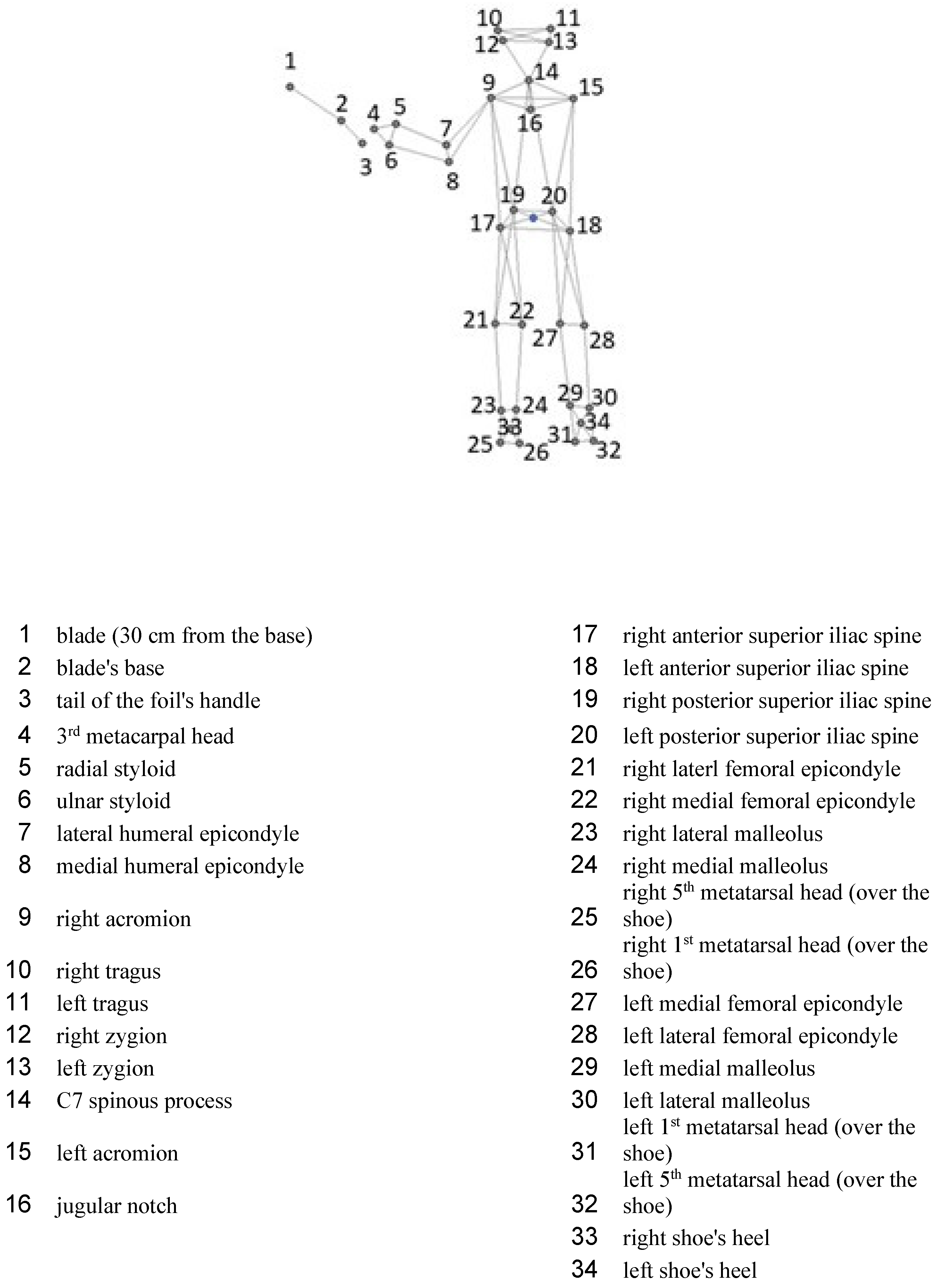
For kinematic measurements, a ten-camera system was employed (BTS SMART DX 7000, BTS Bioengineering, Milan, Italy) with a sampling frequency of 250 Hz. A modified Plug-in Gait set of 34 markers was employed (Figure 1) [14,15].
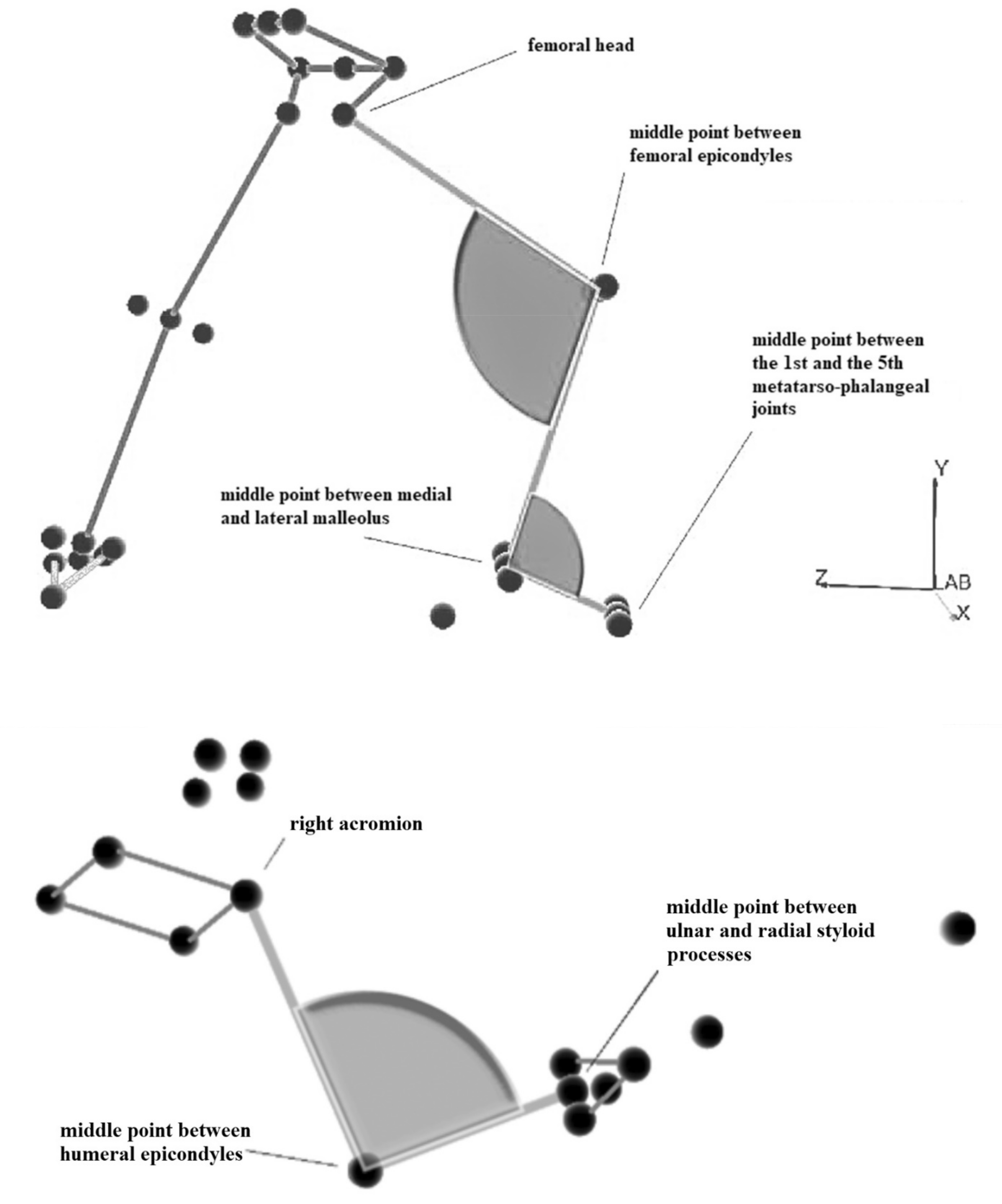
Virtual markers (midpoint between the ASIS (anterior superior iliac spine); midpoint of the ankle, knee, elbow, and wrist; and midpoint between the 1st and the 5th metatarsophalangeal joints) were computed post-acquisition as the midpoint of the 3D coordinates of the left and right side of each joint. Data processing was performed with SMART Tracker software (BTS Bioengineering, Milan, Italy). Volume reference points were set accordingly to ISB norms [16,17]. Three-dimensional points were filtered with a Butterworth low pass filter of 4th order set at 20 Hz [14]. The followings angles and ROMs were measured (Figure 2) using the software SMART Analyzer (BTS Bioengineering, Milan, Italy): “on-guard” position: elbow, anterior and posterior knees and ankles; final lunge position: elbow, knees, and ankles. For the elbow, the maximum extension angle was obtained.
ROMs of the armed elbow, rear ankle and knee, and the four phases of the forward knee, as well as the respective mean angular velocities, were computed.
The start, end, and duration of each event were computed. The start was after the verbal signal of ‘go’. The subjects were instructed to perform the lunge at the maximal possible speed. The lunge distance was defined as the horizontal difference between the posterior heel in the guard position and the anterior heel in the final lunge position. The mean and peak horizontal velocities of ASISm (the middle point between the two ASIS) and of the 3rd metacarpal head marker (3met) were computed along the anterior–posterior direction.

5. Statistics

Data normality was assessed with the Kolmogorov–Smirnov test. All data appeared to be normally distributed. Then, descriptive statistics, bivariate correlation, t-test, and simple regression were performed with lunge speed and lunge distance as dependent variables (IBM SPSS v.25.0, Armonk, NY, USA). According to Austin et al. [18], our number of subjects (15) allowed us to perform linear regression with the number of considered variables (6 ranges of motion, 13 angles, 12 angular velocities, and 6 linear velocities) to estimate regression coefficients, standard errors, and confidence intervals. The significance level was set at p ≤ 0.05. The effect size was calculated using Cohen’s “d”, which was >0.8 for each variable that presented a difference between groups.

6. Results

Anthropometric measurements are reported in Table 2 and Table 3. Height, body weight, and BMI were close to the 50th percentiles of the Italian population for this age range [19].
Significant anthropometric differences between males and females are reported in Table 4. Considering the whole sample, significant differences in anthropometry were found between males and females for FM%, FFM%, and thigh skinfolds. Females showed greater FM% and thigh skinfolds thickness than those of their male counterparts.
Significant differences were found between the left and right side in body segment lengths and CSA in contrast to adult fencers who show a greater CSA of the dominant lower limb [20]. From linear regression of all variables considered, only fat-free mass appeared to be related to lunge speed and distance. The results of the linear regression are reported in Table 5 with FFM (kg) as an independent variable.
FFM was the best predictor of mean horizontal velocity and lunge distance for the hips. The same relationship of lunge speed and distance with anthropometric characteristics was observed in the work of [
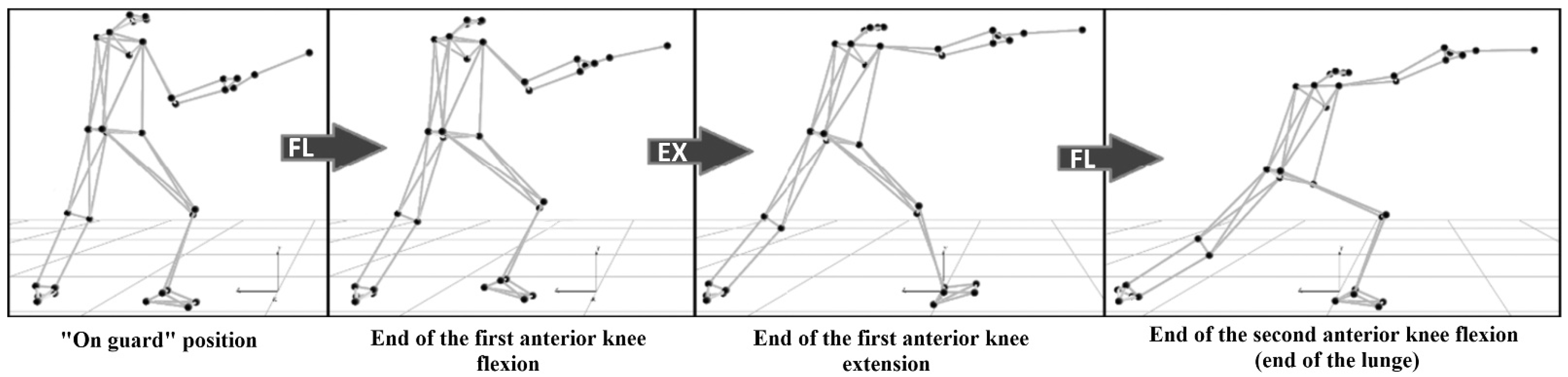
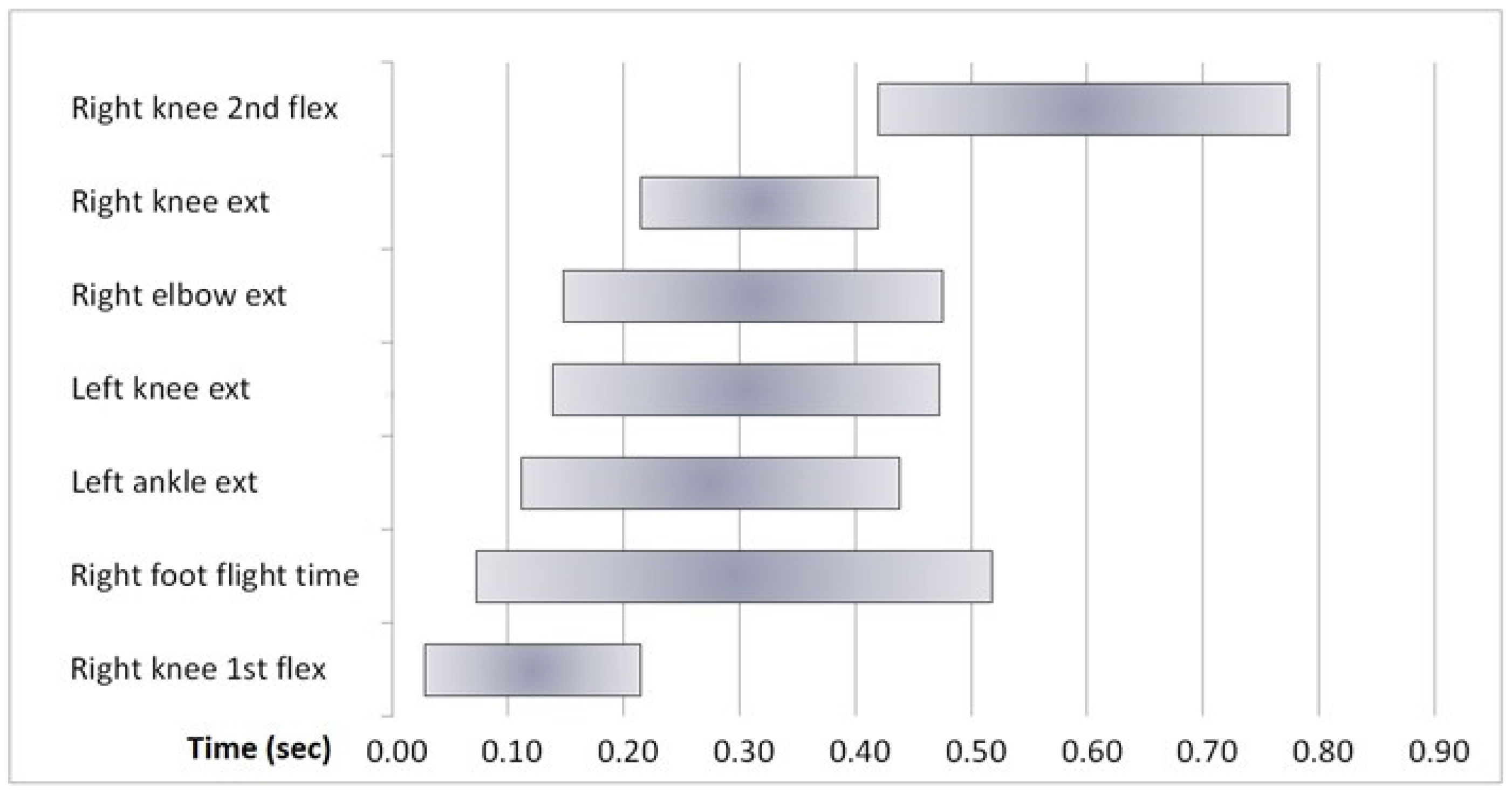
21] on expert fencers. During the lunge, the forward knee performs three different movements: an initial flexion, followed by an extension and a second flexion that brings the lunge to the end (Figure 3). The timing of the lunge action is reported in Figure 4.
The time chart shows the sequences of limbs activation: a distal-to-proximal activation can be observed, with the movement starting with a knee flex; followed by the rear ankle and rear knee extension, elbow extension, and forward knee extension; and ending with a right knee lunge.
Kinematics data are shown in Table 6.

7. Discussion

7.1. Anthropometry

The aims of the study were to compare male and female fencers and to identify the influence, if any, of the subject’s anthropometrics on the lunge. We did not find any asymmetry between body sides in all of the anthropometric measurement performed and in both males and females, except for in the cases of FM%, FFM%, and thigh skinfolds. Females showed greater FM% and thigh skinfold thickness than their male counterparts according to the developmental age considered. While asymmetries were found in adult fencers [21] (forward lower limb muscle and forward arm muscle being more developed), in children, the amount of practice is probably not sufficient to determine asymmetrical development of body segments.
Anthropometry (FFM and body height, but not weight or lower or upper limb heights individually) in our study is the major determinant of lunge velocity and distance. This agrees with the morphology of mesomorph fencers [21] and with lower limb strength (which is proportional to FFM) being the cause of the greatest velocity and distance.

7.2. Kinematics

In the “on-guard” position, the knees were semi-flexed, and the lower limb were nearly perpendicular to the ground. The elbow reached its maximal extension before the final lunge position. Between these two events (on guard and final position), the elbow angle remained almost unchanged. In the final lunge position, the rear knee was almost totally extended, while the anterior knee was flexed. Higher angular velocity was observed during the right knee extension, which is the movement that brings the anterior foot forward after its take-off. The peak velocity of the hand (Vmax 3 met) was observed during the elbow extension and was 1.17 m/s above the mean hand velocity (Vm 3 met) observed in the whole lunge. The anterior superior iliac spine midpoint (ASISm) showed a peak of 0.80 m/s above the mean velocity of the whole lunge.
The forward knee’s first flexion and the following extension angles were lower in our study than those in one observing élite athletes [4]: 20 ± 12° vs. 17 ± 7° and 51± 9° vs. 59 ± 8°, respectively. A similar posterior knee extension was found by Guan et al. [9] in intermediate-level athletes (50.4 ± 9.6° vs. 48 ± 11° in our study), while angular velocities were lower in our sample.
We found a significant correlation between lunge velocity (Vmax ASISm) and the ROM of the rear knee extension (r = 0.56, p = 0.031), as found in a previous study [9] on adult fencers. We found that the ROM of the rear knee extension is influenced by the knee’s angle in the guard position (r = −0.94 and r = −0.85, respectively, with the rear and forward knee angle in the on-guard position; p < 0.001). The more the knees are flexed in the guard position, the wider the extension of the posterior knee. Previous studies [8,9] assert that the lower guard position allows for the pre-tensioning of the thigh muscles and, consequently, a higher lunge horizontal velocity. Moreover, more flexed knees in the guard position allow for a greater extension of the rear knee during the lunge. In summary, a higher lunge velocity is related to a lower guard position, and we hypothesize that young fencers do not have sufficient strength to sustain it. Statistically Significant differences were found in kinematics between males and females. This result is different from that of other studies in which a difference between males and females was found in regard to the lunge action. This difference can be explained by the congenital valgus knee normally found in females [10].
The lunge distance and the hip velocity (Vm ASISm) were mostly correlated with fencers’ height and FFM, the FFM being the major predictor of hip and hand velocities in the regression analysis. As FFM is known to be related to strength, it is not surprising that the fat-free mass is related to the speed and amplitude of the lunge.
It is important to determine the characteristics of young fencers to improv the teaching of properly carrying out the lunge technique. In contrast with previous findings [10], we did not find any difference between males and females in the kinematic variables in the age range considered. In agreement with Tsolakis et al. [21], we can hypothesize that the differences in kinematics that we found by comparing our data with available data on adult experienced fencers were probably due to the normal growth process.
A development of our study could be the examination of subjects when they are fencing against an opponent, as this would facilitate a more realistic environment closer to the competition setting. Furthermore, it would be necessary to study the influence of a target on the lunge kinematics to understand if it is an essential element. A limitation of our study is that we tested one subject at a time and not in competition circumstances, e.g., with an opponent. This may have influenced the kinematics of the lunge. Our group showed that the lunge begins with the lower limb. This point was not clear in previous studies on young fencers [9].

8. Conclusions

Our aims were to assess if any differences exists (in anthropometrics and kinematics of foil lunge) between male and female young fencers; to identify the influence of the subject’s anthropometrics on the lunge; and to determine which parameters among anthropometrics are the major determinants of lunge speed and amplitude. We did not find any asymmetry between body sides in all the anthropometric measurement performed. Body height and fat-free mass content were the major determinants of hip velocity and of the distance achieved in the foil lunge in our group. While fencing is an asymmetrical sport, in this age group, the influence of training on muscle mass remained low: we did not find any side differences between body segments. Anthropometrics are only slightly different between developing male and female fencers. Gender differences in kinematics of the foil lunge in beginner to expert young fencers are not statistically significant. An explanation for this latter result is that with foil fencing being a sport with a high technical content, in this age group, it does not require strength but mostly technique. For the first time, kinematic data on the lunge of young foil fencers were presented. The implications of our study are that when teaching the lunge technique to this age group, a different approach for males and females is not required, and that among anthropometrics, body height and fat-free mass are major factors for the recruitment of young foil fencers. Moreover, at this age, body asymmetry was not detected, suggesting the asymmetric development of body parts, as a result of fencing training, occurs at a later stage of development.

Author Contributions

A.C., designed the study, performed the analysis of the results, and wrote and corrected the paper. A.M., performed the measurements and cooperated in writing the paper. J.W., revised the paper and cooperated in the analysis of the results. All authors have read and agreed to the published version of the manuscript.

Funding

This research received no external funding.

Institutional Review Board Statement

University of Bologna, Ethical Committee, 27 January 2020.

Informed Consent Statement

Informed consent was obtained from all subjects involved in the study.

Data Availability Statement

The data presented in this study are available on request from the corresponding author. The data are not publicly available due to age of the subjects.

Conflicts of Interest

The authors have no conflicts of interests.

References

  1. Aquili, A.; Tancredi, V.; Triossi, T.; De Sanctis, D.; Padua, E.; D’Arcangelo, G.; Melchiorri, G. Performance analysis in saber. J. Strength Cond. Res. 2013, 7, 624–630. [Google Scholar] [CrossRef] [PubMed]
  2. Chen, T.W.L.; Wong, D.W.; Wang, Y.; Ren, S.; Yan, F.; Zhang, M. Biomechanics of fencing sport: A scoping review. PLoS ONE 2017, 2, e0171578. [Google Scholar] [CrossRef] [PubMed]
  3. Hassan, S.E.A.; Klauck, J. Kinematics of lower and upper extremities motions during the fencing lunge: Results and training implications. In Proceedings of the 16th International Symposium on Biomechanics in Sports Conference, Konstanz, Germany, 21–25 July 1998; Volume 1, pp. 170–173. [Google Scholar]
  4. Gholipour, M.; Tabriz, A.; Farahmand, F. Kinematics analysis of lunge fencing using stereophotogrametry. World J. Sport Sci. 2008, 1, 32–37. [Google Scholar]
  5. Frère, J.; Gopfert, B.; Nuesch, C.; Huber, C.; Fischer, M.; Wirz, D.; Friederich, N.F. Kinematical and EMG-classifications of a fencing attack. Int. J. Sport Med. 2011, 32, 28–34. [Google Scholar] [CrossRef] [PubMed]
  6. Morris, N.; Farnsworth, M.; Robertson, D.G.E. Kinetic analyses of two fencing attacks and lunge and fleche. Port. J. Sport Sci. 2001, 11, 343–346. [Google Scholar]
  7. Mullo, F.; Mullineaux, D.; Irwin, G. Use of the kinematic chain in the fencing attacking lunge. In Proceedings of the 33 International Conference on Biomechanics in Sports, Poitiers, France, 29 June–3 July 2015. [Google Scholar]
  8. Bottoms, L.; Greenhalgh, A.; Sinclair, J. Kinematic determinants of weapon velocity during the fencing lunge in experienced épée fencers. Acta Bioeng. Biomech. 2013, 15, 109–113. [Google Scholar] [PubMed]
  9. Guan, Y.; Guo, L.; Wu, N.; Zhang, L.; Warburton, D.E.R. Biomechanical insights into the determinants of speed in the fencing lunge. Eur. J. Sports Sci. 2018, 18, 201–208. [Google Scholar] [CrossRef] [PubMed]
  10. Sinclair, J.; Bottoms, L. Gender differences in the kinetics and lower extremity kinematics of the fencing lunge. Int. J. Perform. Anal. Sport 2013, 13, 440–451. [Google Scholar] [CrossRef]
  11. Marfell-Jones, M.; Olds, T.; Stewart, A.; Lindsay-Carter, L.E. ISAK Manual, International Standards for Anthropometric Assessment; International Society for the Advancement of Kinanthropometry: Lower Hutt, New Zealand, 2011. [Google Scholar]
  12. Martine, T.; Claessens, A.L.; Vlietinck, R.; Marchal, G.; Beunen, G. Accuracy of anthropometric estimation of muscle cross-sectional area of the arm in males. Am. J. Hum. Biol. 1997, 9, 73–86. [Google Scholar] [CrossRef]
  13. Knapik, J.J.; Staab, J.S.; Harman, E.A. Validity of an anthropometric estimate of thigh muscle cross-sectional area. Med. Sci. Sports Exerc. 1996, 28, 1523–1530. [Google Scholar] [CrossRef] [PubMed]
  14. McGinley, J.L.; Baker, R.; Wolfe, R.; Morris, M.E. The reliability of three-dimensional kinematic gait measurements: A systematic review. Gait Posture 2009, 29, 360–369. [Google Scholar] [CrossRef] [PubMed]
  15. Oxford Metrics. Plug-in Gait Reference Guide; Oxford Metrics: Oxford, UK, 2016. [Google Scholar]
  16. Wu, G.; Siegler, S.; Allard, P.; Kirtley, C.; Leardini, A.; Rosenbaum, D.; Whittle, M.; D’Lima, D.D.; Cristofolini, M.; Witte, H. ISB recommendation on definitions of joint coordinate system of various joints for the reporting of human joint motion—Part I: Ankle, hip, and spine. J. Biomech. 2002, 35, 543–548. [Google Scholar] [CrossRef]
  17. Wu, G.; Van der Helm, F.C.; Veeger, H.E.; Makhsous, M.; Van Roy, P.; Anglin, C.; Nagels, J.; Karduna, A.R.; McQuade, K.; Wang, X.; et al. ISB recommendation on definitions of joint coordinate systems of various joints for the reporting of human joint motion--Part II: Shoulder, elbow, wrist and hand. J. Biomech. 2005, 38, 981–992. [Google Scholar] [CrossRef] [PubMed]
  18. Austin, P.C.; Steyerberg, E.W. The number of subjects per variable required in linear regression analyses. J. Clin. Epidemiol. 2015, 68, 627–636. [Google Scholar] [CrossRef] [PubMed] [Green Version]
  19. Cacciari, E.; Milani, S.; Balsamo, A.; Spada, E.; Bona, G.; Cavallo, L.; Cerutti, F.; Gargantini, L.; Greggio, N.; Tonini, G. Italian cross-sectional growth charts for height, weight and BMI (2-20y). J. Endocrinol. Investig. 2006, 29, 581–593. [Google Scholar] [CrossRef] [PubMed]
  20. Turner, A.; Bishop, C.; Chavda, S.; Edwards, M.; Brazier, J.; Kilduff, L.P. Physical characteristics underpinning lunging and change of direction speed in fencing. J. Strength Cond. Res. 2016, 30, 2235–2241. [Google Scholar] [CrossRef] [PubMed]
  21. Tsolakis, C.; Bogdanis, G.C.; Vagenas, G. Anthropometric profile and limb asymmetries in young male and female fencers. J. Hum. Mov. Stud. 2006, 50, 201–215. [Google Scholar]
Figure 1. The model is a modified Plug-In-Gait models of 34 markers placed on relevant body landmarks. Additional virtual internal markers were calculated as joints centroids.
Biomechanics 01 00014 g001
Figure 2. Angles and ranges of motions of lower (above) and upper (below) limb.
Biomechanics 01 00014 g002
Figure 3. Four phases of the lunge (FL = flexion; EX = extension) and forward knee angle table.
Biomechanics 01 00014 g003
Figure 4. Timing of the lunge action (ext: joint extension, flex: joint flexion).
Biomechanics 01 00014 g004
Table 1. Demographics of the subjects.
Age (Years)Height (cm)Weight (kg)
Mean ± SDMean ± SDMean ± SD
Males (n. 8)13.6 ± 3.1162.5 ± 18.553.1 ± 15.3
Female (n. 7)11.7 ± 2152.8 ± 10.746.8 ± 7.2
Table 2. Anthropometrical measurements of the male and female subjects. CSA = cross-sectional area (right = forward; left = rear).
M (N = 7) F (N = 8)
M±SDM±SD
BMI19.73.220.11.5
Fat mass (kg)44.812.935.66.9
Fat mass %84.96.375.85.2
Fat Free mass (kg)8.34.411.22.3
Fat Free mass %0.06.324.20.0
Upper limb length (cm)69.89.165.65.5
Lower limb length (cm)81.510.377.15.3
Trunk length (cm)81.08.375.76.3
Circumf. upper arm (cm)23.53.723.02.1
Biceps skinfold (cm)0.80.51.10.3
Tricep skinfold (cm)1.20.71.60.3
CSA upper arm (cm2)30.28.724.14.6
Table 3. Lengths, circumference, skinfolds, and cross-sectional area (CSA) of male and females (right = forward; left = rear).
M (N = 7)F (N = 8)
RightLeftRightLeft
M±SDM±SDM±SDM±SD
Thigh length (cm)39.24.939.24.837.63.137.73.1
Bi-epicondylar breadth (cm)8.50.70.00.78.20.68.10.0
Thigh circumference (cm)49.97.248.56.851.93.351.33.3
Thigh Skinfold (cm)1.50.61.50.62.70.42.70.4
CSA thigh (cm2)131.932.60.032.6118.318.3118.30.0
Table 4. Anthropometric differences between males and females young fencers. R = forward; L = rear.
M (N = 7)F (N = 8)
M±DSM±DSEffect Sizep
FM%15.16.224.25.11.60.011
FFM%84.86.275.75.11.60.011
Right thigh skinfold (cm)1.540.62.60.42.10.003
Left thigh skinfold (cm)1.530.62.60.42.20.002
Table 5. Linear regression with FFM (kg) as an independent variable. VmASISm: mean horizontal velocity in the lunge of anterior superior iliac spine midpoint.
BSEβp
Vm ASISm0.5160.091 0.000
0.0090.0020.7330.002
Lunge distance0.5310.129 0.001
0.0150.0030.7970.000
Table 6. Angles, linear variables, and linear and angular velocities. ASISm = anterior superior iliac spine midpoint; 3 met = 3rd metacarpal head.
Angles (deg)Mean±SD
On guard
Elbow (deg)9616
Forward knee (deg)12010
Rear knee (deg)12110
Forward ankle (deg)986
Rear ankle (deg)835
During lunge
Elbow extension angle (deg)1515
End of forward knee at 1st flexion (deg)10311
End of forward knee extension (deg)1515
At final lunge position
Elbow (deg)1505
Forward knee (deg)909
Rear knee(deg)1704
Rear ankle (deg)1036
Rear ankle (deg)1165
Range of motion
Elbow extension (deg)5517
Forward knee 1st flexion (deg)177
Forward knee in extension (deg)4711
Forward knee 2nd flexion (deg)598
Rear knee extension (deg)4811
Rear ankle plantar flexion (deg)326
Linear velocities and lunge distance and duration
Mean velocity of ASISm (m/s)0.90.1
Mean velocity of 3met (m/s)1.30.2
Peal Velocity ASISm (m/s)1.70.2
Peak velocity 3 met (m/s)2.40.3
Lunge distance (m)1.10.2
Lunge duration (s)0.70.1
Angular Velocities
Elbow extension (deg/s)180.456.3
Forward knee 1st flexion (deg)87.820.4
Forward knee extension (deg)242.161.3
Forward knee 2nd flexion (deg)179.526.8
Rear knee extension (deg)160.946.9
Rear ankle plantar flexion (deg)108.626.6
Publisher’s Note: MDPI stays neutral with regard to jurisdictional claims in published maps and institutional affiliations.

