Article

Theoretical Three-Dimensional Zinc Complexes with Glutathione, Amino Acids and Flavonoids

by
José Manuel Pérez de la Lastra
1,*,
Celia Andrés-Juan
2,
Francisco J. Plou
3 and
Eduardo Pérez-Lebeña
4
1
Biotechnology of Macromolecules Research Group, Instituto de Productos Naturales y Agrobiología, IPNA, CSIC, 38206 San Cristóbal de la Laguna, Spain
2
CINQUIMA Institute and Department of Organic Chemistry, Facultad de Ciencias, University of Valladolid, 47011 Valladolid, Spain
3
Instituto de Catálisis y Petroleoquímica, CSIC, 28049 Madrid, Spain
4
Sistemas de Biotecnología y Recursos Naturales, 47625 Valladolid, Spain
*
Author to whom correspondence should be addressed.
Submission received: 2 June 2021 / Revised: 21 July 2021 / Accepted: 22 July 2021 / Published: 28 July 2021
(This article belongs to the Special Issue Responses and Defense Mechanisms against Toxic Metals)

Abstract

:
Zinc plays an important role in the regulation of many cellular functions; it is a signaling molecule involved in the transduction of several cascades in response to intra and extracellular stimuli. Labile zinc is a small fraction of total intracellular zinc, that is loosely bound to proteins and is easily interchangeable. At the cellular level, several molecules can bind labile zinc and promote its passage across lipophilic membranes. Such molecules are known as ionophores. Several of these compounds are known in the scientific literature, but most of them can be harmful to human health and are therefore not allowed for medical use. We here performed a theoretical three-dimensional study of known zinc ionophores, together with a computational energetic study and propose that some dietary flavonoids, glutathione and amino acids could form zinc complexes and facilitate the transport of zinc, with the possible biological implications and potential health benefits of these natural compounds. The study is based on obtaining a molecular conformational structure of the zinc complexes with the lowest possible energy content. The discovery of novel substances that act as zinc ionophores is an attractive research topic that offers exciting opportunities in medicinal chemistry. We propose that these novel complexes could be promising candidates for drug design to provide new solutions for conditions and diseases related to zinc deficiency or impairment derived from the dysregulation of this important metal.

1. Introduction

All living forms are compartmentalized. Membranes surround the cells of all organisms and intracellular organelles, which act as a barrier to control the transfer of substances between the inside and outside of these structures. The lipophilic nature of the membranes prevents the passage of charged particles, such as ions. Therefore, the transport of ions must be regulated by specific proteins that are an integral part of the membranes, acting as channels allowing the entry and exit of ions [1]. These proteins maintain asymmetric concentrations of ions and facilitate their selective transport, which leads to the formation of energy that is eventually stored in the form of ATP [2]. Therefore, the control of ion transport and the homeostatic regulation of ion concentration levels across the membrane plays a critical role in sustaining life.
Zinc plays an important role in protein function, being a structural element of many macromolecules. Its importance has been recognized since 1869 when it was found to be necessary for the growth of Aspergillus niger [3] and later was found that it also affected the growth of plants and animals. In humans, Ananda S Prasad first suspected zinc deficiency in 1958 after he, at the request of James A. Halsted, evaluated a patient with iron deficiency. In addition In addition to iron deficiency, the patient appeared ~10 years older and was severely stunted and prepubertal age [4].
As a micronutrient, the importance of zinc has been reflected through the numerous diseases that are associated with its dietary deficiency. For example, zinc deficiency led to growth disorder, cognitive disorder and compromised immune function. Zinc deficiency occurs because of nutritional factors, but also in various disease states (malabsorption, Crohn’s disease, alcoholism, liver cirrhosis, chronic renal disease and other chronically debilitating diseases) [5,6,7]. Indeed, zinc supplementation improved these syndromes, as well as testicular and ovarian dysfunction or growth retardation [8].
Labile zinc is a small fraction of total intracellular zinc that is loosely linked to proteins and easily interchangeable. It is the pool sensitive to chelation and capable of detection by a variety of chemical and genetic sensors, participates in signal transduction pathways. Both free and labile zinc function as a second messenger molecule, modulating the activity of several enzymes and, as a result, signaling and metabolic pathways, as well as cellular activities. When cellular zinc levels are elevated, zinc transporter proteins (ZnT) contribute to modulating the cytoplasmic zinc balance by exporting zinc out to extracellular space or by sequestrating cytoplasmic zinc into intracellular compartments. On the contrary, when cellular zinc is depleted, zinc transporter (Zrt) and Irt-like proteins, known as ZIP proteins, work to increase cytoplasmic zinc concentrations [9,10,11].
At the cellular level, several natural molecules can bind labile zinc and promote its passage across lipophilic membranes. Such molecules are known as ionophores, from the ancient Greek, meaning “ion carriers”. Ionophores are membrane-active compounds that do not depend on active metabolism to exert their biological activity. They are chemical entities that bind reversibly with the ion, forming a supramolecular complex that can diffuse across hydrophobic membranes [12]. This capacity of ionophores to alter the normal ion balance of membranes can be used as a mechanism for inducing cell death, or to eliminate harmful microorganisms or cells with abnormal growth, like tumor cells [13].
Zinc chelators are able to transport zinc cations across the plasma membrane, independently of zinc transporters [14]. In chemistry, zinc is available in the form of inorganic and organic salts. Some inorganic zinc salts are zinc sulfate, zinc chloride, zinc oxide and zinc carbonate, while organic salts include zinc citrate, zinc lactate, zinc acetate, zinc gluconate, zinc stearate, zinc picolinate and zinc ascorbate [15,16]. The ion selectivity of ionophores is a combined function of the energy required for desolvation of the ion and the liganding energy obtained on complexation. For metal binding, thermodynamic stability of complexes is the measure of a metal ion’s tendency to selectively form a certain metal complex and is directly connected to metal-ligand bond energies, which would result in a decrease in the system’s free energy [17,18]. Several molecular characteristics can be correctly represented as a sum of individual bond properties, without the need to explicitly solve any wave equations. The molecular mechanics method refers to the study of such molecular characteristics in terms of non-quantum mechanical models (MM). In a nutshell, MM posits that the energy of a molecular system is made up of five additive, non-interacting components [19,20].
Numerous investigations have demonstrated that flavonoids influence the metabolism of zinc [21]. For example, rats fed with baicalin and rutin for long periods of time had lower liver levels of total zinc, iron and copper, suggesting that certain flavonoids, contain metal-binding motifs, like phytate, may retain these metals and render them inaccessible for absorption. On the other hand, polyphenols can transport zinc across the plasma membrane without relying on cell transport systems like zinc transporters or endocytosis. Dabbagh-Bazarbachi et al. investigated the interactions between a variety of polyphenols and polyphenol metabolites and zinc in mouse hepatocarcinoma Hepa 1–6 cells under physiological conditions [22]. After the addition of quercetin and epigallocatechin-gallate, the zinc-polyphenol chelation complex was formed and transported across the lipid bilayer. When given to cells, these complexes rapidly increase the intracellular zinc pool, which may be detected using various fluorophores such as Zinquin or FluoZin-3 [23]. They concluded that flavonoids could generate water-insoluble membrane-permeant zinc complexes that penetrate the plasma membrane and serve as zinc ionophores.
In order to ascertain whether other natural compounds, such as flavonoids, glutathione and plant amino acids, can serve as new zinc ionophores, we here provide a computational energetic study of the forming zinc complexes. The study is based on obtaining a molecular conformational structure of the zinc complexes with the lowest possible energy content. We also discuss the relevance and biological implications of the formation of such complexes. We hope that this preliminary study could offer new opportunities to prevent zinc deficiency by using nutraceutics, such as amino acids, glutathione or flavonoids, together with zinc supplementation.

2. Results

For initial considerations and validation of our study, we computed the energy and favorable spatial orientation of some well-known zinc ionophores when complexed with zinc cations. The differences in their energetic level, derived from molecular mechanics, is summarized in Table 1. For each complex, we studied different three-dimensional structural conformations, considering those with the lowest possible energy and favorable geometry respect to the internal position of the zinc cation.
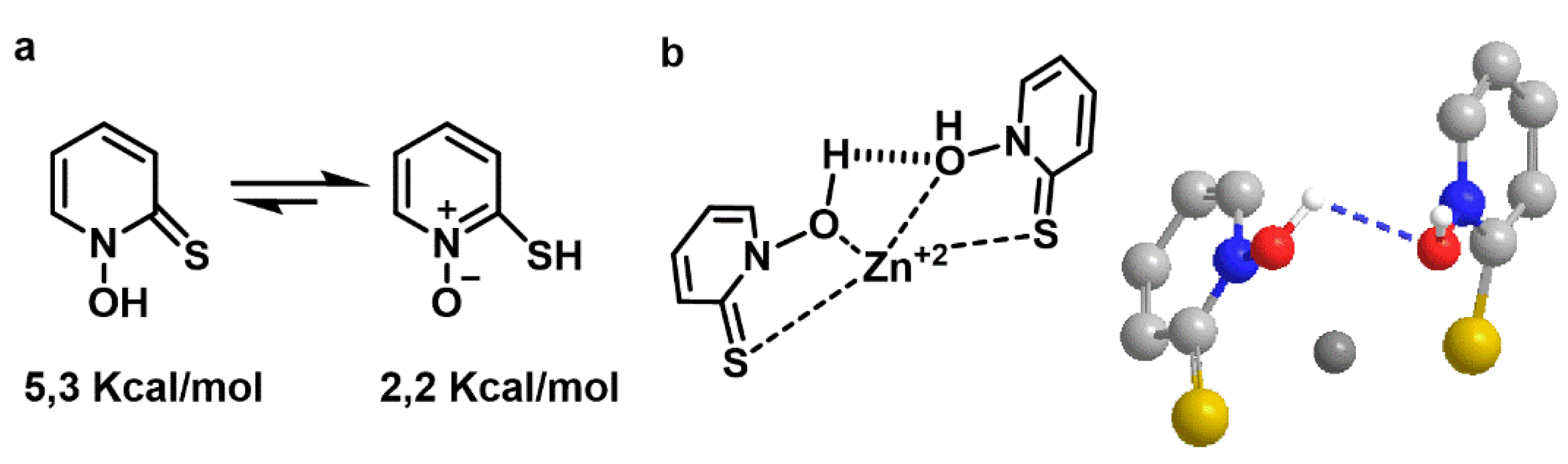
Free pyrithione has two structures with different energies. The left one has an energy of E = 5.3 kcal/mol and the right one is E = 2.2 kcal/mol (the lower the energy, the higher the stability, so the balance is shifted to the right). The zinc-pyrithione complex has an energy of E = −37.2 kcal/mol, which is considerably more stable than free pyrithione (Figure 1). The favorable geometry of the complex and spatial orientation of the molecules, showed that the zinc cation was in an internal position between two adjacent pyrithione molecules, with the polar groups orienting towards the inside.
The efficient shielding of its polar core, which delocalizes the cation charge, may explain the resultant complex’s lipid solubility, consistent with the role of pyrithione as a zinc ionophore (Figure 1).
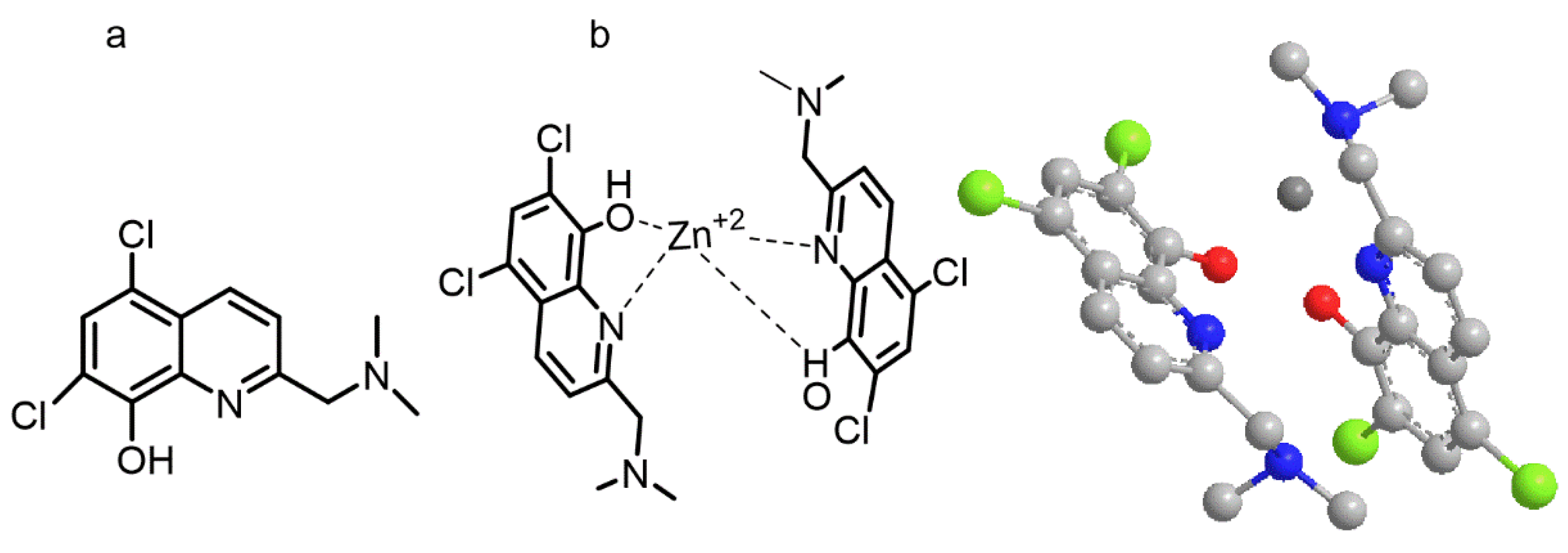
Similarly, the zinc ionophore adopted a disposition around the zinc cation with two molecules of 5,7-Dichloro-2-((dimethylamino) methyl) quinolin-8-ol (PBT2) in opposite orientations (Figure 2). The computed energy for the free form is E = 11.7 kcal/mol whereas for the zinc/PBT2 complex depicted in Figure 2b was E = −110.5 kcal/mol.
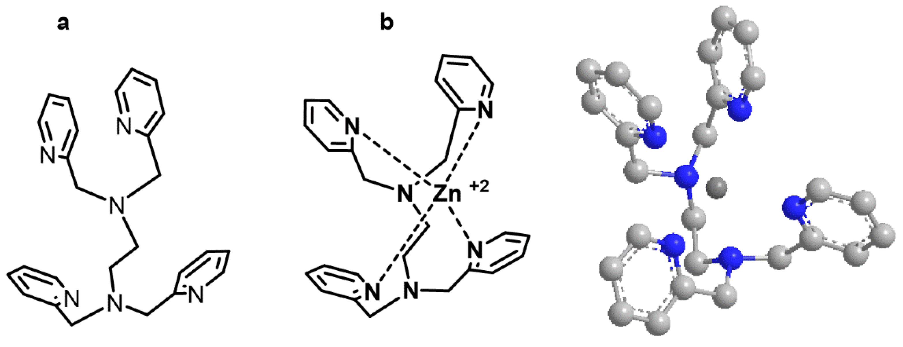
TPEN (N,N,N′,N′-tetrakis(2-pyridinylmethyl)-1,2-ethanediamine), a zinc ionophore without oxygen atoms, has an E = 38.9 kcal/mol for the free form and of E = −126.6 kcal/mol for the complex zinc/TPEN. The spatial orientation of the most favorable complex also showed an internal zinc cation surrounded by four pyridine rings (Figure 3).
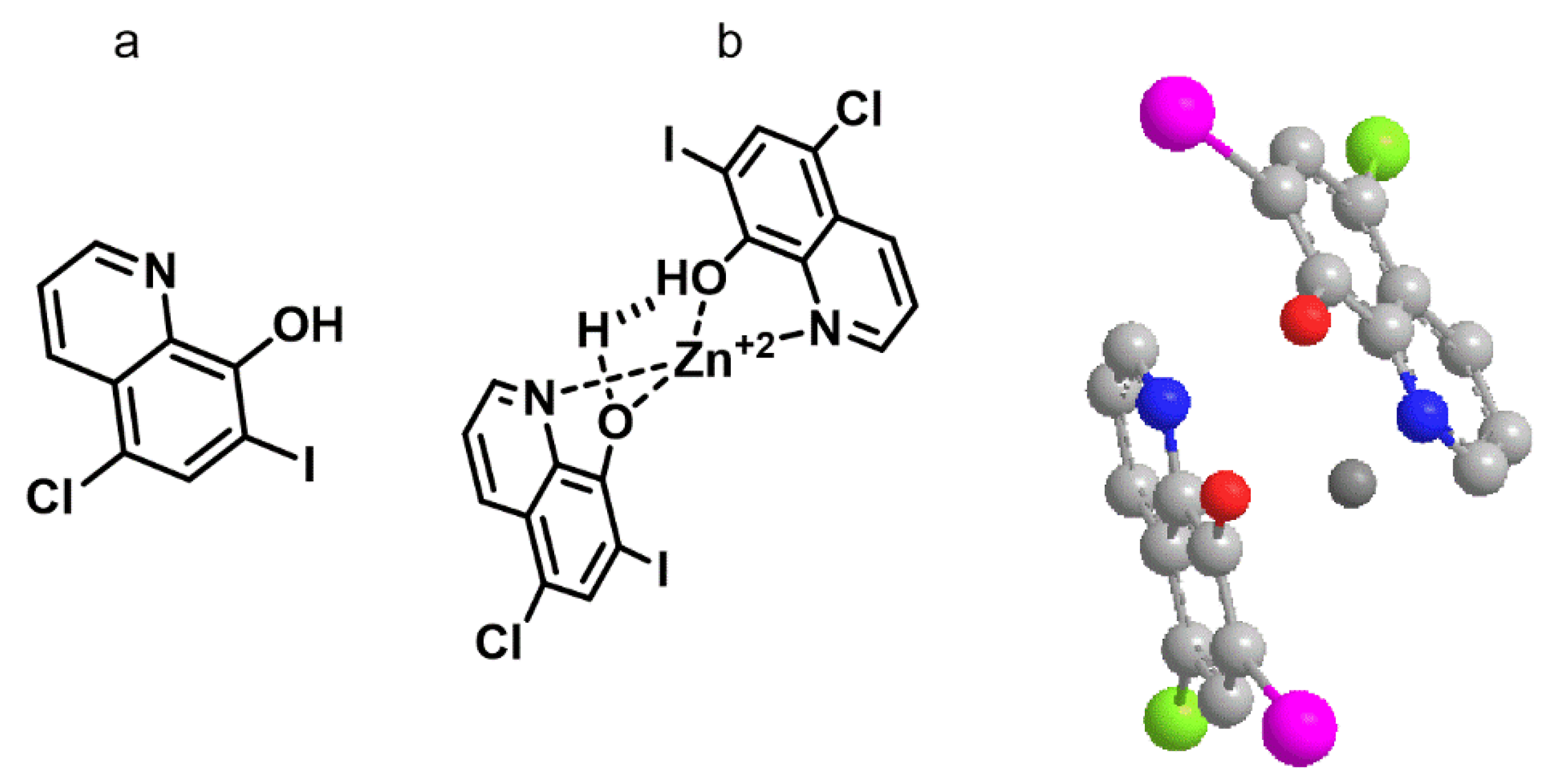
Clioquinol (5-Chloro-7-iodo-8-quinolinol) had an energy of E = 6.9 kcal/mol, whereas for the complex with zinc the energy was of E = −78.4 kcal/mol, indicating the favorable formation of the zinc. The geometry and spatial orientation of the complex showed the internal zinc cation between two adjacent hydroxyquinoline groups in opposite directions and the zinc cation coordinated with oxygen and nitrogen atoms (Figure 4).
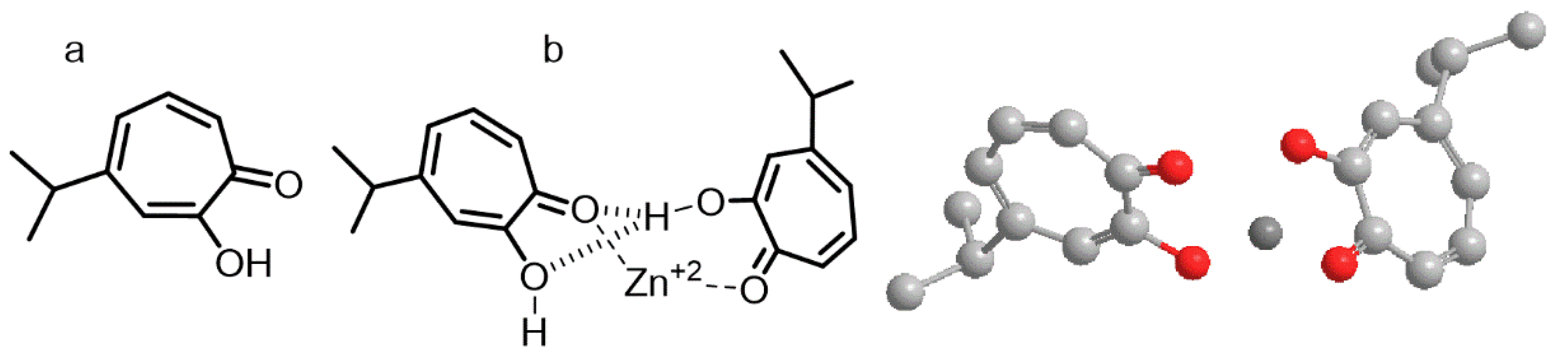
Hinokitiol (2-hydroxycyclohepta-2,4,6-trien-1-one) is structurally related to tropolone (a well-known chelating agents). In our computational analysis, free hinokitiol had an energy of E = 18.1 kcal/mol, the dimer form had an energy of E = 29.5 kcal/mol, whereas the complex hinokitiol/zinc had an energy content of E = −125.6 kcal/mol. The spatial orientation for the lowest energetic form of hinokitiol/zinc showed an internal zinc cation coordinated with oxygen atoms from the hydroxyl and carbonyl groups of the aromatic rings that were orienting their hydrophobic isobutyl groups at opposite ends (Figure 5).
The greatest difference in energy was found for the TPEN molecules, in free form was of 38.9 kcal/mol and complexed with zinc was of E = −126.6 kcal/mol. The lowest shift in the computed molecular mechanical energy was found for pyrithione, which in free form was of E = 2.2 kcal/mol and and in combination with zinc cation, was of E = −37.2 kcal/mol (Table 1).
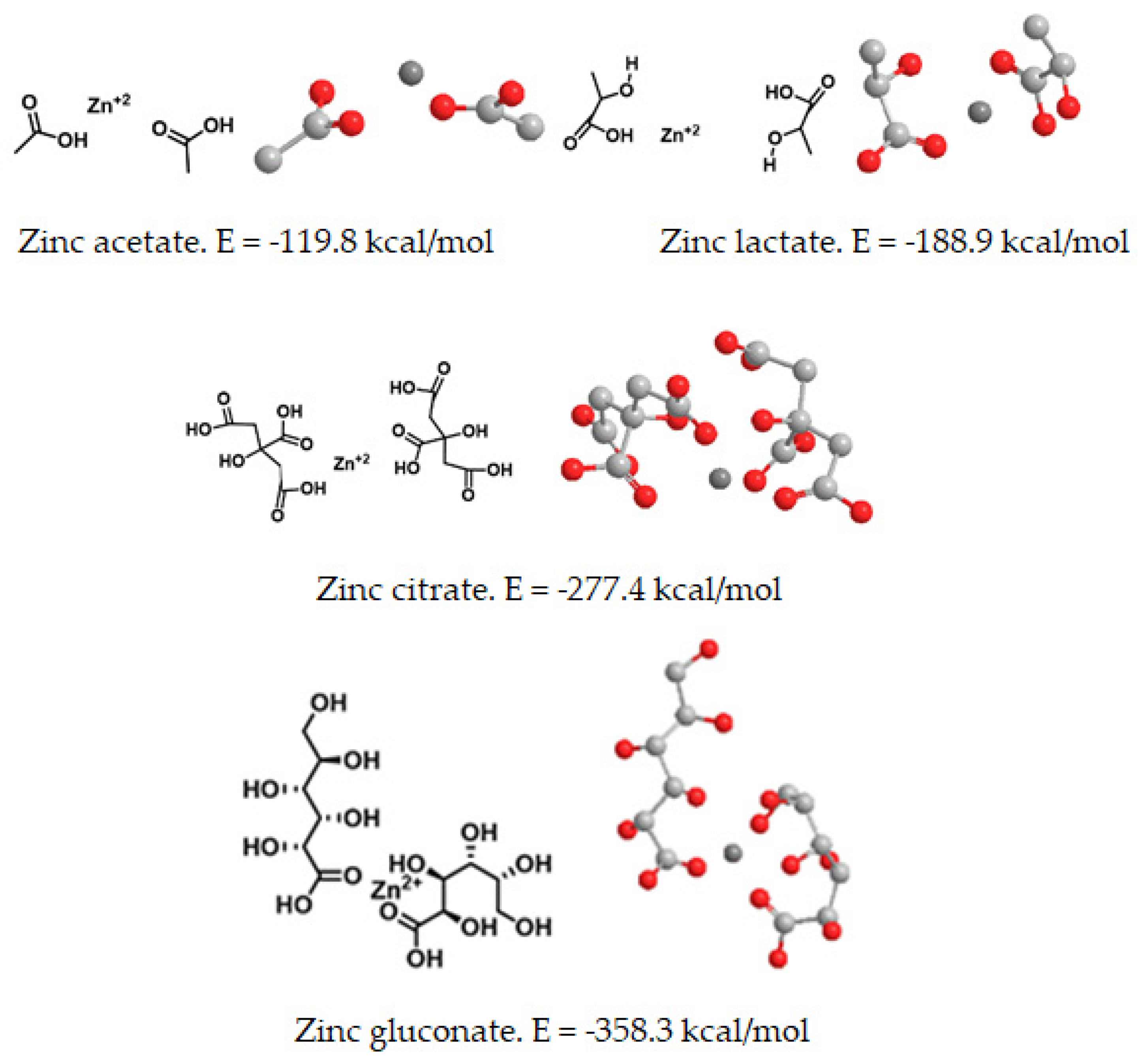
Next, an energetic study was carried out to analyze whether there may be energetically optimized ionophores that are able to chelate zinc, independently of their previous commercial forms (acetate, citrate, gluconate and zinc lactate), which are already on the market. In free forms, the lowest energy (E = −358.3 kcal/mol) was found for zinc gluconate, whereas the highest energy for a free form of zinc salts of organic acids (E = −119.8 kcal/mol) corresponded to zinc acetate. In all organic forms studied, the three-dimensional analysis of the complexes showed an internal zinc cation surrounded by two organic molecules in coordination with oxygen atoms from hydroxyl and carbonyl groups of the organic molecules. (Figure 6).
When combined with an amino acid (tryptophan) or a polyphenol (silybin), the corresponding complexes with zinc showed different energy. The lowest energy was found for the zinc gluconate, whereas the highest energy obtained corresponded to the amino acid and flavonoid complexes with zinc citrate (Table 1, Figure 6). Similar values were obtained for amino acid and flavonoid complexes formed with zinc acetate and zinc lactate (Table 1). Therefore, we decided to use zinc gluconate for further theoretical studies, as it was the most stable form of dietary zinc salts of organic acids, according to our molecular mechanics calculations (Figure 6). The complexes with zinc gluconate showed the lowest energy values, so the formation of these complexes with zinc gluconate were more stable than those formed with other forms of zinc salts of organic acids (Table 2).
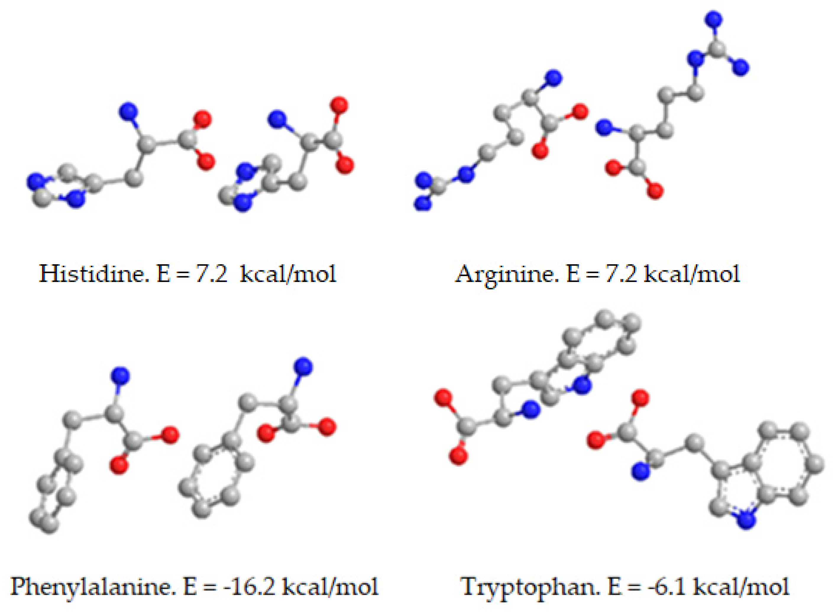
For this reason, we computed the energy and favorable spatial orientation of zinc gluconate when complexed with natural compounds, such as amino acids (phenylalanine, tryptophan, histidine and arginine). The most favorable forms of free amino acids were as dimers with different spatial orientation, according to our energetic calculations (Figure 7). The dimer of amino acid with lowest energy level was arginine (E = −16.7 kcal/mol) whereas the least stable and most energetic dimer of the amino acids studied corresponded to tryptophan (E = −6.1 kcal/mol). The spatial orientations are shown in Figure 7.
When complexes with zinc gluconate, the computed energy and spatial orientation of the studied amino acids indicated the favorable formation of the corresponding ionophores of zinc cation surrounded by two molecules of amino acids and two molecules of gluconate, as shown in Figure 8.
The calculated energy of free zinc gluconate was of E = −358.3 Kcal/mol (Table 1). However, when complexed with the amino acids, the computed energy was lower in all cases studied, indicating that there are favorable mechanical forces that make these complexes more stable and less energetic than in their corresponding free forms. For the amino acids studied, the lowest value of computed mechanical energy was obtained for the complex of tryptophan with zinc gluconate (
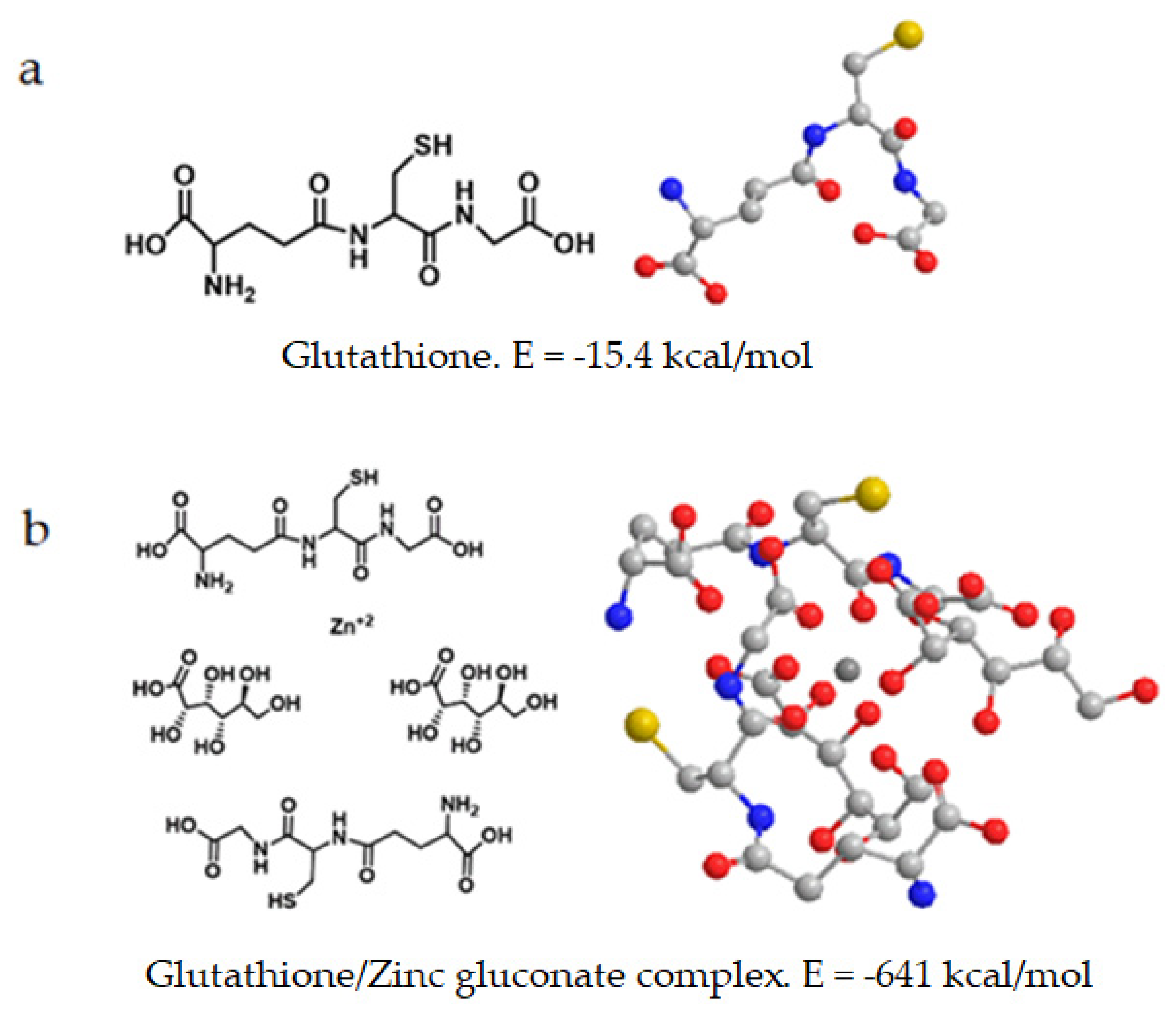
Table 3). The lowest energy of all compounds is obtained for the glutathione/zinc gluconate complex, with −641 kcal/mol.
The computed energy value of glutathione and the spatial orientation of both free form and when complexed with zinc gluconate is shown in Figure 9.
The complex of glutathione with zinc gluconate showed the least energetic value (E = −641 kcal/mol) obtained from the compounds studied (Table 2). The most favorable spatial orientation corresponded to the zinc cation surrounded by four molecules: two outer antiparallel mirrored images of GSH molecules at opposite ends and two inner gluconate molecules with same orientation (Figure 9b).
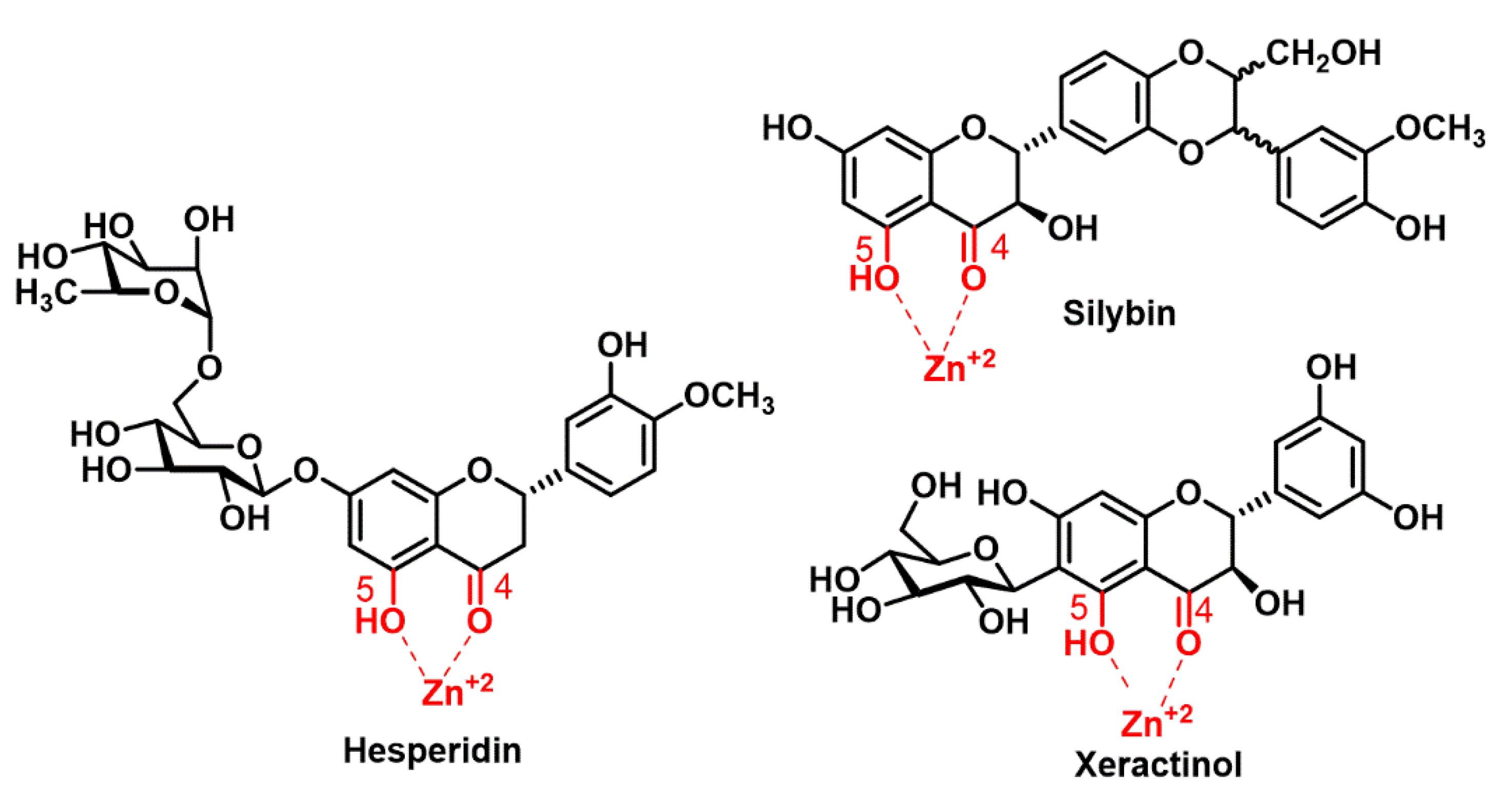
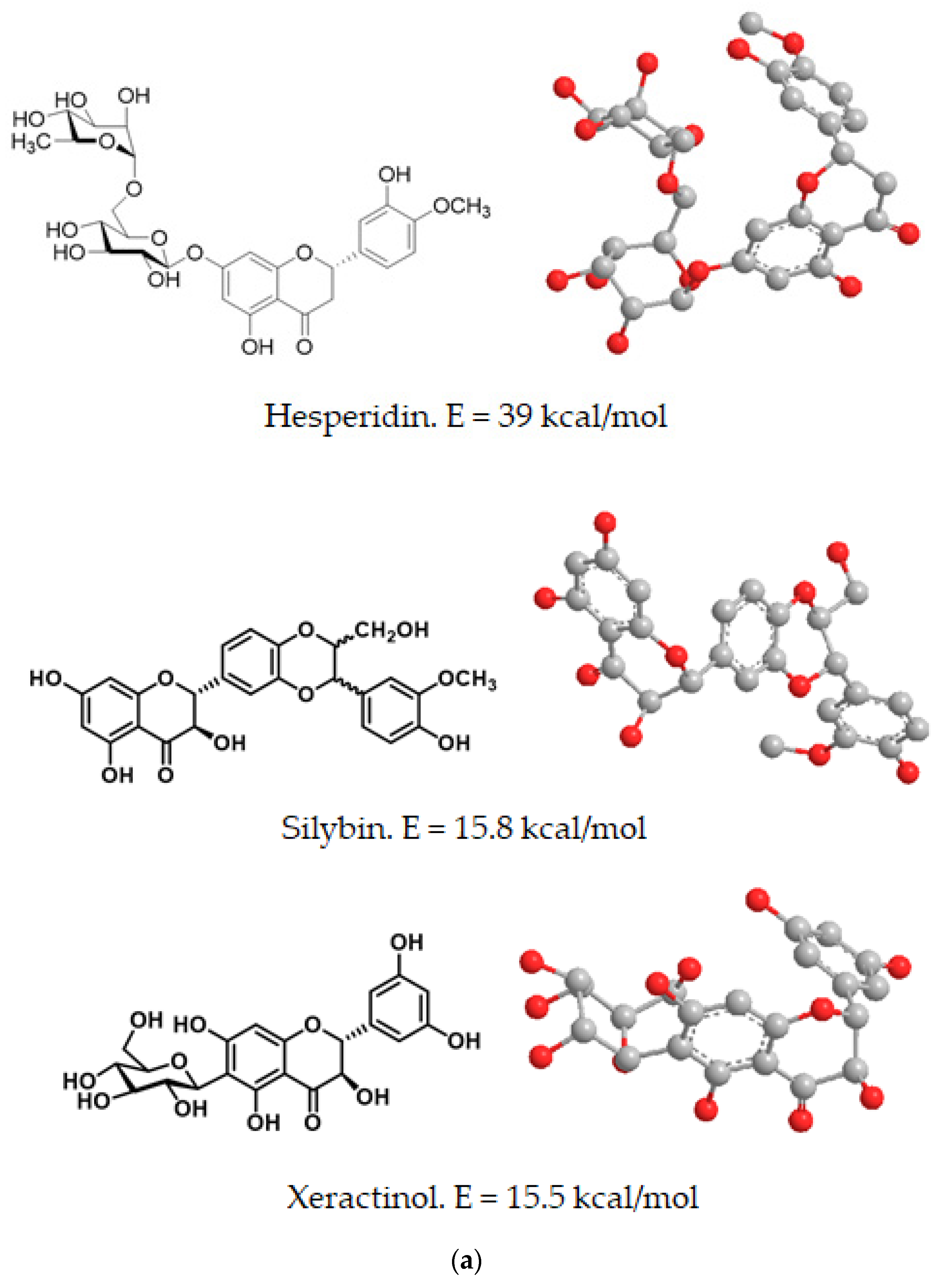
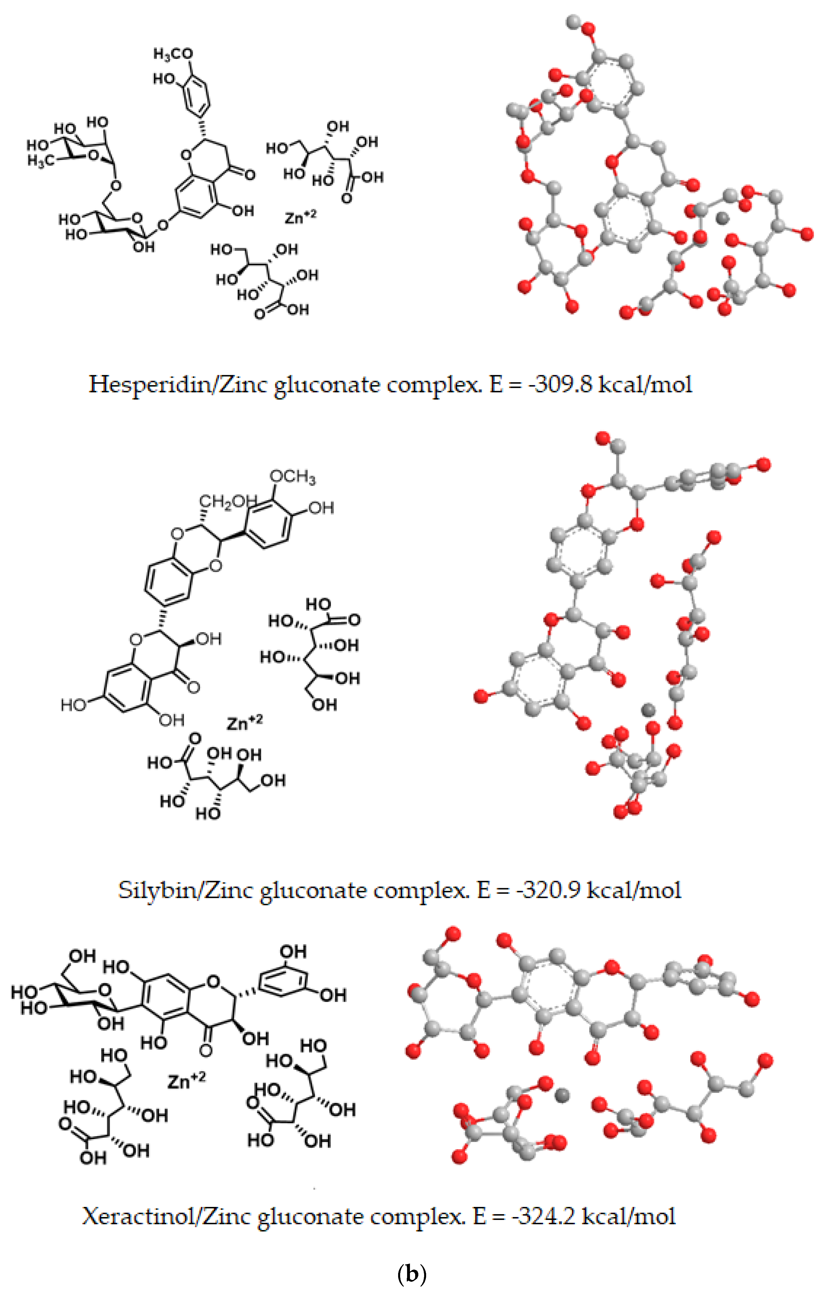
Finally, we performed a computational analysis of the complex of three well known polyphenols (hesperidin, silybin and xeractinol) with zinc gluconate, with the assumption that the high stability of the complexes was due to the formation of a greater number of intramolecular hydrogen bonds between the oxygenated functions of the polyphenol and the zinc cation. The zinc binding sites can be formed through the hydroxyl and carbonyl groups of the polyphenols (Figure 10).
Both in free form and in combination with zinc gluconate, the computed energy content and spatial orientation of dietary polyphenols (hesperidin, silybin and xeractinol), as representatives of the flavonoid family, is shown in in Table 2 and Figure 11.
The lowest energy value, as computed by molecular mechanics and thus most stable form of these complexes was found for the xeractinol/zinc gluconate (E = −324.2 kcal/mol) and the higher value of computed energy (E = −309.81 kcal/mol) was fond for the hesperidin/zinc gluconate complex (Figure 11). In all flavonoid studies, the complexes with zinc gluconate consisted in an internal zinc cation surrounded by two molecules of gluconate and one molecules of flavonoid with their carbonyl or hydroxyl groups facing internally to facilitate the coordination with the internal zinc (Figure 11).

3. Discussion

We here provide a computational energetic analysis of the forming zinc complexes to determine whether natural substances, such as flavonoids, glutathione and plant amino acids, can act as novel zinc ionophores. Our research is focused on the study of molecular conformational structure of the zinc complexes with the lowest amount of energy. This favorable energy content indicates that polyphenols, glutathione and amino acids may form zinc complexes, which might function as novel ionophores. These natural compounds might be attractive candidates for the treatment of diseases caused by zinc deficiency.
The chemistry of ionophore molecules is an important area of supramolecular chemistry [24]. The notion of a metal ionophore consist of a ligand attached to a metal cation in a reversible manner. In the bonded form, the metal complex is generally charge neutral as well as sufficiently hydrophobic to facilitate membrane passage across lipid membranes. Metal cation release can then be activated by either a low local concentration of the metal cation or the presence of a stimulus that favors metal cation release. Due to the reversibility of the metal binding process, the ionophore may repeatedly transport metal cations across membranes [25].
Experimental determinations of the stability constants of ionophore metal-complexes and the corresponding free energies of complexation reactions are time-consuming and expensive tasks. Stable and favorable complexation with metals may improve ionophore bioavailability to cells, being a major critical step in the process of a chemical acting as zinc ionophores [26]. As a result, a theoretical quantitative assessment of complex stability, together with the analysis of the geometry and spatial orientation around the metal cation, may prove to be a valuable complement to experimental investigations, allowing researchers to decrease the number of tests performed and to identify the strategy of “optimization” of known metal binders, among other advantages.
The zinc ion is stereo-chemically inactive. Zinc conformational structure is not important in our study, only as a chelating agent. The zinc ion is highly flexible in terms of coordination numbers, exhibiting coordination dynamics that do not conform to the stereochemical restrictions common to transition metal ions. Additionally, it is distinguished from magnesium (Mg2+) and calcium (Ca2+) because it forms considerably stronger complexes with water and a range of anions and ligands [26]. These features are critical for the proper functioning of its biological activities.
To validate our molecular mechanics approach, we first computed the energy content of several known zinc ionophores (pyrithione, PBT2, TPEN, clioquinol and hinokitiol) both as free molecules and when forming complexes with zinc cation. In all cases studied, the values obtained for the free molecules had positive values and negative values when complexed with zinc cation. In some complex up to 100 kcal/mol lower. This reduction in conformational energy implies the favorable formation of the zinc complexes, as corresponding for zinc ionophores.
Another aspect of potential ionophore molecules is the geometry of the most stable form of the ionophore-metal cation complex [25]. Ionophores are flexible, the final spatial orientation of the complexes includes configurational or conformational remodeling of the ionophore to accommodate the internalization of the metal ion. Ionophores, in general, are molecules having backbones of a range of different topologies that include oxygen or nitrogen atoms that are strategically spaced [25]. The liganding atoms include a variety of functional groups, including ether, alcohol, carboxyl and amide, among others. It is through ion-dipole interaction that the neutral oxygen or nitrogen function as a ligand of cations, which is comparable to the solvation of ions in highly dielectric fluids. For the permeation through the lipophilic environment of the membrane, the polar groups of ionophore complexes must face inward, while the outside of the complex is covered with different hydrocarbon hydrophobic groupings. In our analysis, the most favorable geometry of the complex zinc pyrithione showed the zinc cation in an internal position between two adjacent pyrithione molecules, with their polar groups orienting towards the inside [27]. Similarly, for the zinc ionophore TPEN, the spatial orientation of the most favorable complex with zinc showed an internal zinc cation surrounded by four pyridine rings. The resultant complex’s lipid solubility of ionophores can be explained in part by the efficient shielding of its polar core, which delocalizes the cation charge and in part by the complex’s external compatibility with low-dielectric solvents [28]. Several articles have recently been published that report the utility of theoretical calculations on these kind of chemical interactions [29,30].
In human nutrition, zinc deficiency is a global nutritional problem. Zinc supplementation has been used for the treatment of several diseases, but mainly for its potential ability to attenuate the accumulation of free radicals [31]. The total zinc content of eukaryotic cells is quite high, approximately 200 µM. However, the majority of cellular zinc is firmly bound to proteins [32]. A weak-binding fraction, capable of rapidly triggering ligand exchanges, is represented by the free (bioavailable, rapidly exchangeable, or labile) zinc ion. This labile zinc comprises the major mobile reactive pool of cellular zinc. This pool is not a viable source for incorporation into proteins, as it is loosely bound to low molecular ligands (such as amino acids and citrate). When labile zinc combines with a low-molecular-weight ligands or chelators to be absorbed, solubility is increased [33]. As a result, the chemical form in which this labile zinc is made available is of critical consideration. Amino acids (e.g., histidine, tryptophan) and organic acids (e.g., citrate, gluconate) have been used to improve zinc absorption and have been utilized in zinc supplements to improve zinc bioavailability. In most of these studies usually only one type of Zn2+salt was used rising the question whether different counterions and ligands of Zn2+ may influence its uptake and antioxidative function.
Zinc supplementation has been proposed as an efficacious chemo-preventive agent for some cancers [34]. Zinc can restrict the growth of cancer cells via cell cycle arrest, as well as induction of apoptosis and necrosis [35]. In clinical studies oral supplementation with zinc gluconate resulted in a decreased frequency of infections, production of tumor necrosis factor alpha (TNF-) and oxidative stress in elderly subjects, compared to placebo [36]. Therefore, any strategies that can increase zinc levels in the cancer cells are likely to be effective cancer treatment strategies, having minimal toxicity to normal cells. Accordingly, various complexes between zinc and bioactive molecules have been prepared and explored in the development of new antitumor drugs [25]. Although zinc sulfate is the most widely utilized zinc form, zinc gluconate stabilized with glycine has the same bioavailability and metabolic behavior as zinc sulphate in aqueous solution with the advantages of good solubility, mild flavor and low cost of production [37]. Our theoretical results suggest that zinc gluconate forms the most favorable complex of zinc among four zinc salts of organic acids: zinc gluconate, zinc citrate, zinc acetate and zinc lactate. These organic salts are already available on the market as zinc food supplements. All salt studied have negative energy values of up to E = −358.3 kcal/mol, as in the case of zinc gluconate, indicating that they were acting as zinc complexes. Therefore, zinc gluconate was used as a starting point for its complexation with amino acids, glutathione and flavonoids.
Tryptophan and histidine-rich sites exposed to the solution could be the most likely targets for the first phase of the metallization process by zinc [38]. Zinc complexes with proteinogenic amino acids have been recently proposed as potential candidates for dermatological treatments in the cosmetic and pharmaceutical industries. Theoretically, these derivatives should be better absorbed and safer due to their non-ionic structure and weak influence on the skin pH, which will make them excellent ingredients for dermal products [39]. Our theoretical tree-dimensional study shows that histidine and tryptophan formed stable complexes with zinc gluconate. The complexes obtained with tryptophan or histidine have lower energy values, up to 300 kcal/mol less than in the case of zinc salts of organic acids, indicating the formation of a more stable basal complex than the individual compounds. We observed that more ligands are formed between the zinc cation of the primary ionophores with the amino acids and polyphenols. The decrease in the total energy of the complex might be due to the formation of hydrogen bonds between the hydroxyl and amino groups present in final complex, which results in greater energy stability of the final complex. This is consistent with the finding that, in clinical studies, orally administered zinc-histidine displayed a 3-fold bioavailability, as compared to zinc sulphate [40]. Zinc salts of amino acids may exert neuroprotection. In the brain, zinc-histidine was revealed to be the preferred substrate for membrane transport, suggesting that amino acids play a role in biological zinc complexation [26]. Zinc-histidine has been studied as the most effective yet least toxic form of zinc for the treatment of central nervous system disorders associated with zinc deficiency. The neuroprotective role of zinc supplementation with histidine is related to the attenuation of oxidative stress [41].
Glutathione is a key antioxidant peptide found in plants, animals, fungi and some bacteria and archaea, capable of preventing damage to important cellular components caused by reactive oxygen species (ROS), such as free radicals, peroxides, lipid peroxides, as well as heavy metals [42]. Reduced glutathione GSH is considered a chelating ligand for zinc, as they form various complexes [26]. It is a peptide formed between the carboxyl group of the glutamate side chain and cysteine, where the carboxyl group of this residue is attached by peptide linkage to glycine. Our theoretical result also showed that the complexes obtained with glutathione have much lower energy contents than zinc gluconate.
Flavonoids are antioxidants present in food and play an important role in the defense against oxidative stress [43], which is present in various physiological and pathophysiological processes. The antioxidant activity of phenolic compounds results from a combination of their transition metal chelating and free radical scavenging properties. In addition, they act by inhibiting enzymes such as oxidases, lipoxygenase, myeloperoxidase, nicotinamide adenine dinucleotide phosphate (NADPH) oxidase and xanthine oxidase, preventing the generation of ROS in vivo, as well as organic hydroperoxides [44,45]. For example, zinc and polyphenols play an important antioxidant role in the immune response against SARS-CoV-2 [46]. We first studied the potential interaction of the flavonoids silybin, with zinc salts of organic acids. The energy of silybin, when forming complexes with zinc salts of organic acids showed lower values for zinc lactate and zinc acetate, but slightly higher for zinc gluconate and zinc citrate. This is consistent with previous reports showing that the flavones morin and quercetin interfered with zinc accumulation, which resulted in growth improvement. However, supplemental zinc turned the initially benefic action of morin and quercetin into potential toxicity, which may increase the anticancer potency of these drugs [47]. The complexation with Zn2+ also improved the biological activity of the free rutin. Rutin–zinc has shown cytotoxicity against leukemia, multiple myeloma and melanoma cell lines, but did not show any cytotoxicity against normal cells in vitro. One explanation is that Rutin–zinc complex is more efficient as an antioxidant agent than free rutin [48].
In humans, diabetic polyneuropathy has been documented to improve by taking supplements of zinc/polyphenol [49]. When we consume zinc without any food, iron inhibits the absorption of zinc in our body, with a molar ratio of iron:zinc of 25:1 [16]. The formation of poorly soluble complexes with natural chelating agents, such as inositol hexaphosphate and pentaphosphates (phytic acid), leads to reduced assimilation of zinc. Phytates, present in foods such as cereals, corn and rice, has a very negative effect on zinc absorption [50]. These metal-polyphenol complexes are reported to exhibit better pharmaceutical properties. Some studies indicate that certain individuals should anticipate their zinc status if they routinely eat procyanidin-containing food [51].
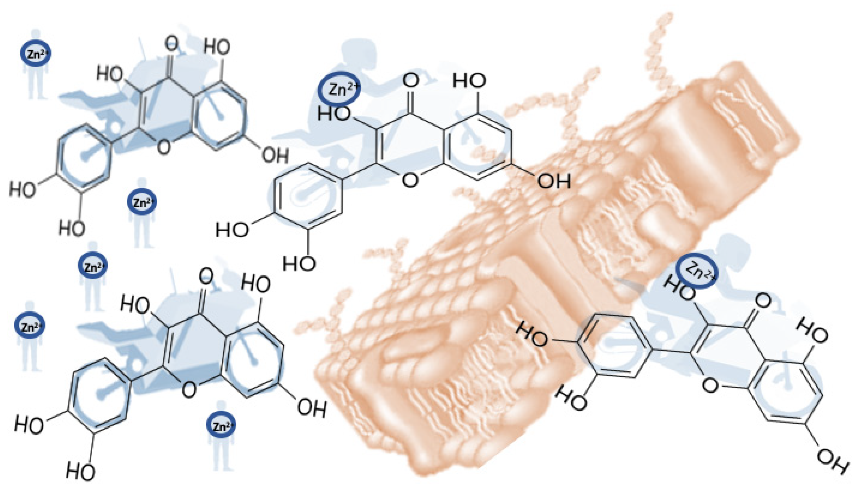
Ionophores are molecules that alter the permeability of biological membranes in response to certain metal ions for which they have an affinity and selectivity. Ionophores form a strong bond with a specific metal ion, shielding it from the effects of the surrounding environment. As a result, the metal ion is more easily transported into the hydrophobic core of the lipid membrane. Thus, ionophores function as a mobile carrier, transporting ions through the hydrophobic environment of cell membranes. We can illustrate the capacity of flavonoids, for example, as zinc ionophores, with a motorcyclist/motorbike interaction model. The biker would represent the zinc cation, which by itself cannot cross the membrane without the help of the motorbike, which, in this case, would be the flavonoid acting as an ionophore (Figure 12).

4. Materials and Methods

In this conformational energy study, the following steps were considered:

4.1. Known Zinc Ionophores as Models for the Study

The following compounds were chosen as known zinc ionophores able to modulate intracellular labile zinc concentrations [52]: pyrithione (PT) (an antimicrobial effective against several pathogens of the genera Streptococcus and Staphylococcus) [27], 5,7-Dichloro-2-((dimethylamino) methyl) quinolin-8-ol (PBT2), N,N,N′,N′-tetrakis(2-pyridinylmethyl)-1,2-ethanediamine (TPEN), 2-hydroxycyclohepta-2,4,6-trien-1-one (hinokitiol) and 5-Chloro-7-iodo-8-quinolinol (clioquinol).

4.2. Molecular Mechanics Calculations

Energy content of different molecules and zinc complexes with polyphenols and amino acids was calculated using ChemBio3D Ultra 16,0 (Perkin-Elmer Inc., Waltham, MA, USA) and Molecular Mechanics calculation method (MM2) optimization method. Calculations were performed to obtain the energy of different conformations of the same 3D-complex, analyzing and exposing those with different energy of formation. Different spatial conformations were estimated for each complex. The lower the energy, the higher the stability. Therefore, the lowest possible energy content was then selected and expressed in kcal/mol.
To calculate the potential energy of the molecule based on force fields, the MM2 method was used. This type of method considered the atoms as a ball containing the nucleus and the fused electrons as a solid sphere without considering any electronic energy term.
The total energy of a molecule was described by the sum of the following interactions:
E Total   =   E Stretching +   E Bending +   E Torsion +   E Non - bonded   inte
MM2 was chosen as a rapid method for determining the geometry, molecular energies, vibrational spectra and enthalpies of formation of stable molecules in their basal state. It was primarily used to determine geometries in large molecules, such as those of biological and pharmaceutical importance, which were currently beyond the reach of more intensive molecular orbital-based methods. For this reason, MM2 was not useful for modelling transition state of chemical processes that involved a large spectrum of experimental parameters, such as force field parameters.

4.3. Compounds under Theoretical Analysis

For theoretical considerations, we studied different zinc salts of organic acids, commonly used for dietary supplements already on the market, such as zinc gluconate, zinc citrate, zinc acetate and zinc lactate. The proteinogenic amino acids: phenylalanine, tryptophan, histidine and arginine; the peptide glutathione (GSH) and the flavonoids: silybin, hesperidin and xeractinol were also studied. To ascertain whether theoretical computation of the zinc complexes was compatible with their potential roles as zinc ionophores, we studied different three-dimensional structural conformations for each complex, considering those with the lowest possible energy and favorable geometry respect to the internal position of the zinc cation. This energy is understood to be derived exclusively from molecular mechanics. The type, strength and specificity of metal ion interaction with ionophores complexing groups should provide a good estimate of the range of lowest energy configurations of free ionophores that can easily complex with metals.

5. Conclusions

Our energetic and three-dimensional analysis of the zinc complexes with amino acids, glutathione and flavonoids studied was compatible with the considerations required for acting as potential zinc ionophores. The study of energy content, binding orientation of zinc cations and three-dimensional disposition of the molecules might be useful for initial theoretical studies of novel molecules to be considered as potential zinc ionophores. Traditional zinc ionophores, such as pyrithione, PBT2, TPEN and clioquinol, are associated with problems of cell toxicity [53,54,55], so their use is very limited or non-existent. Moreover, these molecules are expensive to synthesize, so the use of new ionophores that meet the conditions of safety and ease of obtaining and use are always welcome in the world of medicine. The energetic study suggests that polyphenol, glutathione and amino acids may form zinc complexes, likely acting as new ionophores. We propose that these novel complexes could be promising candidates for drug design to provide new solutions for conditions and diseases related to zinc deficiency or impairment derived from the dysregulation of this important metal. This preliminary study provides new opportunities to prevent zinc deficiency by using polyphenol nutraceutics, together with zinc supplementation. The regulation of intracellular zinc levels is fundamental to the survival of living organisms including humans. Zinc deficiency, particularly in developing countries, is still a widespread nutritional deficiency, due not only to a low intake of this essential trace metal, but also to poor bioavailability.
These chelators form complexes that are transported to the cells and, once at the inside or even at the outside, zinc complexes must dissociate into single compounds due to the low intracellular concentration of free and labile zinc, joining to proteins or peptides. This electrochemical potential depends on the concentration of ions on either side of the membrane, as well as the chemical potential which depends on the concentration of ionophores on either side of the membrane. Further experimental studies are necessary to conclude that the substances, such as amino acids, glutathione and polyphenols, that are harmless and even beneficial to human health, are really acting as zinc ionophores.
We are hoping to stimulate collaborative research interests from medicinal chemistry to develop new strategies and experiments to reinforce this idea.

Author Contributions

Conceptualization, C.A.-J. and E.P.-L.; investigation, C.A.-J. and E.P.-L.; writing—review and editing, C.A.-J.; J.M.P.d.l.L.; E.P.-L. and F.J.P.; supervision, C.A.-J. and J.M.P.d.l.L. All authors have read and agreed to the published version of the manuscript.

Funding

This research was funded by “Junta de Castilla y León”, projects FEDER-VA115P17 and VA149G18); by projects APOGEO (Cooperation Program INTERREG-MAC 2014–2020, with European Funds for Regional Development-FEDER, “Agencia Canaria de Investigación, Innovación y Sociedad de la Información (ACIISI) del Gobierno de Canarias” (project ProID2020010134), CajaCanarias (project 2019SP43); and Spanish Ministry of Economy and Competitiveness (Grant PID2019-105838RB-C31).

Institutional Review Board Statement

Not applicable.

Informed Consent Statement

Not applicable.

Data Availability Statement

Not applicable.

Conflicts of Interest

The authors declare no conflict of interest. The funders had no role in the design of the study; in the collection, analyses, or interpretation of data; in the writing of the manuscript, or in the decision to publish the results.

References

  1. Sanders, C.R.; Mittendorf, K.F. Tolerance to changes in membrane lipid composition as a selected trait of membrane proteins. Biochemistry 2011, 50, 7858–7867. [Google Scholar] [CrossRef] [Green Version]
  2. Müller, V.; Grüber, G. ATP synthases: Structure, function and evolution of unique energy converters. Cell. Mol. Life Sci. CMLS 2003, 60, 474–494. [Google Scholar] [CrossRef] [PubMed]
  3. Steinberg, R.A. A study of some factors influencing the stimulative action of zinc sulphate on the growth of aspergillus niger. I. The effect of the presence of zinc in the cultural flasks. Mem. Torrey Bot. Club 1918, 17, 287–293. [Google Scholar]
  4. Sandstead, H.H. Human Zinc Deficiency: Discovery to Initial Translation. Adv. Nutr. 2013, 4, 76–81. [Google Scholar] [CrossRef] [Green Version]
  5. Roohani, N.; Hurrell, R.; Kelishadi, R.; Schulin, R. Zinc and its importance for human health: An integrative review. J. Res. Med. Sci. 2013, 18, 144–157. [Google Scholar] [PubMed]
  6. Ackland, M.L.; Michalczyk, A. Zinc deficiency and its inherited disorders—A review. Genes Nutr. 2006, 1, 41–49. [Google Scholar] [CrossRef] [Green Version]
  7. Salgueiro, M.; Zubillaga, M.; Lysionek, A.; Sarabia, M.; Caro, R.; Paoli, T.; Hager, A.; Weill, R.; Boccio, J. Zinc as an essential micronutrient: A review. Nutr. Res. 2000, 20, 737–755. [Google Scholar] [CrossRef]
  8. Aggett, P.J.; Harries, J.T. Current status of zinc in health and disease states. Arch. Dis. Child. 1979, 54, 909–917. [Google Scholar] [CrossRef] [Green Version]
  9. Jeong, J.; Eide, D.J. The SLC39 family of zinc transporters. Mol. Asp. Med. 2013, 34, 612–619. [Google Scholar] [CrossRef] [Green Version]
  10. Huang, L.; Tepaamorndech, S. The SLC30 family of zinc transporters—A review of current understanding of their biological and pathophysiological roles. Mol. Asp. Med. 2013, 34, 548–560. [Google Scholar] [CrossRef]
  11. Kambe, T.; Hashimoto, A.; Fujimoto, S. Current understanding of ZIP and ZnT zinc transporters in human health and diseases. Cell. Mol. Life Sci. 2014, 71, 3281–3295. [Google Scholar] [CrossRef]
  12. Pressman, B.C. Biological Applications of Ionophores. Annu. Rev. Biochem. 1976, 45, 501–530. [Google Scholar] [CrossRef]
  13. Ding, W.-Q.; Lind, S.E. Metal ionophores—An emerging class of anticancer drugs. IUBMB Life 2009, 61, 1013–1018. [Google Scholar] [CrossRef]
  14. Maares, M.; Haase, H. A Guide to Human Zinc Absorption: General Overview and Recent Advances of In Vitro Intestinal Models. Nutrients 2020, 12, 762. [Google Scholar] [CrossRef] [PubMed] [Green Version]
  15. Swinkels, J.W.G.M.; Kornegay, E.T.; Verstegen, M.W.A. Biology of Zinc and Biological Value of Dietary Organic Zinc Complexes and Chelates. Nutr. Res. Rev. 2007, 7, 129–149. [Google Scholar] [CrossRef]
  16. Krebs, N.F. Overview of Zinc Absorption and Excretion in the Human Gastrointestinal Tract. J. Nutr. 2000, 130, 1374S–1377S. [Google Scholar] [CrossRef] [Green Version]
  17. Calvin, M.; Wilson, K.W. Stability of Chelate Compounds. J. Am. Chem. Soc. 1945, 67, 2003–2007. [Google Scholar] [CrossRef]
  18. Bjerrum, J. On the Tendency of the Metal Ions toward Complex Formation. Chem. Rev. 1950, 46, 381–401. [Google Scholar] [CrossRef] [PubMed]
  19. Ōsawa, E.; Musso, H. Molecular Mechanics Calculations in Organic Chemistry: Examples of the Usefulness of this Simple Non-Quantum Mechanical Model. Angew. Chem. Int. Ed. Engl. 1983, 22, 1–12. [Google Scholar] [CrossRef]
  20. Clark, T. Does quantum chemistry have a place in cheminformatics. In Molecular Informatics: Confronting Complexity. Proceedings of the Beilstein-Institut Workshop; Beilstein Institute: Bozen, Italy, 2002; pp. 193–208. [Google Scholar]
  21. Refat, M.S.; Hamza, R.Z.; A Adam, A.M.; Saad, H.A.; Gobouri, A.A.; Azab, E.; Al-Salmi, F.A.; Altalhi, T.A.; Khojah, E.; Gaber, A.; et al. Antioxidant, Antigenotoxic, and Hepatic Ameliorative Effects of Quercetin/Zinc Complex on Cadmium-Induced Hepatotoxicity and Alterations in Hepatic Tissue Structure. Coatings 2021, 11, 501. [Google Scholar] [CrossRef]
  22. Dabbagh-Bazarbachi, H.; Clergeaud, G.; Quesada, I.M.; Ortiz, M.; O’Sullivan, C.K.; Fernández-Larrea, J.B. Zinc ionophore activity of quercetin and epigallocatechin-gallate: From hepa 1-6 cells to a liposome model. J. Agric. Food Chem. 2014, 62, 8085–8093. [Google Scholar] [CrossRef] [PubMed]
  23. Karim, M.R.; Petering, D.H. Detection of Zn2+ release in nitric oxide treated cells and proteome: Dependence on fluorescent sensor and proteomic sulfhydryl groups. Metallomics 2016, 9, 391–401. [Google Scholar] [CrossRef] [Green Version]
  24. Tetko, I.V.; Solov’ev, V.P.; Antonov, A.V.; Yao, X.; Doucet, J.P.; Fan, B.; Hoonakker, F.; Fourches, D.; Jost, P.; Lachiche, N.; et al. Benchmarking of Linear and Nonlinear Approaches for Quantitative Structure−Property Relationship Studies of Metal Complexation with Ionophores. J. Chem. Inf. Modeling 2006, 46, 808–819. [Google Scholar] [CrossRef]
  25. Steinbrueck, A.; Sedgwick, A.C.; Brewster, J.T.; Yan, K.-C.; Shang, Y.; Knoll, D.M.; Vargas-Zúñiga, G.I.; He, X.-P.; Tian, H.; Sessler, J.L. Transition metal chelators, pro-chelators, and ionophores as small molecule cancer chemotherapeutic agents. Chem. Soc. Rev. 2020, 49, 3726–3747. [Google Scholar] [CrossRef]
  26. Krężel, A.; Maret, W. The biological inorganic chemistry of zinc ions. Arch. Biochem. Biophys. 2016, 611, 3–19. [Google Scholar] [CrossRef] [Green Version]
  27. Schwartz, J.R. Zinc pyrithione: A topical antimicrobial with complex pharmaceutics. J. Drugs Dermatol. 2016, 15, 140–144. [Google Scholar] [PubMed]
  28. Hussain, G.; Robinson, A.; Bartlett, P. Charge Generation in Low-Polarity Solvents: Poly(ionic liquid)-Functionalized Particles. Langmuir 2013, 29, 4204–4213. [Google Scholar] [CrossRef] [PubMed]
  29. Makrlík, E.; Novák, V.; Vanura, P.; Bour, P. Experimental and theoretical study on complexation of Li+ with lithium ionophore VIII. Mon. Fur Chem. 2013, 144, 1607–1611. [Google Scholar] [CrossRef]
  30. Kaur, K. Computer Simulation Studies on Metal-Ionophore Interactions. Master’s Thesis, Thapar University, Patiala, India, 2009. [Google Scholar]
  31. Santos, H.O.; Teixeira, F.J. Use of medicinal doses of zinc as a safe and efficient coadjutant in the treatment of male hypogonadism. Aging Male 2020, 23, 669–678. [Google Scholar] [CrossRef]
  32. Wang, Y.; Weisenhorn, E.; MacDiarmid, C.W.; Andreini, C.; Bucci, M.; Taggart, J.; Banci, L.; Russell, J.; Coon, J.J.; Eide, D.J. The cellular economy of the Saccharomyces cerevisiae zinc proteome. Metallomics 2018, 10, 1755–1776. [Google Scholar] [CrossRef]
  33. Singh, J.; Srivastav, A.N.; Singh, N.; Singh, A. Stability Constants of Metal Complexes in Solution. IntechOpen 2019. [Google Scholar] [CrossRef]
  34. Prasad, A.S.; Beck, F.W.J.; Snell, D.C.; Kucuk, O. Zinc in Cancer Prevention. Nutr. Cancer 2009, 61, 879–887. [Google Scholar] [CrossRef]
  35. Franklin, R.B.; Costello, L.C. The important role of the apoptotic effects of Zinc in the development of cancers. J. Cell. Biochem. 2009, 106, 750–757. [Google Scholar] [CrossRef] [PubMed] [Green Version]
  36. Bao, B.; Prasad, A.S.; Beck, F.W.J.; Snell, D.; Suneja, A.; Sarkar, F.H.; Doshi, N.; Fitzgerald, J.T.; Swerdlow, P. Zinc supplementation decreases oxidative stress, incidence of infection, and generation of inflammatory cytokines in sickle cell disease patients. Transl. Res. 2008, 152, 67–80. [Google Scholar] [CrossRef] [PubMed]
  37. Ishihara, K.; Yamanami, K.; Takano, M.; Suzumura, A.; Mita, Y.; Oka, T.; Juneja, L.R.; Yasumoto, K. Zinc Bioavailability Is Improved by the Micronised Dispersion of Zinc Oxide with the Addition of L-Histidine in Zinc-Deficient Rats. J. Nutr. Sci. Vitaminol. 2008, 54, 54–60. [Google Scholar] [CrossRef] [Green Version]
  38. Trzaskowski, B.; Adamowicz, L.; Deymier, P.A. A theoretical study of zinc(II) interactions with amino acid models and peptide fragments. JBIC J. Biol. Inorg. Chem. 2008, 13, 133–137. [Google Scholar] [CrossRef] [PubMed]
  39. Abendrot, M.; Chęcińska, L.; Kusz, J.; Lisowska, K.; Zawadzka, K.; Felczak, A.; Kalinowska-Lis, U. Zinc(II) Complexes with Amino Acids for Potential Use in Dermatology: Synthesis, Crystal Structures, and Antibacterial Activity. Molecules 2020, 25, 951. [Google Scholar] [CrossRef] [Green Version]
  40. Pavlica, S.; Gebhardt, R. Comparison of uptake and neuroprotective potential of seven zinc-salts. Neurochem. Int. 2010, 56, 84–93. [Google Scholar] [CrossRef]
  41. Williams, R.J.; Spencer, J.P.E.; Goni, F.M.; Rice-Evans, C.A. Zinc–histidine complex protects cultured cortical neurons against oxidative stress-induced damage. Neurosci. Lett. 2004, 371, 106–110. [Google Scholar] [CrossRef]
  42. Kurutas, E.B. The importance of antioxidants which play the role in cellular response against oxidative/nitrosative stress: Current state. Nutr. J. 2016, 15, 71. [Google Scholar] [CrossRef] [Green Version]
  43. Juan, C.A.; Pérez de la Lastra, J.M.; Plou, F.J.; Pérez-Lebeña, E. The Chemistry of Reactive Oxygen Species (ROS) Revisited: Outlining Their Role in Biological Macromolecules (DNA, Lipids and Proteins) and Induced Pathologies. Int. J. Mol. Sci. 2021, 22, 4642. [Google Scholar] [CrossRef]
  44. Nagao, A.; Seki, M.; Kobayashi, H. Inhibition of Xanthine Oxidase by Flavonoids. Biosci. Biotechnol. Biochem. 1999, 63, 1787–1790. [Google Scholar] [CrossRef]
  45. Maraldi, T. Natural Compounds as Modulators of NADPH Oxidases. Oxidative Med. Cell. Longev. 2013, 2013, 271602. [Google Scholar] [CrossRef] [Green Version]
  46. Pérez de la Lastra, J.M.; Andrés-Juan, C.; Plou, F.J.; Pérez-Lebeña, E. Impact of Zinc, Glutathione, and Polyphenols as Antioxidants in the Immune Response against SARS-CoV-2. Processes 2021, 9, 506. [Google Scholar] [CrossRef]
  47. Ruta, L.L.; Farcasanu, I.C. Interaction between polyphenolic antioxidants and Saccharomyces cerevisiae cells defective in heavy metal transport across the plasma membrane. Biomolecules 2020, 10, 1512. [Google Scholar] [CrossRef]
  48. Ikeda, N.E.A.; Novak, E.M.; Maria, D.A.; Velosa, A.S.; Pereira, R.M.S. Synthesis, characterization and biological evaluation of Rutin–zinc(II) flavonoid -metal complex. Chem. Biol. Interact. 2015, 239, 184–191. [Google Scholar] [CrossRef]
  49. Bădescu, L.; Ciocoiu, M.; Bădescu, C.; Bădescu, M. Chapter 7—Diabetic Neuropathy Modulation by Zinc and/or Polyphenol Administration. In Nutritional Modulators of Pain in the Aging Population; Watson, R.R., Zibadi, S., Eds.; Academic Press: Cambridge, MA, USA, 2017; pp. 87–100. [Google Scholar] [CrossRef]
  50. Arsenault, J.E.; Brown, K.H. Zinc intake of US preschool children exceeds new dietary reference intakes. Am. J. Clin. Nutr. 2003, 78, 1011–1017. [Google Scholar] [CrossRef] [PubMed] [Green Version]
  51. Kim, E.-Y.; Pai, T.-K.; Han, O. Effect of Bioactive Dietary Polyphenols on Zinc Transport across the Intestinal Caco-2 Cell Monolayers. J. Agric. Food Chem. 2011, 59, 3606–3612. [Google Scholar] [CrossRef] [Green Version]
  52. Xue, J.; Moyer, A.; Peng, B.; Wu, J.; Hannafon, B.N.; Ding, W.-Q. Chloroquine is a zinc ionophore. PLoS ONE 2014, 9, e109180. [Google Scholar] [CrossRef] [PubMed] [Green Version]
  53. Huntington Study Group Reach2HD Investigators. Safety, tolerability, and efficacy of PBT2 in Huntington’s disease: A phase 2, randomised, double-blind, placebo-controlled trial. Lancet Neurol. 2015, 14, 39–47. [Google Scholar] [CrossRef]
  54. Zhu, B.; Wang, J.; Zhou, F.; Liu, Y.; Lai, Y.; Wang, J.; Chen, X.; Chen, D.; Luo, L.; Hua, Z.C. Zinc Depletion by TPEN Induces Apoptosis in Human Acute Promyelocytic NB4 Cells. Cell. Physiol. Biochem. 2017, 42, 1822–1836. [Google Scholar] [CrossRef] [PubMed]
  55. Arbiser, J.L.; Kraeft, S.-K.; van Leeuwen, R.; Hurwitz, S.J.; Selig, M.; Dickersin, G.R.; Flint, A.; Byers, H.R.; Chen, L.B. Clioquinol-Zinc Chelate: A Candidate Causative Agent of Subacute Myelo-Optic Neuropathy. Mol. Med. 1998, 4, 665–670. [Google Scholar] [CrossRef] [PubMed] [Green Version]
Figure 1. Energy content of the two structures of free pyrithione (a). Binding sites and spatial orientation with zinc cations (b). Light grey balls represent carbon atoms. Blue balls represent nitrogen atoms. Non linked dark grey balls represent zinc cations. Red balls represent oxygen atoms. Yellow balls represent sulfur atoms.
Stresses 01 00011 g001
Figure 2. Structure (a) and three-dimensional study (b) of PBT2 (5,7-Dichloro-2-((dimethylamino) methyl) quinolin-8-ol). Light grey balls represent carbon atoms. Blue balls represent nitrogen atoms. Non linked dark grey balls represent zinc cations. Red balls represent oxygen atoms. Green balls represent chlorine atoms.
Stresses 01 00011 g002
Figure 3. Structure (a) and three-dimensional study (b) of TPEN (N,N,N′,N′-tetrakis (2-pyridinylmethyl)-1,2-ethanediamine). Light grey balls represent carbon atoms. Blue balls represent nitrogen atoms. Non linked dark grey balls represent zinc cations.
Stresses 01 00011 g003
Figure 4. Structure (a) and three-dimensional study (b) of Clioquinol, 5-Chloro-7-iodo-8-quinolinol. Light grey balls represent carbon atoms. Blue balls represent nitrogen atoms. Non linked dark grey balls represent zinc cations. Red balls represent oxygen atoms. Purple balls represent iodine atoms. Green balls represent chlorine atoms.
Stresses 01 00011 g004
Figure 5. Structure (a) and three-dimensional study (b) of Hinokitiol (2-hydroxycyclohepta-2,4,6-trien-1-one). Light grey balls represent carbon atoms. Non linked dark grey balls represent zinc cations. Red balls represent oxygen atoms.
Stresses 01 00011 g005
Figure 6. Energy and most favorable spatial orientation of zinc acetate, zinc lactate, zinc citrate and zinc gluconate. Light grey balls represent carbon atoms. Red balls represent oxygen atoms. Non-linked dark grey balls represent zinc cations.
Stresses 01 00011 g006
Figure 7. Energy content and most favorable spatial orientation of the dimers of proteinogenic amino acids (phenylalanine, tryptophan, histidine and arginine) computed by molecular mechanics. Light grey balls represent carbon atoms. Blue balls represent nitrogen atoms. Red balls represent oxygen atoms.
Stresses 01 00011 g007
Figure 8. Energy content and most favorable spatial orientation of zinc gluconate complexes with proteinogenic amino acids (phenylalanine, tryptophan, histidine and arginine). Light grey balls represent carbon atoms. Blue balls represent nitrogen atoms. Non linked dark grey balls represent zinc cations. Red balls represent oxygen atoms.
Stresses 01 00011 g008
Figure 9. Energy content and most favorable spatial orientation of free glutathione (a) and when complexed with zinc gluconate (b). Light grey balls represent carbon atoms. Blue balls represent nitrogen atoms. Non linked dark grey balls represent zinc cations. Red balls represent oxygen atoms and yellow balls represent sulfur atoms.
Stresses 01 00011 g009
Figure 10. Intramolecular hydrogen bonds between the oxygenated functions of the polyphenols silybin, hesperidin and xeractinol in combination with the zinc cation.
Stresses 01 00011 g010
Figure 11. Energy content and spatial orientation of polyphenols silybin, xeractinol and hesperidin both in free forms (a) or in complexes with zinc gluconate (b). Light grey balls represent carbon atoms. Red balls represent oxygen atoms. Non linked dark grey balls represent zinc cations.
Stresses 01 00011 g011aStresses 01 00011 g011b
Figure 12. Biker/motorcycle analogy model of flavonoids acting as zinc ionophores. The metal binding event permits the ionophore to traffic metal cations across the hydrophobic environment of cell membranes. In the bound state, the zinc cations (biker) and flavonoids (motorbikes) are sufficiently hydrophobic to permit passage across lipid membranes, where the zinc ions would not be soluble.
Stresses 01 00011 g012
Table 1. Molecular mechanics computed energy E of free forms of zinc ionophores and when forming complexes with zinc cations.
Zinc IonophoreFree FormComplexed with Zinc
Pyrithione2.2 kcal/mol−37.2 kcal/mol
PBT211.7.1 kcal/mol−110.5 kcal/mol
TPEN38.9 kcal/mol−126.6 kcal/mol
Clioquinol6.9 kcal/mol−78.4 kcal/mol
Hinokitiol18.1 kcal/mol−125.6 kcal/mol
Table 2. Molecular mechanics computed energy E of free forms of zinc salts of organic acids and when forming complexes with the amino acid tryptophan and the flavonoid silybin.
Zinc Salts of Organic AcidsFree FormComplexed with TryptophanComplexed with Silybin
Zinc gluconate−358.3 kcal/mol−457.3 kcal/mol−320.9 kcal/mol
Zinc citrate−277.4 kcal/mol−257.4 kcal/mol−185.8 kcal/mol
Zinc acetate−119.8 kcal/mol−419.6 kcal/mol−304 kcal/mol
Zinc lactate−188.9 kcal/mol−412.9 kcal/mol−307.3 kcal/mol
Table 3. Molecular mechanics computed energy E of free forms of natural compounds, such as amino acids, glutathione and polyphenols and when forming complexes with zinc gluconate.
CompoundFree FormComplexed with Zinc Gluconate
Phenylalanine−16.2 kcal/mol−414.1 kcal/mol
Tryptophan−6.1 kcal/mol−457.3 kcal/mol
Histidine7.2 kcal/mol−450.5 kcal/mol
Arginine−16.7 kcal/mol−403.2 kcal/mol
Glutathione−15.4 kcal/mol−641 kcal/mol
Silybin15.8 kcal/mol−320.9 kcal/mol
Hesperidin39 kcal/mol−309.8 kcal/mol
Xeractinol15.5 kcal/mol−324.2 kcal/mol
Publisher’s Note: MDPI stays neutral with regard to jurisdictional claims in published maps and institutional affiliations.

