Article

MicroCT as a Useful Tool for Analysing the 3D Structure of Lichens and Quantifying Internal Cephalodia in Lobaria pulmonaria

by
Julia Gerasimova
1,2,
Bernhard Ruthensteiner
3 and
Andreas Beck
1,2,4,*
1
Botanische Staatssammlung München, SNSB-BSM, D-80638 Munich, Germany
2
Systematsche Botanik und Mykologie, University of Munich, D-80638 Munich, Germany
3
Zoologische Staatssammlung München, SNSB-ZSM, D-81247 Munich, Germany
4
GeoBio-Center, University of Munich, D-80333 Munich, Germany
*
Author to whom correspondence should be addressed.
Submission received: 20 May 2021 / Revised: 5 July 2021 / Accepted: 6 July 2021 / Published: 13 July 2021

Abstract

:
High-resolution X-ray computer tomography (microCT) is a well-established technique to analyse three-dimensional microstructures in 3D non-destructive imaging. The non-destructive three-dimensional analysis of lichens is interesting for many reasons. The examination of hidden structural characteristics can, e.g., provide information on internal structural features (form and distribution of fungal-supporting tissue/hypha), gas-filled spaces within the thallus (important for gas exchange and, thus, physiological processes), or yield information on the symbiont composition within the lichen, e.g., the localisation and amount of additional cyanobacteria in cephalodia. Here, we present the possibilities and current limitations for applying conventional laboratory-based high-resolution X-ray computer tomography to analyse lichens. MicroCT allows the virtual 3D reconstruction of a sample from 2D X-ray projections and is helpful for the non-destructive analysis of structural characters or the symbiont composition of lichens. By means of a quantitative 3D image analysis, the volume of internal cephalodia is determined for Lobaria pulmonaria and the external cephalodia of Peltigera leucophlebia. Nevertheless, the need for higher-resolution tomography for more detailed studies is emphasised. Particular challenges are the large sizes of datasets to be analysed and the high variability of the lichen microstructures.

Graphical Abstract

1. Introduction

The availability of non-destructive 3D examination technologies and increases in personal computer hardware and software enables various 3D graphical applications, such as surface visualisations of biological specimens. This approach indirectly recalled an old morphological method back to life, namely the investigation of soft part anatomy by a light microscopical serial section analysis [1]. The correlative examination of a small biological specimen offers many advantages in analysing, visualising, and validating results. The topographical correlation of high-resolution areas to the morphology of the whole specimen provides significant benefits for understanding the overall organisation of the sample [2].
Lichens comprise a large morphological diversity and appear as one organism, but in fact, they represent a symbiosis of at least two primary partners, a fungus (mycobiont) and an alga (photobiont), and many additional microorganisms of still unknown function [3,4,5,6,7]. In most cases, the fungal partner forms the major part and outer side of the lichen thallus and is often judged to be the predominant symbiont [8]. It positions the photoautotrophic algal partner in such a way to ensure optimal illumination and an increasing water supply while still facilitating a gas exchange, especially in the highly developed foliose and fruticose lichens. The mycobiont produces lichen secondary substances to protect the photobiont against damage caused by high irradiation or UV light ([8,9,10,11] and references therein). The non-invasive determination of the internal morphology and anatomy of these fascinating life forms will help to understand their organisation and functioning better.
Species recognition has important implications for assessing the diversity and ecological and biogeographical patterns [12]. Based on phylogenetic and population studies, many cases have shown that numerous distinct lineages may be hidden under a single species name [12,13,14,15,16,17,18]. In these cases, microCT may be a beneficial tool, providing a complementation to other morphological examinations. It allows the visualisation and analysis of internal lichen microstructures, which could not be observed using routine light microscopy alone or can only be seen partially (with the need of preparing cross-sections).
In this proof of principle study, we intend to demonstrate the workflow for examining external and internal structures of lichens using datasets obtained from microCT. We used six samples of lichens from different genera and various growth forms: Bacidia rubella (Hoffm.) A. Massal., Evernia divaricata (L.) Ach., Hypogymnia physodes (L.) Nyl., Lobaria pulmonaria (L.) Hoffm., Peltigera leucophlebia (Nyl.) Gyeln., and Xanthoria parietina (L.) Th. Fr. The two lichens Lobaria and Peltigera have a green alga as the primary photobiont and incorporate nitrogen-fixing cyanobacteria into gall-like structures, the cephalodia. These are located either on the thallus surface (external cephalodia) or in its interior (internal cephalodia) [19]. Cephalodia have an essential ecological function: incorporated cyanobacteria fix atmospheric nitrogen, leading to a relatively high organic nitrogen content and, thus, enabling the colonisation of extremely oligotrophic habitats [9,20]. Consequently, these lichens contribute significant amounts of organic nitrogen to these ecosystems [21,22,23]. Lobaria pulmonaria is a tripartite lichen that contains the green alga Symbiochloris reticulata (Tschermak-Woess) Škaloud, Friedl, A. Beck & Dal Grande as the primary photobiont and Nostoc sp. in the internal cephalodia [24,25,26,27]. However, the structure and amount of these internal cephalodia are, so far, unknown. For this reason, we selected the internal and external cephalodia of L. pulmonaria and P. leucophlebia, respectively, for further analysis and quantification in this proof of principle study. The volumes of the segmented cephalodia of L. pulmonaria and P. leucophlebia relative to the size of the whole thallus were measured for the first time. Moreover, the shape and size of the internal cephalodia of L. pulmonaria were examined.

2. Materials and Methods

2.1. Taxon Sampling

We used six samples of lichens from different genera representing various growth forms: crustose, foliose, and fruticose (Table 1).

2.2. CT Acquisition

Lichen samples (Table 1) were stained with 1% iodine in 100% ethanol for approximately 24 h to enhance the contrast [28]. Dried lichen thalli were then directly glued with contact cement to a glass holder or put into a gelatin capsule glued to the glass holder. CT acquisition was performed with a Phoenix Nanotom M CT scanner (GE Measurement and Control, Wunstorf, Germany) using a voltage of 80 kV and a current of 325 mA while using a tungsten (“standard”) target. Projection images (1700–1800) were taken during a 360° rotation at a total duration ranging from 71 to 90 min. Voxel size ranged from 1.2 to 1.6 μm. The 16-bit dataset generated by reconstruction (with phoenix datos|x 2.2 software) was cropped and converted to 8 bit at a histogram adjustment prior to the other graphical processes.

2.3. Image Processing Using Amira Software

Data examination and analysis was performed using AMIRA® software v. 6.4 (Thermo Fisher Scientific, Hillsboro, OR, USA). It is a powerful, multifaceted software platform for visualising, manipulating, and understanding research data from many image modalities, including CT techniques [29]. The tools we applied are indicated in italics with a capital letter.

2.3.1. Visualisation of Lichen Structure Performed by Volume Rendering and Clipping Tools

We used OrthoSlice and Volume Rendering (Volren) modules to visualise the lichen image stack. OrthoSlices were combined with a specifically adapted Grayscale Colormap with standard grey values and a transparency function where the opacity can be set between 0 and 255. The adjustment of transparency allowed for the observation of internal lichen structures. The Volren module used for volume rendering provided a 3D visualisation of the whole sample dataset. The Volren module provided several options for adjustment: shaded and classical texture-based Volume Rendering (VRT), Maximum Intensity Projection (MIP), and Digitally Reconstructed Radiograph (DRR) (for more details, see reference [29]). To find the most appropriate one, we tested all the techniques and finally selected the option of VRT. Its virtual lighting effects enabled a better visualisation of fine details, which fit for our lichen objects best. The Specular lighting effect was enabled (Figure 1, Figure 2, Figure 3 and Figure 4) as the most suitable for better visualisation of the thalli surface.
We applied the Clipping tool to examine the internal structures of the lichen thalli. We could quickly explore the data images by screening through multiple orthographic sections, which provided us with easy access and observation of the internal parts of lichen thalli. After Clipping, the Volren image (3D) was complemented with OrthoSlices (2D) for a better visualisation and examination of the internal surfaces (Figure 3).

2.3.2. Segmentation

Segmentation (separation of parts of the lichen tissue) was the most time-consuming step. It was mainly performed following the procedures outlined by Ruthensteiner [1], including a description and explanation of the Segmentation tools. A further detailed description of the applied techniques can be found in reference [29].
As an initial step of Segmentation, we applied Thresholding to separate the object (lichen tissue) from the background (air) by classifying the voxels into the foreground and background voxels based on their intensities or voxel values [29].
Segmentation of the internal and external lichen structures was performed manually (subjective decision), since we could not acquire an automated workflow. When trying automatic tools, some parts of interest were not included, or vice versa; the false attributes were labelled as well. Thus, at manual Segmentation, approximately every 10th section was labelled and followed by Interpolation to facilitate the segmentation process by obtaining relatively smooth material surfaces right away [1]. When necessary, we carried out additional correction steps by labelling missed parts followed by an Interpolation step until the complete labelling of the selected structure was achieved.

2.3.3. Measurements of Segmented Structures Performed by Label Analysis

After Segmentation, we applied Label Analysis to measure the volume of the whole lichen sample and the segmented cephalodia. Label Analysis computes a group of measurements on each component (Material) of the input label field dataset and calculates the resulting values [29], such as Volume 3D: Volume of the component (mm3). After Label Analysis, we calculated the cephalodia volume relative to the whole thalli volume.

3. Results

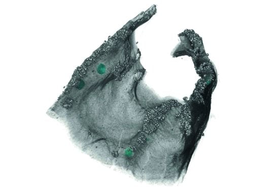
3.1. Morphological Observation

The microCT volumes allowed the detailed observation and examination of both the external and internal components of lichen thalli. The majority of lichens, including many crustose species, develop internally stratified thalli: upper cortex, photobiont layer, medulla, and lower cortex [19]. MicroCT resolved the overall structure of all the lichens investigated in this study (Figure 1A,C,E, Figure 2A,C,E and Figure 3). Figure 3 shows virtual sections of the stratified foliose thalli of Xanthoria parietina and Hypogymnia physodes differentiated in the upper cortex, medulla, and lower cortex. For Xanthoria parietina, the structure of the fruiting bodies (apothecia) is discernible in Figure 1A,B and Figure 3A. In Hypogymnia physodes, the typical hollow part between the medulla and lower cortex is prominent (Figure 1C,D and Figure 3B). Virtual sections through the foliose Lobaria pulmonaria (Figure 1E,F and Figure 3C) and Peltigera leucophlebia (Figure 2A,B and Figure 3D) reveal that the lower surfaces of these species are covered by hyphae forming a dense, felt-like, woolly mat, the tomentum. A cross-section through the thallus of the fruticose Evernia divaricata shows the characteristic loosely woven medulla (Figure 2C,D and Figure 3F). Figure 2E,F and Figure 3F illustrate a section through the thallus and apothecium of the crustose lichen Bacidia rubella.

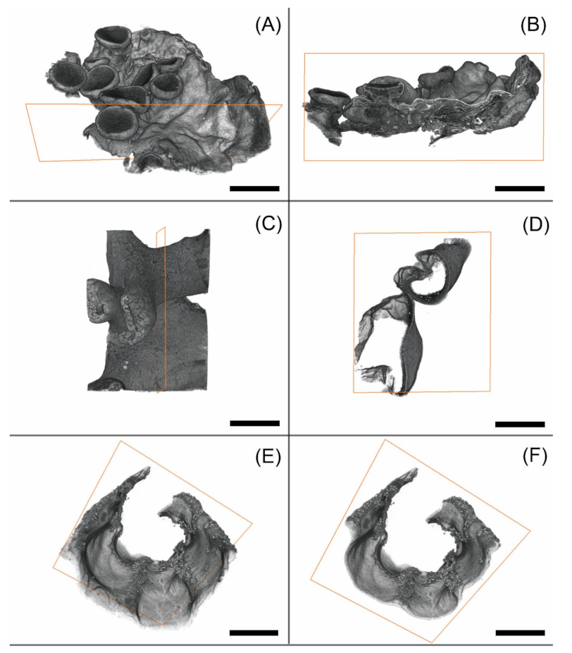
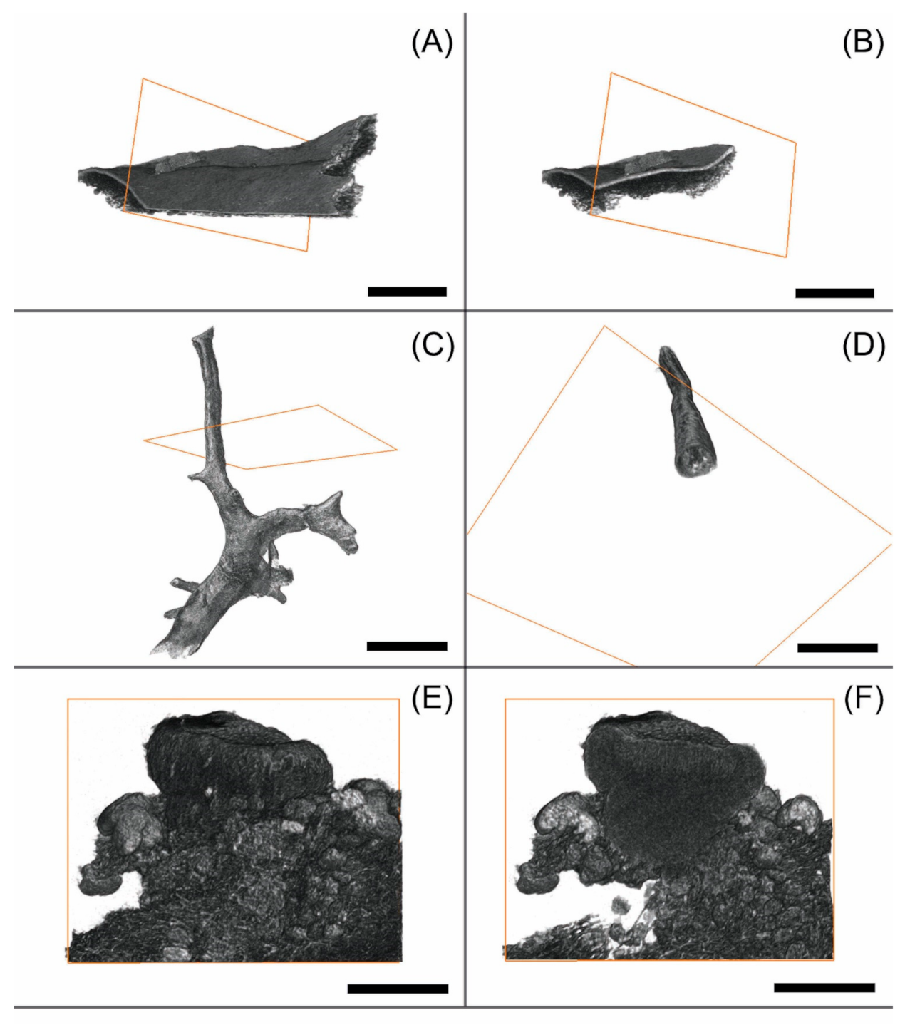
3.2. Observation of Microstructures

We examined the shape and number of external and internal cephalodia in Peltigera leucophlebia and Lobaria pulmonaria, respectively. Figure 4A depicts the characteristic appressed, convex, wart-like shaped cephalodia of P. leucophlebia with diameters 80–100 × 90–210 µm. We examined ± globose-shaped internal cephalodia in L. pulmonaria, with diameters 50–250 × 60–280 µm (Figure 4B–D).

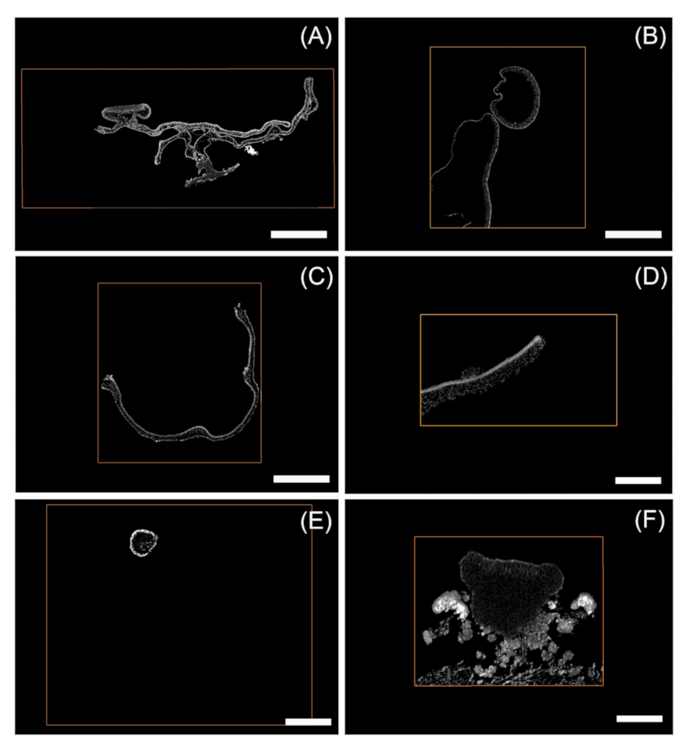
3.3. Segmentation Results and Label Analysis

After the Segmentation process, we applied Label Analysis to assess the volume of the thalli samples and segmented cephalodia. Subsequently, we calculated the volume of all cephalodia relative to the whole thalli volume.
The L. pulmonaria sample had a total volume of 2.091 mm3 (Table 2) and nine cephalodia of volumes ranging from 0.000046 to 0.004525 mm3. The total cephalodia volume of 0.153 mm3 corresponds to 0.73% of the whole thallus volume.
The size of the P. leucophlebia sample had a total volume of 0.200 mm3 (Table 3) and four cephalodia of volumes from 0.000201 to 0.000873 mm3. The total cephalodia volume of 0.0019 mm3 corresponds to 0.97% of the whole thallus volume.

4. Discussion

4.1. Pros and Cons in the Visualisation of Lichens Using MicroCT

Previous studies with microCT applications mainly focused on animal or biomedical data, e.g., references [30,31,32,33,34]. For lichens, examination using X-ray tomography was done very rarely—namely, to estimate the relative mass contribution of lichen symbionts in Stereocaulon sorediiferum Hue [35]. MicroCT analysis is a beneficial tool in detailed morphological investigations. For visual examinations, a variety of settings provided by Amira software allow changing or tweaking the appearance of different components of lichen thalli. MicroCT can also be combined with a light microscopic technique [31], complementing the examination of morphological characteristics.
Most importantly, microCT imaging allows the non-destructive exploration of both the external and the internal structures of lichen thalli simultaneously. To this end, we used Volume Rendering (Volren) by applying the Grey Colormap, VRT, and Specular lighting effects. Moreover, the adjustment of transparency (Figure 4C,D), selective imaging of individual components using Segmentation (Figure 4), or clipping off parts of surfaces (Figure 1, Figure 2 and Figure 3) help to explore the internal lichen structures.
Consequently, a microCT analysis improved the understanding of the structural and functional features of lichens. Separation of the main morphological components depicted in Figure 1, Figure 2 and Figure 3 (viz., cortex, medulla, and cephalodia) was possible but only related to the fungal component. Thus, the resolution was insufficient to visualise the algal layer with the current setup and depict the algal cells, which appeared as a continuum with the upper cortex. Other approaches, such as staining and embedding (in resin) or applying a higher resolution to smaller thallus areas at microCT acquisition need to be tested for a more detailed examination of the algal cells. These techniques may lead to an enhanced resolution of the microstructure.
Some lichens are very hardy organisms and can even survive conditions in outer space [36]. It is therefore likely that they can withstand the X-ray treatment in the CT instrument and resume growing afterwards. If this proves right, it will pave the way for developmental studies of 3D visualisation of one and the same lichen thallus at different points in time. Lichen developmental studies would greatly benefit from such investigations.
The microCT technique may also advance the knowledge on trapped particles or lichen secondary metabolites—often deposited in crystals—in the thallus. Given the voxel size of 1.25–1.65, particles/crystals larger than 4 µm (2.5× voxel size) can routinely be detected. The MicroCT analysis of these components will be useful for ecological studies investigating the effect of the lichen secondary metabolites, e.g., in herbivory, or the amount of substrate erosion by particle uptake.

4.2. Segmentation and Statistical Analysis of External and Internal Cephalodia

Most cephalodia are morphologically simple globose, sacculate, lobate, or coralloid structures that differ from the rest of the vegetative thallus by their photosynthetic partner and, thus, coloration [19]. The number of cephalodia is ecologically important, because the incorporated cyanobacteria bind atmospheric nitrogen, resulting in a comparatively high organic nitrogen content of the lichen thallus and, in consequence, contribute significant amounts of organic nitrogen to forests and, especially, to oligotrophic ecosystems [21,22,23]. Cornejo and Scheidegger [25] studied the ontogeny of cephalodia structures in L. pulmonaria, indicating the active incorporation of the cyanobacteria into the lichen thallus. The application of the Segmentation tool allowed us to examine formerly hardly accessible internal cephalodia. Thus, using a microCT analysis, we depicted the form of the internal cephalodia of L. pulmonaria for the first time. Our study could clearly demonstrate that L. pulmonaria is characterised by having ± globose-shaped internal cephalodia (Figure 4B–D). Moreover, we surveyed the external cephalodia of P. leucophlebia as convex and oblong with narrowed ends (so-called peltate cephalodia), which corresponds to previous studies [37].
To further demonstrate the power of the microCT analysis in lichenology, we disclosed the number and distribution of cephalodia within/on the surface of the lichen sample. This was followed by measuring the volume of all cephalodia and their relation to the whole lichen sample (see above). We could clearly demonstrate that the internal cephalodia of L. pulmonaria have a volume ranging between 0.046 and 4.5 × 10⁻³ mm3. In comparison, volumes of the external P. leucophlebia cephalodia are smaller and range between 0.2 and 0.87 × 10⁻³ mm3. These data considerably complement our knowledge of the morphological and quantitative characteristics of these lichens.
So far, we have performed a manual Segmentation of cephalodia—the most time-consuming step in whole-image processing. Future studies may include automatised (machine learning-based) approaches, which may significantly improve the workflow in terms of efficiency and accuracy. This will allow for the analysis of larger quantities of lichen thalli, facilitating the comparison of lichens grown under different ecological regimes and allowing to investigate the algal layer.

Author Contributions

Conceptualisation, A.B.; formal analysis, J.G.; funding acquisition, A.B.; investigation, J.G.; methodology, B.R. and A.B.; project administration, A.B.; software, B.R.; supervision, B.R. and A.B.; validation, J.G., B.R. and A.B.; writing—original draft, J.G. and A.B.; and writing—review and editing, J.G., B.R. and A.B. All authors have read and agreed to the published version of the manuscript.

Funding

This research was funded by the Staatliche Naturwissenschaftliche Sammlungen Bayerns, grant SNSBinnovativ to A.B.

Institutional Review Board Statement

Not applicable.

Informed Consent Statement

Not applicable.

Data Availability Statement

Not applicable.

Acknowledgments

We thank the government of the administrative district of Swabia for the sampling permit. J.G. was supported by a BAYHOST fellowship from the Bayerische Staatsministerium für Bildung und Kultus, Wissenschaft und Kunst.

Conflicts of Interest

The authors declare no conflict of interest.

Ethical Statements

The lichens used in this study were collected in Bavaria, Germany. Bacidia rubella, Evernia divaricata, Hypogymnia physodes, Lobaria pulmonaria, and Peltigera leucophlebia were collected under permit 55.3-8645-2/35 of the government of the administrative district of Swabia. Xanthoria parietina was collected in Upper Bavaria outside protected areas, and thus, no permit was required. Voucher specimens were deposited in the Botanische Staatssammlung München under the herbarium numbers given in the Materials and Methods section.

References

  1. Ruthensteiner, B. Soft Part 3D visualisation by serial sectioning and computer reconstruction. Zoosymposia 2008, 1, 61–100. [Google Scholar] [CrossRef]
  2. Handschuh, S.; Baeumler, N.; Schwaha, T.; Ruthensteiner, B. A correlative approach for combining microCT, light and transmission electron microscopy in a single 3D scenario. Front. Zool. 2013, 10, 44. [Google Scholar] [CrossRef] [PubMed] [Green Version]
  3. Farrar, J.F. The lichen as an ecosystem: Observation and experiment. In Lichenology: Progress and Problems; Brown, D.H., Hawksworth, D.L., Bailey, R.H., Eds.; Academic Press: London, UK, 1976; pp. 385–406. [Google Scholar]
  4. Werth, S. How does the lichen symbiosis work? Journey into the unknown. Fritschiana 2017, 85, 48–49. [Google Scholar]
  5. Mark, K.; Laanisto, L.; Bueno, C.G.; Niinemets, Ü.; Keller, C.; Scheidegger, C. Contrasting co-occurrence patterns of photobiont and cystobasidiomycete yeast associated with common epiphytic lichen species. New Phytol. 2020, 227, 1362–1375. [Google Scholar] [CrossRef]
  6. Tzovaras, B.G.; Segers, F.H.I.D.; Bicker, A.; Dal Grande, F.; Otte, J.; Anvar, S.Y.; Hankeln, T.; Schmitt, I.; Ebersberger, I. What is in Umbilicaria pustulata? A metagenomic approach to reconstruct the holo-genome of a lichen. Genome Biol. Evol. 2020, 12, 309–324. [Google Scholar] [CrossRef] [PubMed]
  7. Grube, M. Lichens growing greenhouses en miniature. Microbial Cell 2021, 8, 65–68. [Google Scholar] [CrossRef] [PubMed]
  8. Honegger, R. The Symbiotic Phenotype of Lichen-Forming Ascomycetes and Their Endo- and Epibionts. In The Mycota IX—Fungal Associations; Hock, B., Ed.; Springer: Berlin/Heidelberg, Germany, 2012; pp. 287–339. [Google Scholar]
  9. Büdel, B.; Scheidegger, C. Thallus morphology and anatomy. In Lichen Biology; Nash, T.H., III, Ed.; Cambridge University Press: Cambridge, UK, 1996; pp. 24–36. [Google Scholar]
  10. Eisenreich, W.; Knispel, N.; Beck, A. Advanced methods for the study of the chemistry and the metabolism of lichens. Phytochem. Rev. 2011, 10, 445–456. [Google Scholar] [CrossRef]
  11. Kuhn, V.; Geisberger, T.; Huber, C.; Beck, A.; Eisenreich, W. A facile in vivo procedure to analyse metabolic pathways in intact lichens. New Phytol. 2019, 224, 1657–1667. [Google Scholar] [CrossRef]
  12. Singh, G.; Grande, F.D.; Divakar, P.K.; Otte, J.; Leavitt, S.D.; Szczepańska, K.; Crespo, A.; Rico, V.J.; Aptroot, A.; Cáceres, M.E.D.S.; et al. Coalescent–based species delimitation approach uncovers high cryptic diversity in the cosmopolitan lichen–forming fungal genus Protoparmelia (Lecanorales, Ascomycota). PLoS ONE 2015, 10, e0124625. [Google Scholar] [CrossRef]
  13. Altermann, S.; Leavitt, S.D.; Goward, T.; Nelsen, M.P.; Lumbsch, H.T. How do you solve a problem like Letharia? A new look at cryptic species in lichen-forming fungi using Bayesian clustering and SNPs from multilocus sequence data. PLoS ONE 2014, 9, e97556. [Google Scholar] [CrossRef]
  14. Kraichak, E.; Lücking, R.; Aptroot, A.; Beck, A.; Dornes, P.; John, V.; Lendemer, J.C.; Nelsen, M.P.; Neuwirth, G.; Nutakki, A.; et al. Hidden diversity in the morphologically variable script lichen (Graphis scripta) complex (Ascomycota, Ostropales, Graphidaceae). Org. Divers. Evol. 2015, 15, 447–458. [Google Scholar] [CrossRef]
  15. Frolov, I.; Vondrák, J.; Fernández-Mendoza, F.; Wilk, K.; Khodosovtsev, A.; Halici, H.G. Three new, seemingly–cryptic species in the lichen genus Caloplaca (Teloschistaceae) distinguished in two–phase phenotype evaluation. Ann. Bot. Fenn. 2016, 53, 243–262. [Google Scholar] [CrossRef] [Green Version]
  16. Leavitt, S.D.; Esslinger, T.L.; Divakar, P.K.; Crespo, A.; Lumbsch, H.T. Hidden diversity before our eyes: Delimiting and describing cryptic lichen-forming fungal species in camouflage lichens (Parmeliaceae, Ascomycota). Fungal Biol. 2016, 120, 1374–1391. [Google Scholar] [CrossRef] [Green Version]
  17. Boluda, C.G.; Rico, V.J.; Divakar, P.K.; Nadyeina, O.; Myllys, L.; McMullin, R.T.; Zamora, J.C.; Scheidegger, C.; Hawksworth, D.L. Evaluating methodologies for species delimitation: The mismatch between phenotypes and genotypes in lichenised fungi (Bryoria sect. Implexae, Parmeliaceae). Persoonia 2019, 42, 75–100. [Google Scholar] [CrossRef] [Green Version]
  18. Del-Prado, R.; Buaruang, K.; Lumbsch, H.T.; Crespo, A.; Divakar, P.K. DNA sequence-based identification and barcoding of a morphologically highly plastic lichen forming fungal genus (Parmotrema, Parmeliaceae) from the tropics. Bryologist 2019, 122, 281–291. [Google Scholar] [CrossRef]
  19. Nash, T.H. (Ed.) Lichen Biology, 2nd ed.; Cambridge University Press: Cambridge, UK, 2008. [Google Scholar]
  20. Lawrey, J.D. Biological role of lichen substances. Bryologist 1986, 89, 111–122. [Google Scholar] [CrossRef]
  21. Knops, J.M.H.; Nash, T.H., III; Boucher, V.L.; Schlesinger, W.H. Mineral cycling and epiphytic lichens: Implications at the ecosystem level. Lichenologist 1991, 23, 309–321. [Google Scholar] [CrossRef]
  22. Knowles, R.D.; Pastor, J.; Biesboer, D.D. Increased soil nitrogen associated with dinitrogen-fixing, terricolous lichens of the genus Peltigera in northern Minnesota. Oikos 2006, 114, 37–48. [Google Scholar] [CrossRef]
  23. Asplund, J.; Wardle, D.A. How lichens impact on terrestrial community and ecosystem properties. Biol. Rev. 2017, 92, 1720–1738. [Google Scholar] [CrossRef] [PubMed]
  24. Rose, F.; Purvis, O.W. Lobaria (Schreb.) Hoffm. 1796. In Lichens of Great Britain and Ireland; Smith, C.W., Aptroot, A., Coppins, B.J., Fletcher, A., Gilbert, O.L., James, P.W., Wolseley, P.A., Eds.; MPG Books Group: London, UK, 2009; pp. 560–562. [Google Scholar]
  25. Cornejo, C.; Scheidegger, C. New morphological aspects of cephalodium formation in the lichen Lobaria pulmonaria (Lecanorales, Ascomycota). Lichenologist 2013, 45, 77–87. [Google Scholar] [CrossRef] [Green Version]
  26. Škaloud, P.; Friedl, T.; Hallmann, C.; Beck, A.; Dal Grande, F. Taxonomic revision and species delimitation of coccoid green algae currently assigned to the genus Dictyochloropsis (Trebouxiophyceae, Chlorophyta). J. Phycol. 2016, 52, 599–617. [Google Scholar] [CrossRef]
  27. Grimm, M.; Grube, M.; Schiefelbein, U.; Zühlke, D.; Bernhardt, J.; Riedel, K. The Lichens’ Microbiota, Still a Mystery? Front. Microbiol. 2021, 12, 623839. [Google Scholar] [CrossRef] [PubMed]
  28. Metscher, B.D. MicroCT for developmental biology: A versatile tool for high-contrast 3D imaging at histological resolutions. Dev. Dyn. 2009, 238, 632–640. [Google Scholar] [CrossRef]
  29. Thermo Scientific. User’s Guide Amira Software 2019. Available online: https://assets.thermofisher.com/TFS-Assets/MSD/Product-Guides/users-guide-amira-software-2019.pdf (accessed on 9 March 2021).
  30. Handschuh, S.; Beisser, C.J.; Ruthensteiner, B.; Metscher, B.D. Microscopic dual-energy CT (microDECT): A flexible tool for multichannel ex vivo 3D imaging of biological specimens. J. Microsc. 2017, 67, 3–26. [Google Scholar] [CrossRef] [PubMed]
  31. Karreman, M.A.; Ruthensteiner, B.; Mercier, L.; Schieber, N.L.; Solecki, G.; Winkler, F.; Goetz, J.G.; Schwab, Y. Find your way with X-Ray: Using microCT to correlate in vivo imaging with 3D electron microscopy. Methods Cell Biol. 2017, 140, 277–301. [Google Scholar] [PubMed]
  32. Jacob, D.E.; Ruthensteiner, B.; Trimby, P.; Henry, H.; Martha, S.O.; Leitner, J.; Otter, L.M.; Scholz, J. Architecture of Anoteropora latirostris (Bryozoa, Cheilostomata) and implications for their biomineralisation. Sci. Rep. 2019, 9, 11439. [Google Scholar] [CrossRef]
  33. Schwaha, T.; Ruthensteiner, B.; Melzer, R.R.; Asami, T.; Páll-Gergely, B. Three phyla—Two type specimens—One shell: History of a snail shell revealed by modern imaging technology. J. Zool. Syst. Evol. Res. 2019, 57, 527–533. [Google Scholar] [CrossRef]
  34. Orhan, K. (Ed.) Micro-Computed Tomography (micro-CT) in Medicine and Engineering; Springer: Berlin/Heidelberg, Germany, 2020. [Google Scholar]
  35. Kosugi, M.; Shizuma, R.; Moriyama, Y.; Koike, H.; Fukunaga, Y.; Takeuchi, A.; Uesugi, K.; Suzuki, Y.; Imura, S.; Kudoh, S.; et al. Ideal osmotic spaces for chlorobionts or cyanobionts are differentially realized by lichenized fungi. Plant Physiol. 2014, 166, 337–348. [Google Scholar] [CrossRef] [Green Version]
  36. Brandt, A.; de Vera, J.P.; Onofri, S.; Ott, S. Viability of the lichen Xanthoria elegans and its symbionts after 18 months of space exposure and simulated mars conditions on the ISS. Int. J. Astrobiol. 2015, 14, 411–425. [Google Scholar] [CrossRef] [Green Version]
  37. Goward, T.; Goffinet, B.; Vitikainen, O. Synopsis of the genus Peltigera (lichenized Ascomycetes) in British Columbia, with a key to the North American species. Canad. J. Bot. 1995, 73, 91–111. [Google Scholar] [CrossRef]
Figure 1. MicroCT renderings of lichen samples showing an overall view (A,C,E) and stratified thallus in a cross-section through the thallus (B,D,F) and apothecia (B), as provided by the Clipping tool. The orange rectangle indicates the cutting plane. (A,B) Xanthoria parietina (M-0304310), (C,D) Hypogymnia physodes (M-0304295), and (E,F) Lobaria pulmonaria (M-0304308). Scale: 1 mm (A,B,E,F) and 0.25 mm (C,D).
Applmicrobiol 01 00015 g001
Figure 2. MicroCT renderings of lichen samples showing an overall view (A,C,E) and thalli in a cross-section (B,D,F) and apothecia (F), as provided by the Clipping tool. The orange rectangle indicates the cutting plane. (A,B) Peltigera leucophlebia (M-0304309), (C,D) Evernia divaricata (M-0304299), and (E,F) Bacidia rubella (M-0304302). Scale: 0.5 mm (A,B), 1 mm (C,D), and 0.25 mm (E,F).
Applmicrobiol 01 00015 g002
Figure 3. 2D OrthoSlice image sections, providing a view from the front (xy) of the cutting plane. (A) Xanthoria parietina, (B) Hypogymnia physodes, (C) Lobaria pulmonaria, (D) Peltigera leucophlebia, (E) Evernia divaricata, and (F) Bacidia rubella. Scale: 1 mm (A,C), 0.5 mm (E), and 0.25 mm (B,D,F).
Applmicrobiol 01 00015 g003
Figure 4. Positions of cephalodia in the lichen thalli after identification using the Segmentation tool. (A) Peltigera leucophlebia (overview, cephalodia coloured). (B) Lobaria pulmonaria (overview, cephalodia coloured). (C) L. pulmonaria (close-up, cephalodia uncoloured). (D) L. pulmonaria (close-up, cephalodia coloured). Internal surfaces are shown in a transparent mode and highlighted in cyan blue in A, B and D. Scale 1 mm (A), 2 mm (B), and 0.5 mm (C,D).
Applmicrobiol 01 00015 g004
Table 1. Lichen samples used in this study with CT acquisition parameters and dataset details.
NameHerbarium NumberGrowth FormVoxel Size (µm)X, Y, and Z Voxel Dimensions of 8-Bit DatasetsNumber of ProjectionsDuration of CT Acquisition
Bacidia rubellaM-0304302crustose1.249922085 × 1425 × 2262180076 min
Evernia divaricataM-0304299fruticose1.406181107 × 1332 × 2193170071 min
Hypogymnia physodesM-0304295foliose1.295942931 × 2875 × 2234180090 min
Xanthoria parietinaM-0304310foliose1.653751104 × 2451 × 2298180076 min
Lobaria pulmonariaM-0304308foliose1.606582080 × 2290 × 2200180076 min
Peltigera leucophlebiaM-0304309foliose1.610111901 × 2439 × 1820180076 min
Table 2. Label Analysis of the cephalodia of Lobaria pulmonaria. Volume 3D: Volume of the object (10−3 mm3).
MaterialVolume 3D (10−3 mm3)
Whole sample (thallus)2091
Cephalodium 14.525
Cephalodium 23.527
Cephalodium 33.202
Cephalodium 42.913
Cephalodium 50.722
Cephalodium 60.249
Cephalodium 70.059
Cephalodium 80.054
Cephalodium 90.046
Sum cephalodia15.30 (0.73%)
Table 3. Label Analysis of the cephalodia of Peltigera leucophlebia. Volume 3D: Volume of the object (10−3 mm3).
MaterialVolume 3D (10−3 mm3)
Whole sample (thallus)200
Cephalodium 10.873
Cephalodium 20.507
Cephalodium 30.366
Cephalodium 40.201
Sum cephalodia1.947 (0.97%)
Publisher’s Note: MDPI stays neutral with regard to jurisdictional claims in published maps and institutional affiliations.

