Article

Effect of Oxygen Partial Pressure on Crystal Structure, Oxygen Vacancy, and Surface Morphology of Epitaxial SrTiO3 Thin Films Grown by Ion Beam Sputter Deposition

by
Gasidit Panomsuwan
1,* and
Nagahiro Saito
2
1
Department of Materials Engineering, Faculty of Engineering, Kasetsart University, Bangkok 10900, Thailand
2
Department of Chemical Systems Engineering, Graduate School of Engineering, Nagoya University, Nagoya 464-8603, Japan
*
Author to whom correspondence should be addressed.
Submission received: 1 June 2021 / Revised: 3 August 2021 / Accepted: 4 August 2021 / Published: 2 September 2021

Abstract

:
Epitaxial SrTiO3 (STO) thin films were grown on (001)-oriented LaAlO3 (LAO) substrates at 800 °C by an ion beam sputter deposition (IBSD). Oxygen partial pressure (PO2) was varied at 1.5 × 10⁻⁵, 1.5 × 10⁻⁴, and 1.5 × 10⁻³ Torr during the growth. The effects of PO2 on crystal structure, oxygen vacancy, and surface morphology of the STO films were investigated and are discussed to understand their correlation. It was found that PO2 played a significant role in influencing the crystal structure, oxygen vacancy, and surface morphology of the STO films. All STO films grew on the LAO substrates under a compressive strain along an in-plane direction (a- and b-axes) and a tensile strain along the growth direction (c-axis). The crystalline quality of STO films was slightly improved at higher PO2. Oxygen vacancy was favorably created in the STO lattice grown at low PO2 due to a lack of oxygen during growth and became suppressed at high PO2. The existence of oxygen vacancy could result in a lattice expansion in both out-of-plane and in-plane directions due to the presence of Ti3+ instead of Ti4+ ions. The surface roughness of the STO films gradually decreased and was nearly close to that of the bare LAO substrate at high PO2, indicating a two-dimensional (2D) growth mode. The results presented in this work provide a correlation among crystal structure, oxygen vacancy, and surface morphology of the epitaxial STO films grown by IBSD, which form a useful guideline for further study.

1. Introduction

Epitaxial strontium titanate (SrTiO3, STO) thin films have drawn considerable attention as an essential component in various electronic devices, including capacitors [1], tunable phase shifters [2], sensors [3], non-volatile memory [4], memristors [5], and electromechanical actuators [6], owing to their high dielectric constant, low loss, large dielectric tunability, and tunable optical properties. So far, epitaxial STO films have been successfully grown on various types of substrates by a variety of methods, such as pulsed laser deposition (PLD) [7,8], molecular beam epitaxy (MBE) [9,10], metal-oxide chemical vapor deposition (MOCVD) [11,12], radio-frequency (RF) magnetron sputtering [13,14], and ion beam sputter deposition (IBSD) [15,16,17]. All methods have their own advantages and disadvantages, so the selection of a growth method depends on which aspect is focused on. For example, for film quality, MBE is the most popular method for growing epitaxial films. Several studies have reported on the growth of homoepitaxial and heteroepitaxial STO films, which all showed high crystalline quality with precise control in atomic or unit-cell levels [9,10,18]. However, the small growing area and high cost are significant disadvantages of MBE that limit to the laboratory, and it is difficult to expand MBE to conventional manufacturing processes. For the aspect of the manufacturing process, the IBSD is a viable alternative method to the conventional RF sputtering since it offers several advantages, such as independent control of growth parameters, isolated plasma chamber, minimizing plasma effect to the substrate, and low operating pressure [19,20,21]. Although growing films may be possibly exposed to high-energetic sputtered atoms or ions during growth, this effect can be solved by rationally optimizing growth conditions. According to the literature to date, information on the growth of STO films by IBSD is very limited compared to other methods.
In our previous works, we studied the effects of growth temperature and growth time on the crystal structure and morphology of the epitaxial STO films grown by IBSD [16,17]. It was found that the growth temperature significantly influenced the crystal structure and growth mode. With an increase in growth temperature, the STO lattice shrank, and the surface morphology was transformed from three-dimensional (3D) to two-dimensional (2D) growth modes [16]. For the effect of growth time, STO films became thicker with a strain relaxation at a longer growth time [17]. However, there has not yet been a detailed investigation on the effect of partial oxygen pressure (PO2) on the properties of STO films grown by IBSD. PO2 is known as a critical growth parameter used to control the level of oxygen vacancy densities in oxide films [22,23,24,25]. The level of non-stoichiometry or oxygen vacancy density in STO films plays a vital role in tuning their conductivity [22,23], dielectricity [26], ferroelectricity [27], and photoluminescence [28]. In addition to oxygen vacancy, the change in PO2 also affects the crystal structure, lattice strain, and surface morphology, which vary broadly depending on the growth methods and growth parameters [22,23,24,25,26,27,28,29]. It is therefore essential to elucidate and understand the effect of PO2 on the change in the crystal structure, strain behavior, surface morphology, and oxygen vacancy of epitaxial STO films grown by the IBSD method.
In this work, STO thin films were epitaxially grown on the (001)-oriented LaAlO3 (LAO) substrates by an ion beam sputter deposition (IBSD) method under different PO2 ranging from 1.5 × 10⁻⁵ to 1.5 × 10⁻³ Torr. The effect of PO2 on the crystal structure, lattice strain, oxygen vacancy, and surface morphology of the STO films was investigated and analyzed by X-ray diffraction (XRD), X-ray photoelectron spectroscopy (XPS), and atomic force microscopy (AFM), respectively. The correlation among all properties obtained in this work was further evaluated and is discussed in detail.

2. Materials and Methods

2.1. Growth of STO Films by IBSD

The (001)-oriented LAO single crystal substrates (MTI Corporation, Richmond, CA, USA) were cut into a square shape of approximately 10 mm × 10 mm × 0.5 mm. LAO substrates were cleaned with acetone and ethanol in an ultrasonicator for 10 min each. To obtain a clear step-and-terrace structure, they were then etched with hydrochloric acid (HCl, conc. 37%) for 10 min at room temperature, followed by repeatedly rinsing with ultrapure water [30].
A stoichiometric STO pellet (Furuuchi Chemical Corporation, Tokyo, Japan), purity 99.9%, 50 mm diameter) was used as a target. The LAO substrates were attached to a heater block of substrate holder with a mechanical clamping at the corners. The substrate-to-target distance was set at 50 mm. The angle between the ion beam and the target surface was 45°. A schematic diagram of the IBSD system is depicted in Figure 1. The growth chamber was initially evacuated to a base pressure of 2 × 10⁻⁶ Torr. Before growing the STO films, the LAO substrates were annealed at 850 °C for 30 min under an oxygen pressure of 1.5 × 10⁻⁵ Torr to obtain a carbon-free surface. Subsequently, the substrate temperature was reduced and kept constant at 800 °C for growing the films. Argon (Ar) gas was introduced to an EMIS-201 ion source (ARIOS Inc., Tokyo, Japan) at a pressure of 1.5 × 10⁻⁴ Torr. Oxygen gas was simultaneously introduced near the LAO substrates during growth. PO2 was varied at 1.5 × 10⁻⁵, 1.5 × 10⁻⁴, and 1.5 × 10⁻³ Torr. An MP-201 microwave power generator (ARIOS Inc., frequency = 2.45 GHz) supplied a power output of 180 W to an ion source to generate plasma. The Ar ions from the ion source were accelerated by a voltage of 1.8 kV and passed through a window, which resulted in a well-confined ion beam with a diameter of 3 mm sputtering the STO target. Growth time was fixed at 60 min. The STO films on the LAO substrates were post-annealed at 800 °C for 30 min and cooled to room temperature under the same environment. The growth rate under these conditions was approximately 0.35 nm min−1. The STO films grown at the PO2 of 1.5 × 10⁻⁵, 1.5 × 10⁻⁴, and 1.5 × 10⁻³ Torr were designated as STO-1, STO-2, and STO-3, respectively.

2.2. Characterization

The structural properties of the STO films were analyzed by high-resolution X-ray diffraction (HRXRD, ω−2θ scan), grazing-incidence in-plane X-ray diffraction (GIXRD, 2θχ scan), rocking curve measurement (ω scan), and ϕ-scan, using a SmartLab X-ray diffractometer (Rigaku, Tokyo, Japan) equipped with a Ge(220) × 2 bound monochromator and Cu Kα radiation (λ = 0.15418 nm). An X-ray tube power was operated at 45 kV and 200 mA. The step size for the HRXRD ω−2θ scan and rocking curve ω scan was 0.0012°, while that of the GIXRD 2θχ scan and ϕ-scan was 0.01° and 0.12°, respectively. The bonding state and chemical composition were examined on a Kratos AXIS X-ray photoelectron spectrometer (Shimadzu Corporation, Kanagawa, Japan) equipped with a monochromatic Mg Kα radiation (1253.6 eV) as an excitation source. The emission current and anode voltage were operated at 10 mA and 12 kV, respectively. Binding energy (BE) was calibrated using the C 1s peak (284.5 eV). The STO films were cleaned by Ar etching for 1 min to remove contamination on the surface before measurement. AFM images were taken on a SPA-300HV instrument equipped with an SPI-3800N controller (Seiko Instruments Inc., Chiba, Japan).

3. Results and Discussion

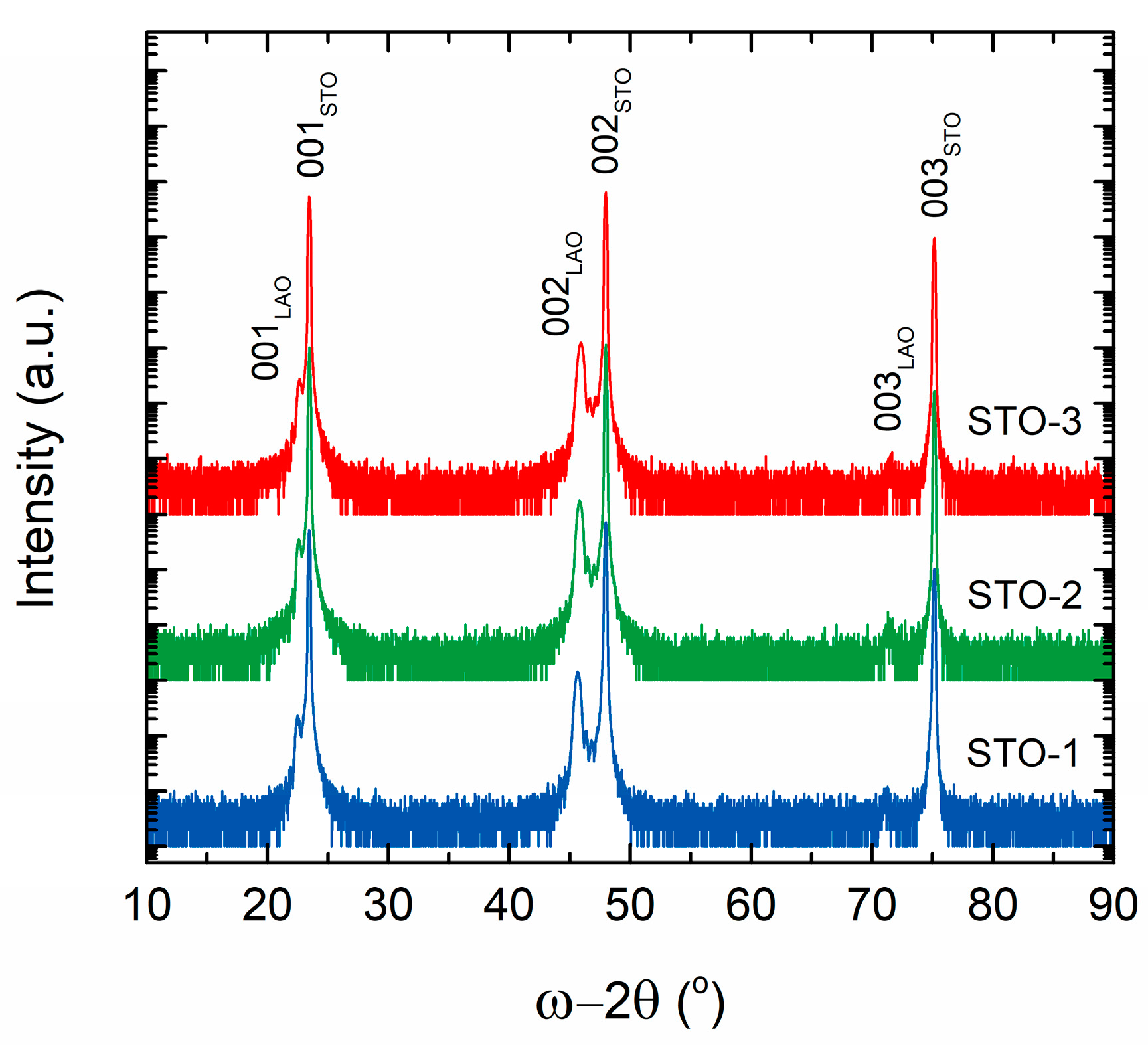
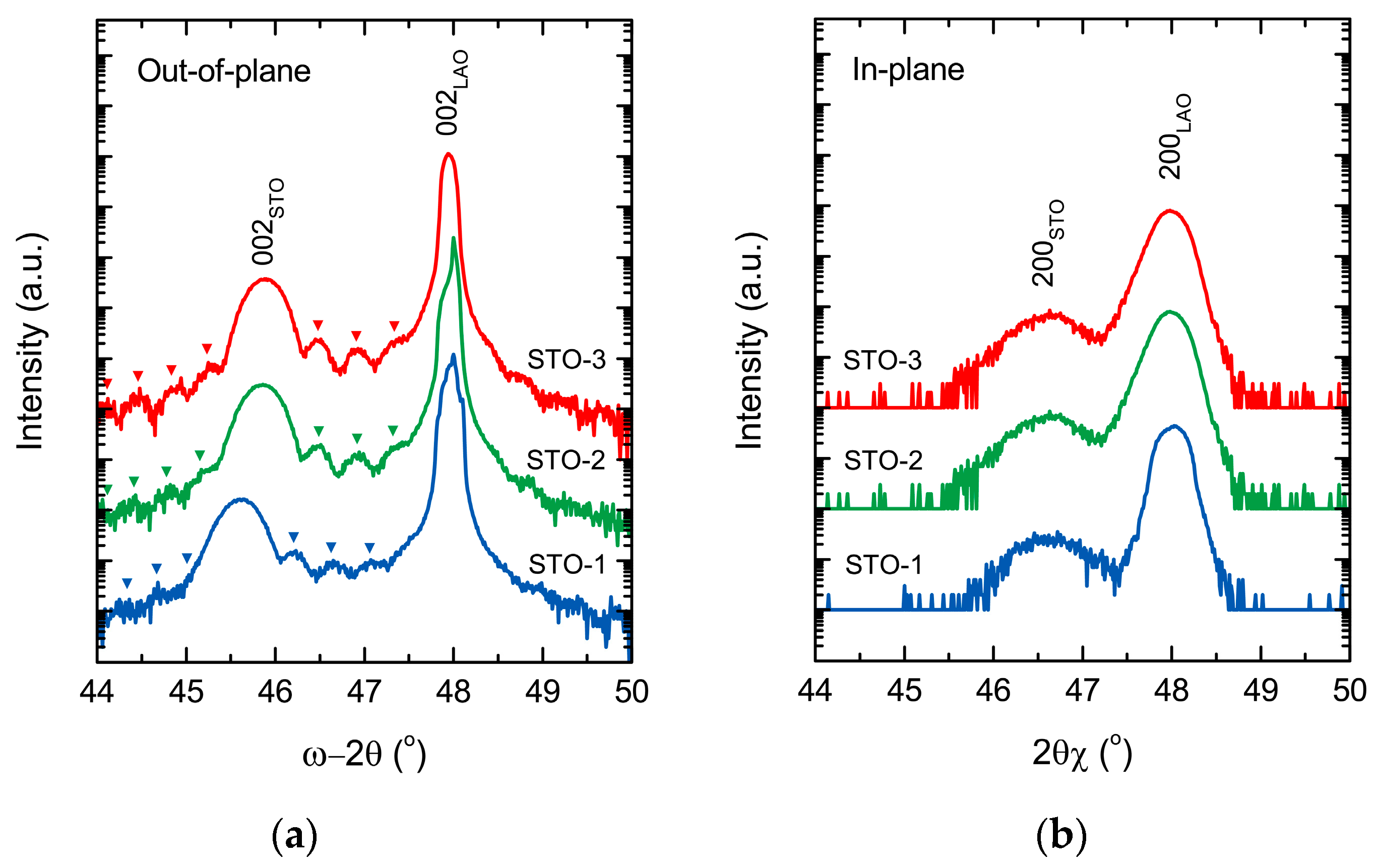
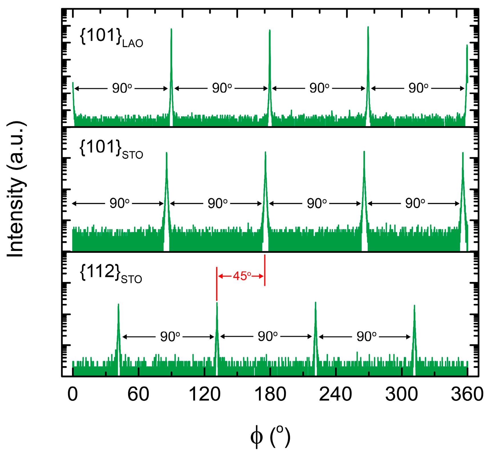
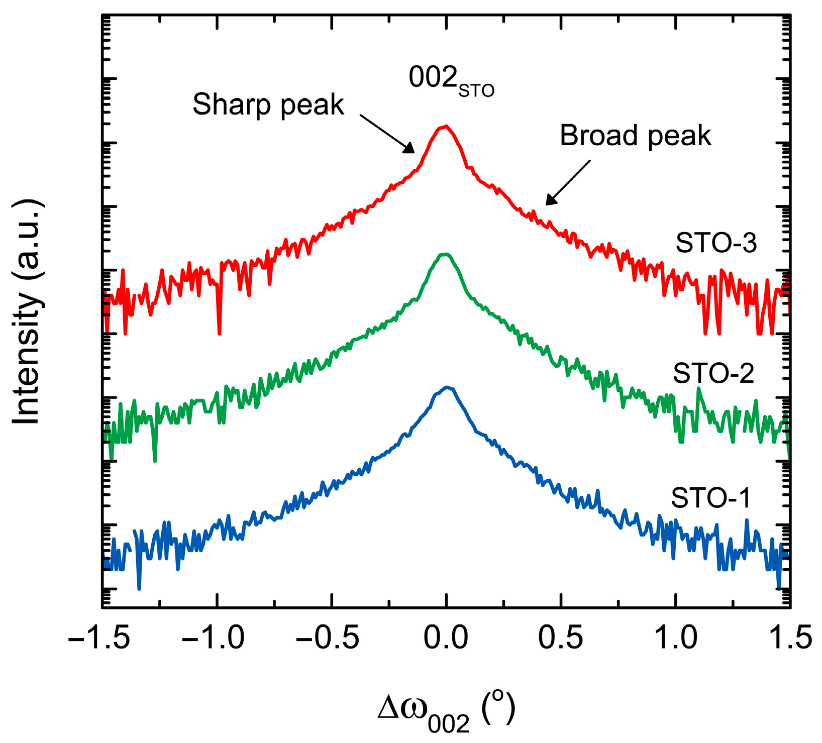
3.1. Structural Properties

The HRXRD spectra of all STO films in Figure 2 show only the 00lSTO and 00lLAO reflections (l = 1, 2, and 3) which originated from the STO film and LAO substrate, respectively. There was no observation of reflections from other crystallographic orientations and undesirable phases, indicating that all STO films were single-phase and highly (001)-oriented orientation or normal to the substrate surface. Moreover, the intensity of 00lSTO reflections was nearly identical for all samples, implying an almost similar thickness and film crystalline quality.
Figure 3a reveals the enlarged HRXRD spectra around the 002STO reflection in the 2θ range of 44°–50°. As the PO2 increased, the 002STO peak shifted towards a higher 2θ angle, indicating a decrease in the out-of-plane c-lattice constant. The oscillation pattern, so-called “Pendellösung fringes”, was evident on both sides of the 002STO reflection (marked with a triangle). The visible Pendellösung fringes indicate that the STO films possessed high crystal quality, low defect density, and smooth film/substrate interface. Film thickness (t) can be estimated from the oscillation period using the following formula:
t = λ 2 ( sin θ n + 1 sin θ n )  
where θn+1 and θn are the two adjacent maxima in the oscillations, λ is the X-ray wavelength. The thickness of all films was approximately 21 nm (growth rate ≈ 0.35 nm min−1), with no significant change with increasing PO2. This result suggested that increased PO2 had a negligible influence on the growth rate of STO films on the LAO substrate by IBSD under this operating condition.
The GIXRD measurement was further employed to examine the in-plane lattice constant (a). We assume that the in-plane lattice constants along a- and b-axes are equal. The GIXRD spectra were recorded on all STO films by fixing ϕ at a certain angle for the (200)STO plane. The scattering vector is perpendicular to the rubbing direction and parallel to the substrate surface. The grazing incidence angle (ω) was fixed at 0.1°, corresponding to a penetration depth of about 4 nm [17]. Figure 3b shows the GIXRD spectra around 200STO reflection of the STO films grown at different PO2. With increasing PO2, the 200STO reflection gradually shifted toward a higher 2θ angle, indicating a decrease of in-plane lattice constant. Based on the HRXRD and GIXRD spectra, the out-of-plane and in-plane lattice constants of the STO films can be calculated by Bragg’s law using the 002STO and 200STO reflections, respectively. The out-of-plane lattice constant was 0.3972 nm for STO-1 and decreased to 0.3951 nm for STO-3. Similarly, the in-plane lattice constant slightly decreased from 0.3895 nm for STO-1 to 0.3888 nm for STO-3. The expansion of both the in-plane and out-of-plane lattice constants at low PO2 was caused by the transformation from Ti4+ to Ti3+ states induced by oxygen vacancy in the STO lattice since the radius of Ti3+ ion (0.069 nm) is larger than that of Ti4+ ion (0.064 nm) [23]. The presence of oxygen vacancy and Ti3+ state in the STO lattice was confirmed by XPS measurements and discussed in Section 3.2.
Owing to a lattice mismatch of about 3% between the LAO substrate (aLAO = 0.3790 nm) and bulk STO (aSTO = 0.3905 nm), the STO films grew on the LAO substrate under in-plane compressive stress, resulting in a contraction of in-plane lattice constant and elongation of out-of-plane lattice constant of the STO unit-cell due to the effect of Poisson’s ratio. The STO films grown at high PO2 exhibited less tetragonal distortion, as reflected by a decrease in tetragonality (c/a) (Table 1). The in-plane lattice strain (εa = (εa − εSTO)/εSTO) was found between −0.0044 and −0.0026, whereas the out-of-plane lattice strain (εc = (εc − εSTO)/εSTO) was higher in the range of 0.0118–0.0172. Note that negative and positive values of lattice strain refer to contraction and expansion of the lattice constant, respectively. Besides the effect of lattice mismatch, the difference in thermal expansion coefficient between the STO film and LAO substrate also induced the tensile in-plane strain on the STO during cooling to room temperature. However, the strain induced by a lattice mismatch was more predominant than that by thermal expansion mismatch. Therefore, we can discard this effect from the consideration. The lattice constants and lattice strains along the in-plane lattice and out-of-plane directions and tetragonality (c/a) of all samples are summarized in Table 1.
To confirm the epitaxial orientation of the STO film on the LAO substrate, a ϕ-scan from 0° to 360° was recorded on the {101}STO (2θ ≈ 32.3°, χ = 45°), {101}LAO (2θ ≈ 33.4°, χ = 45°), and {112}STO planes (2θ ≈ 57.6°, χ = 35.3°) of STO-2, as shown in Figure 4. Four sharp peaks separated by 90° (four-fold symmetry) were clearly observed for all ϕ-scans. The peaks of the ϕ-scans recorded on the {101}STO and {101}LAO planes appeared at almost the same position but those of the {112}STO plane were shifted by 45° with respect to the {101}STO plane. These results confirm that the crystal lattice of STO films had an epitaxial cube-on-cube orientation on the (001)-oriented LAO substrate. The epitaxial orientation relationship between the STO film and the LAO substrate can be expressed as follows: (001)STO || (001)LAO and [100]STO || [100]LAO.
The rocking curve measurement (ω scan) recorded on the 002STO reflection was carried out to evaluate the crystalline quality of the STO films, as shown in Figure 5. The rocking curve for all STO films revealed a similar characteristic feature, which consisted of a sharp central peak lying on top of a broad peak. The sharp peak indicated good alignment of the STO film with respect to the LAO substrate along the c-axis (high-crystal quality region), while the broad peak arose from the film misalignment (low-crystal quality region) due to the existence of dislocations and local relaxation of the film. This characteristic curve is typically found in the heteroepitaxial growth of oxide thin films reported elsewhere [31,32,33,34]. The full-width at half-maximum (FWHM) of the sharp central peak was about 0.1231°, 0.1192°, and 0.1143° for STO-1, STO-2, and STO-3, respectively, which decreased at higher PO2. The FWHM of the broad peak was approximately 0.50° for all films. Although a slight variation of FWHM values was observed in the PO2 range investigated, it can suggest that the STO films had a slightly higher degree of crystallinity or epitaxial improvement at higher PO2.
The rocking curve FWHM values were larger than those of homoepitaxial STO films grown on (001) STO substrate by PLD and MBE, which were reported by Lee et al. (0.023°) [35] and Jalan et al. (0.009°) [36], respectively. Comparing the heteroepitaxial STO films on the LAO substrate grown by PLD, Ambwani et al. reported that the rocking curve FWHM was widely distributed between 0.05° and 0.40° [24], while Lu et al. observed about 0.18°–0.19° [37]. Moreover, the FWHM values of the STO films grown by MOCVD and RF magnetron sputtering were found to be 0.36° [11] and 0.20°–0.70° [13], respectively. This comparison indicates that the crystalline quality of heteroepitaxial STO films in this work was lower than that of homoepitaxial STO films but higher than or comparable to heteroepitaxial STO films grown by other methods. Furthermore, the rocking curve broadening can be used to estimate the total dislocation densities (Ddis) in the films using the following formula [38,39]:
D dis β 2 4.36 b 2   ,
where β is the FWHM of the rocking curve, and b is the length of the Burger vector of the corresponding dislocation (b = a〈100〉) [40]. With increased PO2, the Ddis in the high crystalline-quality region (sharp peak) was found to decrease from 7 × 10⁸ for STO-1 to 6 × 108 cm2 for STO-3. For the Ddis in the low crystalline-quality region (broad peak), it was estimated in the order of 1 × 1010 cm−2.

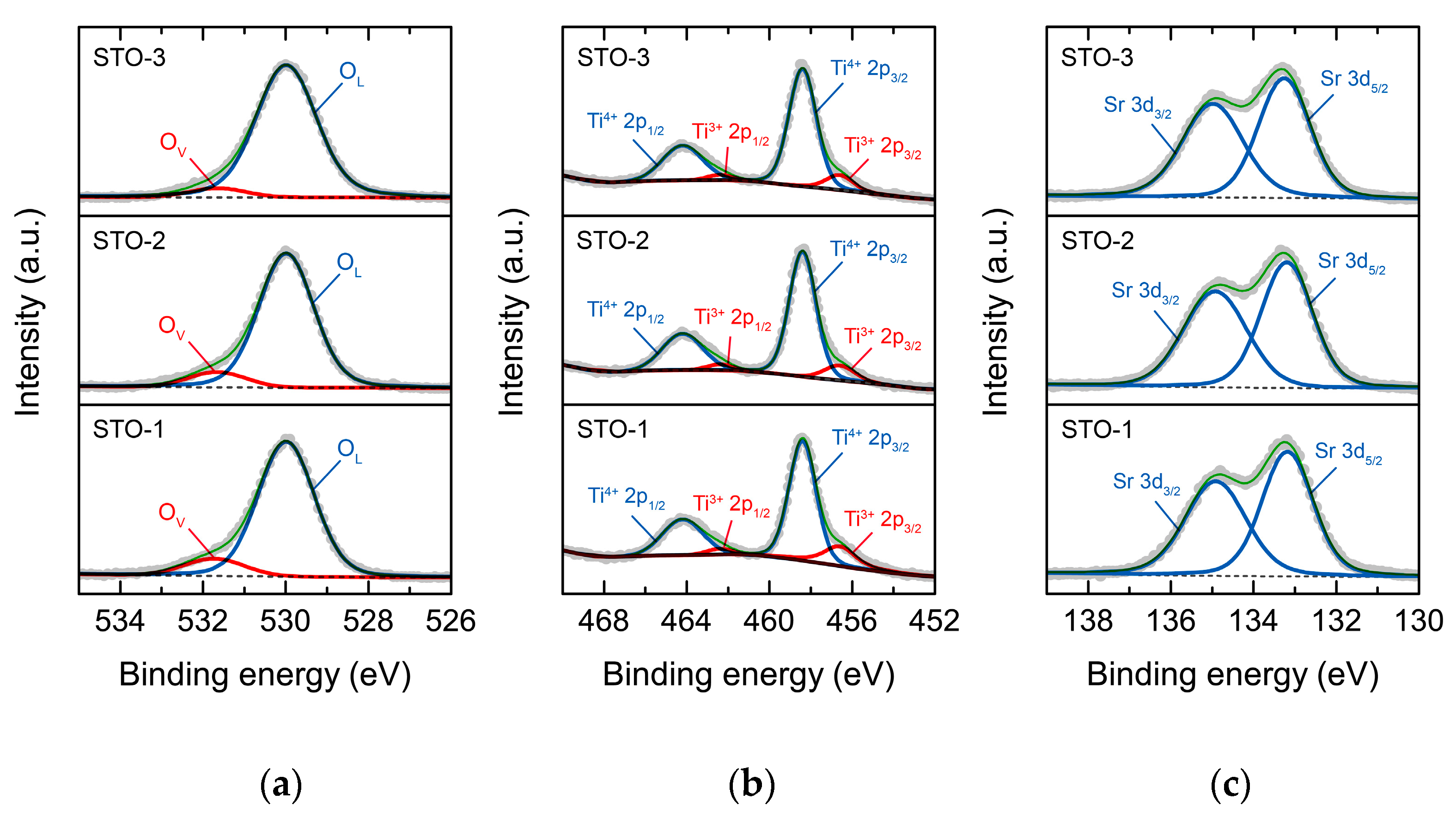
3.2. Surface Chemistry and Oxygen Vacancy

XPS measurement was further conducted to examine the Ti ionic state and oxygen vacancies in the STO films. The XPS O 1s spectra (Figure 6a) were resolved into two peaks. The lower binding energy at 529.8 eV represents the oxygen in the STO lattice (OL), while the higher binding energy at 531.6 eV is assigned to absorbed oxygen species, which is related to the presence of oxygen vacancies (OV) [41,42]. It is seen that the OV/OL ratio decreased slightly at higher PO2. The XPS Ti 2p spectra showed the Ti4+ 2p1/2 and Ti4+ 2p3/2 as the major states at 464.2 and 458.4 eV, respectively (Figure 6b). In addition, the small shoulder peaks of Ti3+ 2p1/2 and Ti3+ 2p3/2 state induced by oxygen vacancy appeared at a lower binding energy of 462.4 eV and 456.6 eV [43,44]. Similarly, the Ti3+/Ti4+ ratio was reduced at high PO2. The coexistence of both Ti3+ and OV peaks confirms the presence of oxygen vacancy in the STO films since when one oxygen vacancy is created in the lattice, two Ti4+ ions will transform into two Ti3+ ions to attain charge equilibrium. At higher PO2 pressure, the oxygen vacancies in the STO lattice decreased, as reflected by the suppression of Ti3+ and OV peaks. The Sr 3d5/2 and Sr 3d3/2 peaks (Figure 6c) were unchanged with increasing PO2, indicating the similar ionic state of Sr. Although the PO2 increased up to 1.5 × 10⁻³ Torr for STO-3, oxygen vacancy still existed in the film. This could be due to the escape of oxygen atoms in the lattice from the films during growth at high temperature, thus creating the oxygen vacancy (OO VO2+ + 2e + ½O2, where OO and VO represent oxygen ions at the normal site and an oxygen vacancy, respectively) [45].
The XPS peak intensities after background subtraction were used to determine the chemical composition using the following equation [23]:
C i I i / S i I i / S i ,
where Ci, Ii, and Si are the concentration, peak area, and atomic sensitivity factor of the element i, respectively. The atomic sensitivity factors of Sr 3d, Ti 2p, and O 1s are 1.48, 1.80, and 0.66, respectively [46]. The composition ratio of Sr:Ti:O deduced from Sr 3d, Ti 2p, and O 1s XPS data of all the films were deviated from the stoichiometric value, as summarized in Table 2. The Sr/Ti ratio of all films was higher than 1, indicating the Ti deficiency in the STO films. There was no significant difference in Sr/Ti ratio as the PO2 increased. The O composition was estimated to be less than stoichiometry, which was reduced at lower PO2 due to the high level of oxygen vacancy in the STO lattice.

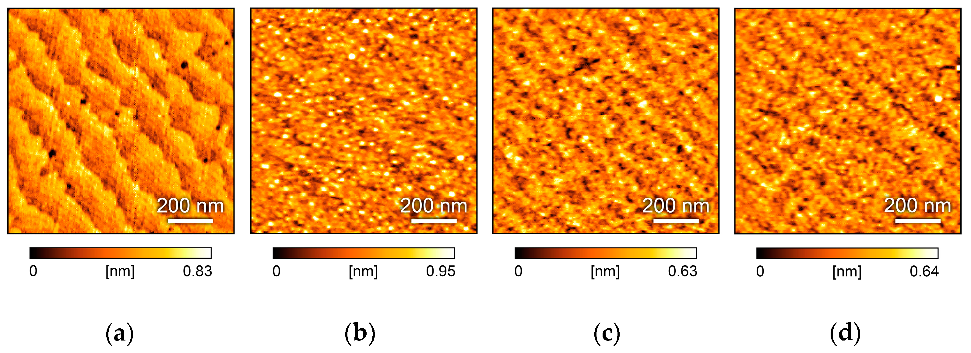
3.3. Surface Morphology

The AFM topography images (2 μm × 2 μm) of the STO films grown at different PO2 are presented in Figure 7. The LAO substrate showed a relatively flat and smooth surface with a well-defined step and terrace (Figure 7a). After growing, the step became blurred while the terrace was rougher due to the coverage of STO films (Figure 7b–d). The average root-mean-square (RMS) roughness deduced from five scan areas was 0.11 ± 0.02, 0.08 ± 0.02, and 0.08 ± 0.01 nm for STO-1, STO-2, and STO-3, respectively. The RMS roughness of STO-2 and STO-3 was close to that of bare LAO substrate (0.08 ± 0.01 nm). This surface feature confirmed that the STO films grew on the LAO substrates with a two-dimensional (2D) growth mode (i.e., either the step-flow mode or the layer-by-layer growth mode). The RMS roughness of the STO films gradually decreased with increasing PO2. At low PO2, the sputtered atoms arrived the substrate with high kinetic energy, making it easy to form small particles on the substrate surface and cause a rough terrace. Yet as PO2 increased, the sputtered atoms were blocked by collision with oxygen molecules. This caused a partial loss of kinetic energy of sputtered atoms to the substrate, which may lead to a smoother surface.

4. Conclusions

The epitaxial STO films were successfully grown on the LAO substrates by the IBSD method at various PO2. All STO films grew on the LAO substrates under a compressive strain along the in-plane direction (a- and b-axes) and a tensile strain along the growth direction (c-axis), indicating a tetragonal distortion. The oxygen vacancies favored creating an STO lattice at low PO2, as confirmed by the formation of Ti3+ ionic state and OV peak and were suppressed at higher PO2. The existence of oxygen vacancies resulted in an expansion of the STO unit cell and also lowered the crystalline quality. The surface roughness of the STO films became flatter and smoother, close to bare LAO substrate at a higher PO2, indicating a 2D growth mode. The present study provides a greater understanding of the effect of PO2 on crystal structure, oxygen vacancy, and surface morphology and their correlation with the epitaxial STO films grown by IBSD. Moreover, these results are a useful reference for optimizing the growth of other epitaxial oxide thin films by IBSD.

Author Contributions

Conceptualization, G.P., N.S.; methodology, G.P.; validation, G.P.; formal analysis, G.P.; investigation, G.P.; resources, N.S.; data curation, G.P.; writing—original draft preparation, G.P.; writing—review and editing, G.P., N.S.; visualization, G.P.; supervision, N.S.; project administration, G.P., N.S.; funding acquisition, G.P., N.S. All authors have read and agreed to the published version of the manuscript.

Funding

This research was funded by Grant-in-Aid for Young Researcher from the Global Center of Excellence (GCOE) at Nagoya University and the Faculty of Engineering, Kasetsart University.

Institutional Review Board Statement

Not applicable.

Informed Consent Statement

Not applicable.

Data Availability Statement

Not applicable.

Acknowledgments

The authors would like to thank all members of the Saito laboratory for their suggestions and help in this work.

Conflicts of Interest

The authors declare no conflict of interest.

References

  1. Takashima, H.; Wang, R.; Kasai, N.; Shoji, A. Preparation of parallel capacitor of epitaxial SrTiO3 film with a single-crystal-like behavior. Appl. Phys. Lett. 2003, 83, 2883–2885. [Google Scholar] [CrossRef]
  2. Hao, J.H.; Luo, Z.; Gao, J. Effects of substrate on the dielectric and tunable properties of epitaxial SrTiO3 thin films. J. Appl. Phys. 2006, 100, 114107. [Google Scholar] [CrossRef] [Green Version]
  3. Hara, T.; Ishiguro, T.; Wakiya, N.; Shinozaki, K. Oxygen sensing properties of SrTiO3 thin films. Jpn. J. Appl. Phys. 2008, 47, 7486–7489. [Google Scholar] [CrossRef]
  4. Niu, G.; Yin, S.; Saint-Girons, G.; Gautier, B.; Leoeur, P.; Pillard, V.; Hollinger, G.; Vilquin, B. Epitaxy of BaTiO3 thin film on Si(0 01) using a SrTiO3 buffer layer for non-volatile memory application. Microelectron. Eng. 2011, 88, 1232–1235. [Google Scholar] [CrossRef]
  5. Rieck, J.L.; Hensling, F.V.E.; Dittmann, R. Trade-off between variability and retention of memristive epitaxial SrTiO3 devices. APL Mater. 2021, 9, 021110. [Google Scholar] [CrossRef]
  6. Biasotti, M.; Pellegrino, L.; Buzio, R.; Bellingeri, E.; Bernini, C.; Siri, A.S.; Marré, D. Fabrication and electromechanical actuation of epitaxial SrTiO3 (001) microcantilevers. J. Micromech. Microeng. 2013, 23, 035031. [Google Scholar] [CrossRef]
  7. Khodan, A.N.; Guyard, S.; Contour, J.-P.; Crété, D.-G.E.; Jacquet, K.; Bouzehouane, K. Pulsed laser deposition of epitaxial SrTiO3 films: Growth, structure and functional properties. Thin Solid Film 2007, 515, 6411–6432. [Google Scholar] [CrossRef]
  8. Groenendijk, D.J.; Gariglio, S. Sequential pulsed laser deposition of homoepitaxial SrTiO3 thin films. J. Appl. Phys. 2016, 120, 225307. [Google Scholar] [CrossRef] [Green Version]
  9. Niu, F.; Wessels, B.W. Surface and interfacial structure of epitaxial SrTiO3 thin films on (001) Si grown by molecular beam epitaxy. J. Cryst. Growth 2007, 300, 509–518. [Google Scholar] [CrossRef]
  10. Lapano, J.; Brahlek, M.; Zhang, L.; Roth, J.; Pogrebnyakov, A.; Engel-Herbert, R. Scaling growth rates for perovskite oxide virtual substrates on silicon. Nat. Commun. 2019, 10, 2464. [Google Scholar] [CrossRef] [PubMed] [Green Version]
  11. Gilbert, S.R.; Wessel, B.W.; Studebaker, D.B.; Marks, T.J. Epitaxial growth of SrTiO3 thin films by metaloraganic chemical vapor deposition. Appl. Phys. Lett. 1995, 66, 3298–3300. [Google Scholar] [CrossRef]
  12. Chen, J.; Ito, A.; Goto, T. High-speed epitaxial growth of SrTiO3 transparent thick films composed of close-packed nanocolumns using laser chemical vapor deposition. Vacuum 2020, 177, 109424. [Google Scholar] [CrossRef]
  13. Wang, X.; Helmersson, U.; Madsen, L.D.; Ivanov, I.P.; Münger, P. Composition, structure, and dielectric tunability of epitaxial SrTiO3 thin films grown by radio frequency magnetron sputtering. J. Vac. Sci. Technol. A 1999, 17, 564–570. [Google Scholar] [CrossRef]
  14. Wang, X.; Olafsson, S.; Sandström, P.; Helmersson, U. Growth of SrTiO3 thin films on LaAlO3(001) substrates; the influence of growth temperature on composition, orientation, and surface morphology. Thin Solid Film 2000, 360, 181–186. [Google Scholar] [CrossRef]
  15. Panomsuwan, G.; Takai, O.; Saito, N. Fabrication and characterization of epitaxial SrTiO3/Nb-doped SrTiO3 superlattices by double ECR ion beam sputter deposition. Vacuum 2013, 89, 35–39. [Google Scholar] [CrossRef]
  16. Panomsuwan, G.; Takai, O.; Saito, N. Effect of growth temperature on structural and morphological evolution of epitaxial SrTiO3 thin films grown on LaAlO3 (001) substrates by ion beam sputter deposition. Vacuum 2014, 109, 175–179. [Google Scholar] [CrossRef]
  17. Panomsuwan, G.; Saito, N. Thickness-dependent strain evolution of epitaxial SrTiO3 thin films grown by ion beam sputter deposition. Cryst. Res. Technol. 2018, 53, 1799211. [Google Scholar] [CrossRef]
  18. Mazet, L.; Yang, S.M.; Kalinin, S.V.; Schamm-Chardon, S.; Dubourdieu, C. A review of molecular beam epitaxy of ferroelectric BaTiO3 films on Si, Ge and GaAs substrates and their applications. Sci. Technol. Adv. Mater. 2015, 16, 036005. [Google Scholar] [CrossRef] [PubMed] [Green Version]
  19. Manova, D.; Gerlach, J.W.; Mändl, S. Thin films deposition using energetic ions. Materials 2010, 3, 4109–4141. [Google Scholar] [CrossRef]
  20. Feder, R.; Frost, F.; Neumann, H.; Bundesmann, C.; Rauschenbach, B. Systematic investigations of low energy Ar ion beam sputtering of Si and Ag. Nucl. Instrum. Methods Phys. Res. Sect. B Beam Interact. Mater. At. 2013, 317, 137–142. [Google Scholar] [CrossRef]
  21. Becker, M.; Gies, M.; Polity, A.; Chatterjee, S.; Klar, P.J. Materials processing using radio-frequency ion-sources: Ion-beam sputter-deposition and surface treatment. Rev. Sci. Instrum. 2019, 90, 023901. [Google Scholar] [CrossRef]
  22. Ohly, C.; Hoffmann-Eifert, S.; Guo, X.; Schubert, J.; Waser, R. Electrical conductivity of epitaxial SrTiO3 thin films as a function of oxygen partial pressure and temperature. J. Am. Ceram. Soc. 2006, 89, 2845–2852. [Google Scholar] [CrossRef]
  23. Cai, H.L.; Wu, X.S.; Gao, J. Effect of oxygen content on structural and transport properties in SrTiO3–x thin films. Chem. Phys. Lett. 2009, 467, 313–317. [Google Scholar] [CrossRef]
  24. Ambwani, P.; Xu, P.; Haugstad, G.; Jeong, J.S.; Deng, R.; Mkhoyan, K.A.; Jalan, B.; Leighton, C. Defects, stoichiometry, and electronic transport in SrTiO3-depilayers: A high pressure oxygen sputter deposition study. J. Appl. Phys. 2016, 120, 055704. [Google Scholar] [CrossRef] [Green Version]
  25. Tyunina, M.; Rusevich, L.L.; Kotomin, E.A.; Pacherova, O.; Kocourek, T.; Dejneka, A. Epitaxial growth of perovskite oxide films facilitated by oxygen vacancies. J. Mater. Chem. C 2021, 9, 1693–1700. [Google Scholar] [CrossRef]
  26. Trabelsi, H.; Bejar, M.; Dhahri, E.; Valente, M.A.; Graca, M.P.F. Oxygen-vacancy-related giant permittivity and ethanol sensing response in SrTiO3–δ ceramics. Phys. E Low-Dimens. Syst. Nanostructures 2019, 108, 317–325. [Google Scholar] [CrossRef]
  27. Kang, K.T.; Seo, H.I.; Kwon, O.; Lee, K.; Bae, J.-S.; Chu, M.-W.; Chae, S.C.; Kim, Y.; Choi, W.S. Ferroelectricity in SrTiO3 epitaxial thin films via Sr-vacancy-induced tetragonality. Appl. Surf. Sci. 2020, 499, 143930. [Google Scholar] [CrossRef]
  28. Xu, W.; Yang, J.; Bai, W.; Tang, K.; Zhang, Y.; Tang, X. Oxygen vacancy induced photoluminescence and ferromagnetism in SrTiO3 thin films by molecular beam epitaxy. J. Appl. Phys. 2013, 114, 154106. [Google Scholar] [CrossRef]
  29. Pai, Y.-Y.; Tylan-Tyler, A.; Irvin, P.; Levy, J. Physics of SrTiO3-based heterostructures and nanostructures: A review. Rep. Prog. Phys. 2018, 81, 036503. [Google Scholar] [CrossRef] [PubMed] [Green Version]
  30. Ohnishi, T.; Takahashi, K.; Nakamura, M.; Kawasaki, M.; Yoshimoto, M.; Koinuma, H. A-site layer terminated perovskite substrate: NdGaO3. Appl. Phys. Lett. 1999, 74, 2531. [Google Scholar] [CrossRef]
  31. Jin, S.; Gao, G.; Wu, W.; Zhou, X. Effect of angular-distortion-induced strain on structural and transport properties of epitaxial La0.7Sr0.3MnO3 thin films. J. Phys. D Appl. Phys. 2007, 40, 305–309. [Google Scholar] [CrossRef] [Green Version]
  32. Biegalski, M.D.; Fong, D.D.; Eastman, J.A.; Fuoss, P.H.; Streiffer, S.K.; Heeg, T.; Schubert, J.; Tian, W.; Nelson, C.T.; Pan, X.Q.; et al. Critical thickness of high structural quality SrTiO3 films grown on orthorhombic (101) DyScO3. J. Appl. Phys. 2008, 104, 114109. [Google Scholar] [CrossRef] [Green Version]
  33. Lee, H.G.; Kim, Y.; Hwang, S.; Kim, G.; Kang, T.D.; Kim, M.; Kim, M.; Noh, T.W. Double-layer buffer template to grow commensurate epitaxial BaBiO3 thin films. APL Mater. 2016, 4, 126106. [Google Scholar] [CrossRef] [Green Version]
  34. Andrei, F.; Ion, V.; Bîrjega, R.; Dinescu, M.; Enea, N.; Pantelica, D.; Mihai, M.D.; Maraloiu, V.-A.; Teodorescu, V.S.; Marcu, I.-C.; et al. Thickness-dependent photoelectrochemical water splitting properties of self-assembled nanostructured LaFeO3 perovskite thin films. Nanomaterials 2016, 11, 1371. [Google Scholar] [CrossRef] [PubMed]
  35. Lee, S.A.; Jeong, H.; Woo, S.; Hwang, J.-Y.; Choi, S.-Y.; Kim, S.-D.; Choi, M.; Roh, S.; Yu, H.; Hwang, J.; et al. Phase transitions via selective elemental vacancy engineering in complex oxide thin films. Sci. Rep. 2016, 6, 23649. [Google Scholar] [CrossRef] [PubMed] [Green Version]
  36. Jalan, B.; Engel-Herbert, R.; Wright, N.J.; Stemmer, S. Growth of high-quality SrTiO3 films using a hybrid molecular beam epitaxy approach. J. Vac. Sci. Technol. 2009, 27, 461–464. [Google Scholar] [CrossRef]
  37. Lu, P.; Jia, Q.X.; Findikoglu, A.T. Effects of homo-epitaxial LaAlO3 layer on microstructural properties of SrTiO3 films grown on LaAlO3 substrates. Thin Solid Film 1999, 348, 38–43. [Google Scholar] [CrossRef]
  38. Ayer, J.E. The measurement of threading dislocation densities in semiconductor crystals by X-ray diffraction. J. Cryst. Growth 1994, 135, 71–77. [Google Scholar] [CrossRef]
  39. Huang, S.R.; Lu, X.; Barnett, A.; Opila, R.L.; Mogili, V.; Tanner, D.A.; Nakahara, S. Characterization of the microstructure of GaP films grown on {111} Si by liquid phase epitaxy. ACS Appl. Mater. Interfaces 2014, 6, 18626–18634. [Google Scholar] [CrossRef]
  40. Zhai, Z.Y.; Li, X.Z.; Zhi, S.S.; Wu, X.S.; Hao, J.H.; Gao, J. Dislocation density in SrTiO3 film grown on DyScO3 by pulse laser ablation. Surf. Rev. Lett. 2007, 4, 779–782. [Google Scholar] [CrossRef]
  41. Li, G.; Bai, Y.; Wu, S.; Zhang, W. Variation in photocatalytic activity of SrTiO3 (100) single-crystal thin films with different substrates and annealing atmosphere. Sci. Adv. Mater. 2013, 5, 746–768. [Google Scholar] [CrossRef]
  42. Wang, X.; Zhang, C.; Zang, G.; Lv, S.; Li, L. Effect of doping content of Pr ions on oxygen vacancies in SrTiO3 films. J. Alloys Compd. 2015, 637, 277–280. [Google Scholar] [CrossRef]
  43. Shkabko, A.; Aguirre, M.H.; Marozau, I.; Lippert, T.; Chou, Y.-S.; Douthwaite, R.E.; Weidenkaff, A. Synthesis and transport properties of SrTiO3−xNy/SrTiO3−δ layered structures produced by microwave-induced plasma nitridation. J. Phys. D: Appl. Phys. 2009, 42, 145202. [Google Scholar] [CrossRef] [Green Version]
  44. Pal, P.; Kumar, P.; Aswin, V.; Dogra, A.; Joshi, A.G. Chemical potential shift and gap-state formation in SrTiO3−δ revealed by photoemission spectroscopy. J. Appl. Phys. 2014, 116, 053704. [Google Scholar] [CrossRef] [Green Version]
  45. Wang, C.C.; Lei, C.M.; Wang, G.J.; Sun, X.H.; Li, T.; Huang, S.G.; Wang, H.; Li, Y.D. Oxygen-vacancy-related dielectric relaxations in SrTiO3 at high temperatures. J. Appl. Phys. 2013, 113, 094103. [Google Scholar] [CrossRef]
  46. Wagner, C.D.; Davis, L.E.; Zeller, M.V.; Taylor, J.A.; Raymond, R.M.; Gale, L.H. Empirical atomic sensitivity factors for quantitative analysis by electron spectroscopy for chemical analysis. Surf. Interface Anal. 1981, 3, 211–225. [Google Scholar] [CrossRef]
Figure 1. Schematic diagram illustrating the IBSD system used to grow the STO films in this work.
Oxygen 01 00007 g001
Figure 2. HRXRD ω−2θ scans of the STO films grown on LAO substrates at different PO2.
Oxygen 01 00007 g002
Figure 3. (a) Enlarged HRXRD ω−2θ scans at around the 002STO reflection, and (b) GIXRD 2θχ scans at around the 200STO reflection of the STO films grown on LAO substrates at various PO2.
Oxygen 01 00007 g003
Figure 4. ϕ-scan of the representative STO-2 film recorded on {101}LAO, {10}STO, and {112}STO.
Oxygen 01 00007 g004
Figure 5. Rocking curve ω scans recorded on the 002STO reflection for all STO films.
Oxygen 01 00007 g005
Figure 6. High-resolution XPS spectra of STO-1, STO-2, and STO-3 recorded on: (a) O 1s, (b) Ti 2p, and (c) Sr 3d core levels.
Oxygen 01 00007 g006
Figure 7. AFM topography images (2 μm × 2 μm) of: (a) bare LAO substrate, (b) STO-1, (c) STO-2, and (d) STO-3.
Oxygen 01 00007 g007
Table 1. In-plane lattice constants (a), out-of-plane lattice constant (c), in-plane lattice strain (εa), out-of-plane lattice strain (εc), and tetragonality (c/a) of the STO films grown on the LAO substrates at various PO2.
Samplea (nm)c (nm)c/aεaεc
STO-10.38950.39721.0198−0.00260.0172
STO-20.38920.39531.0157−0.00330.0123
STO-30.38880.39511.0162−0.00440.0118
Table 2. Sr:Ti:O, Sr/Ti, OV/OL, and Ti3+/Ti4+ ratios obtained from the quantitative XPS analysis of the STO films grown on the LAO substrates at various PO2.
SampleSr:Ti:OSr/TiOV/OLTi3+/Ti4+
STO-11:0.90:2.921.110.130.23
STO-21:0.91:2.871.100.110.16
STO-31:0.89:2.821.120.060.13
Publisher’s Note: MDPI stays neutral with regard to jurisdictional claims in published maps and institutional affiliations.

