


Biomolecules 2024, 14(1), 110; https://0-doi-org.brum.beds.ac.uk/10.3390/biom14010110 - 15 Jan 2024
Viewed by 802
Abstract
►
Show Figures
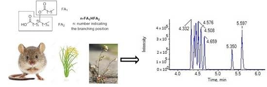
Hydroxy fatty acids (HFAs) constitute a class of lipids, distinguished by the presence of a hydroxyl on a long aliphatic chain. This study aims to expand our insights into HFA bioactivities, while also introducing new methods for asymmetrically synthesizing unsaturated and saturated HFAs.
[...] Read more.
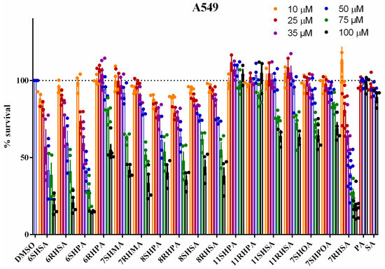
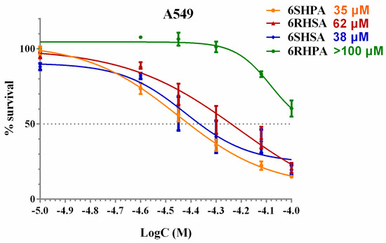
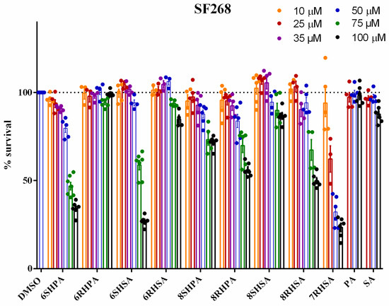
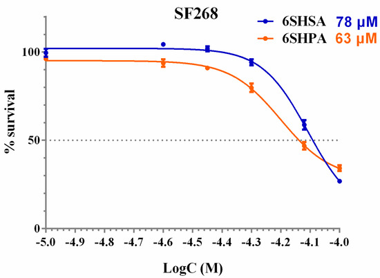
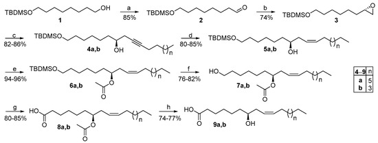
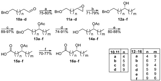
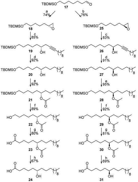
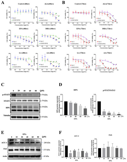
Hydroxy fatty acids (HFAs) constitute a class of lipids, distinguished by the presence of a hydroxyl on a long aliphatic chain. This study aims to expand our insights into HFA bioactivities, while also introducing new methods for asymmetrically synthesizing unsaturated and saturated HFAs. Simultaneously, a procedure previously established by us was adapted to generate new HFA regioisomers. An organocatalytic step was employed for the synthesis of chiral terminal epoxides, which either by alkynylation or by Grignard reagents resulted in unsaturated or saturated chiral secondary alcohols and, ultimately, HFAs. 7-(S)-Hydroxyoleic acid (7SHOA), 7-(S)-hydroxypalmitoleic acid (7SHPOA) and 7-(R)- and (S)-hydroxymargaric acids (7HMAs) were synthesized for the first time and, together with regioisomers of (R)- and (S)-hydroxypalmitic acids (HPAs) and hydroxystearic acids (HSAs), whose biological activity has not been tested so far, were studied for their antiproliferative activities. The unsaturation of the long chain, as well as an odd-numbered (C17) fatty acid chain, led to reduced activity, while the new 6-(S)-HPA regioisomer was identified as exhibiting potent antiproliferative activity in A549 cells. 6SHPA induced acetylation of histone 3 in A549 cells, without affecting acetylated α-tubulin levels, suggesting the selective inhibition of histone deacetylase (HDAC) class I enzymes, and was found to inhibit signal transducer and activator of transcription 3 (STAT3) expression.
Full article
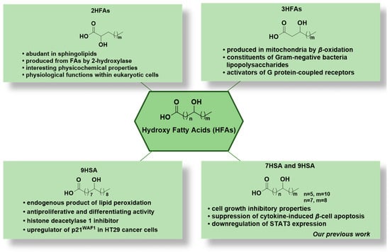
Figure 1
