
Biophysica 2024, 4(2), 195-206; https://0-doi-org.brum.beds.ac.uk/10.3390/biophysica4020014 - 12 Apr 2024
Abstract
►
Show Figures
Past anti-bacterial use of bacteriophages (phage therapy) is already well reviewed as a potential therapeutic response to the emergence of multidrug-resistant, pathogenic bacteria. Phage therapy has been limited by the following. (1) The success rate is too low for routine use and Food
[...] Read more.
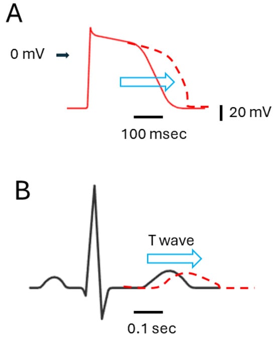
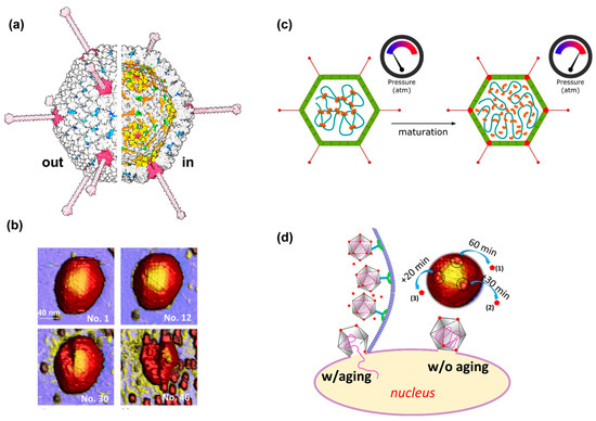
Past anti-bacterial use of bacteriophages (phage therapy) is already well reviewed as a potential therapeutic response to the emergence of multidrug-resistant, pathogenic bacteria. Phage therapy has been limited by the following. (1) The success rate is too low for routine use and Food and Drug Administration (FDA) approval. (2) Current strategies of routine phage characterization do not sufficiently improve the success rate of phage therapy. (3) The stability of many phages at ambient temperature is not high enough to routinely store and transport phages at ambient temperature. In the present communication, we present new and previous data that we interpret as introductory to biophysically and efficiently transforming phage therapy to the needed level of effectiveness. Included are (1) procedure and preliminary data for the use of native gel electrophoresis (a low-cost procedure) for projecting the therapy effectiveness of a newly isolated phage, (2) data that suggest a way to achieve stabilizing of dried, ambient-temperature phages via polymer embedding, and (3) data that suggest means to increase the blood persistence, and therefore the therapy effectiveness, of what would otherwise be a relatively low-persistence phage.
Full article
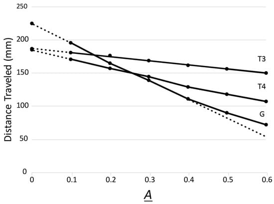
Figure 1


