


Brain Sci. 2023, 13(11), 1598; https://0-doi-org.brum.beds.ac.uk/10.3390/brainsci13111598 - 17 Nov 2023
Cited by 1 | Viewed by 941
Abstract
►
Show Figures
A common neurobiological mechanism in several neurodevelopmental disorders, including fragile X syndrome (FXS), is alterations in the balance between excitation and inhibition in the brain. It is thought that in the hippocampus, as in other brain regions, FXS is associated with increased excitability
[...] Read more.
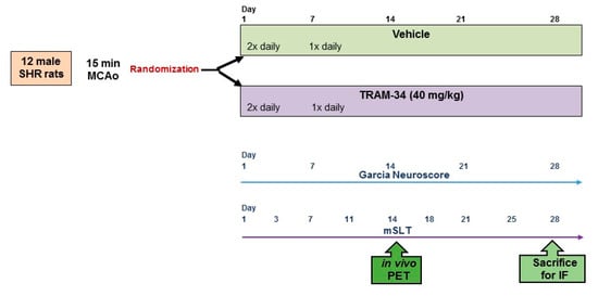
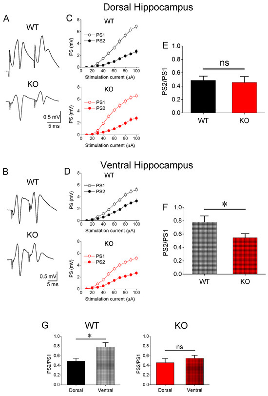
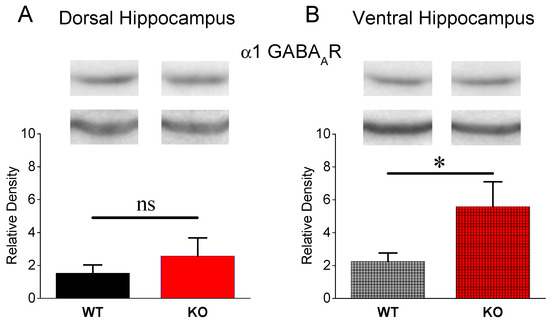
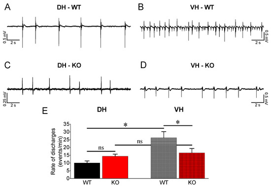
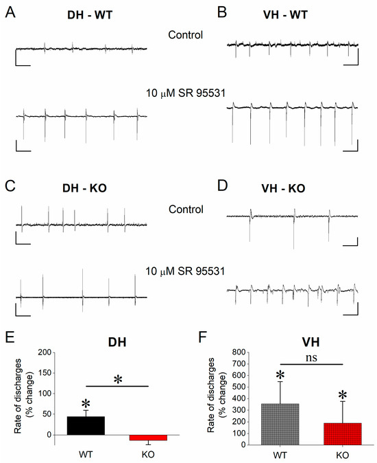
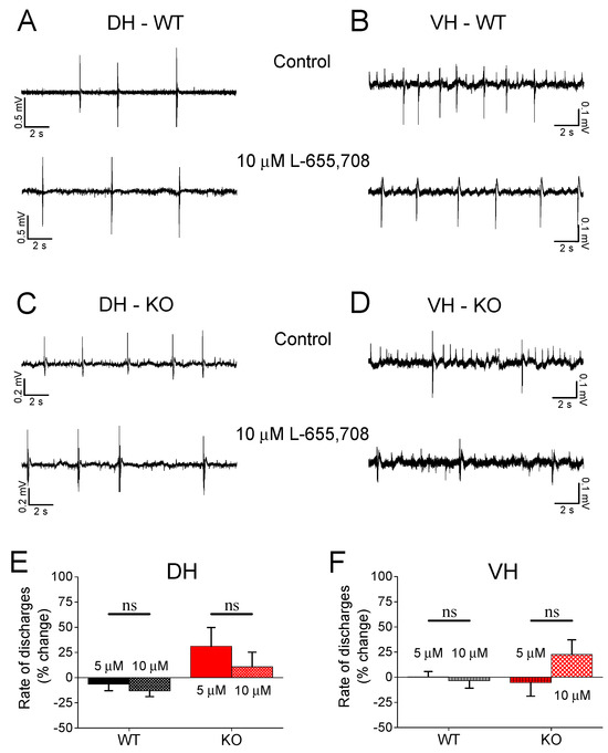
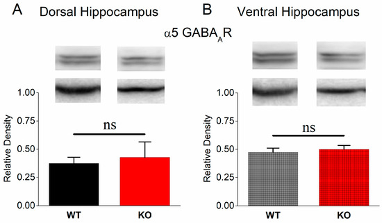
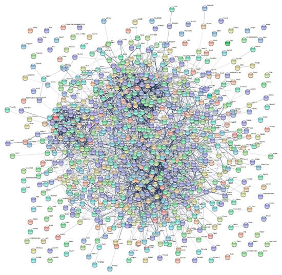
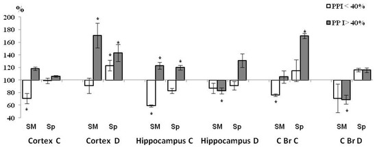
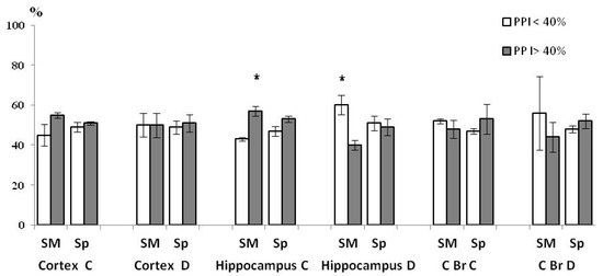
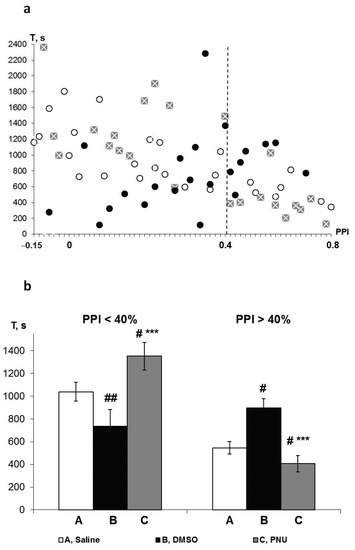
A common neurobiological mechanism in several neurodevelopmental disorders, including fragile X syndrome (FXS), is alterations in the balance between excitation and inhibition in the brain. It is thought that in the hippocampus, as in other brain regions, FXS is associated with increased excitability and reduced inhibition. However, it is still not known whether these changes apply to both the dorsal and ventral hippocampus, which appear to be differently involved in neurodegenerative disorders. Using a Fmr1 knock-out (KO) rat model of FXS, we found increased neuronal excitability in both the dorsal and ventral KO hippocampus and increased excitatory synaptic transmission in the dorsal hippocampus. Interestingly, synaptic inhibition is significantly increased in the ventral but not the dorsal KO hippocampus. Furthermore, the ventral KO hippocampus displays increased expression of the α1GABAA receptor subtype and a remarkably reduced rate of epileptiform discharges induced by magnesium-free medium. In contrast, the dorsal KO hippocampus displays an increased rate of epileptiform discharges and similar expression of α1GABAA receptors compared with the dorsal WT hippocampus. Blockade of α5GABAA receptors by L-655,708 did not affect epileptiform discharges in any genotype or hippocampal segment, and the expression of α5GABAA receptors did not differ between WT and KO hippocampus. These results suggest that the increased excitability of the dorsal KO hippocampus contributes to its heightened tendency to epileptiform discharges, while the increased phasic inhibition in the Fmr1-KO ventral hippocampus may represent a homeostatic mechanism that compensates for the increased excitability reducing its vulnerability to epileptic activity.
Full article
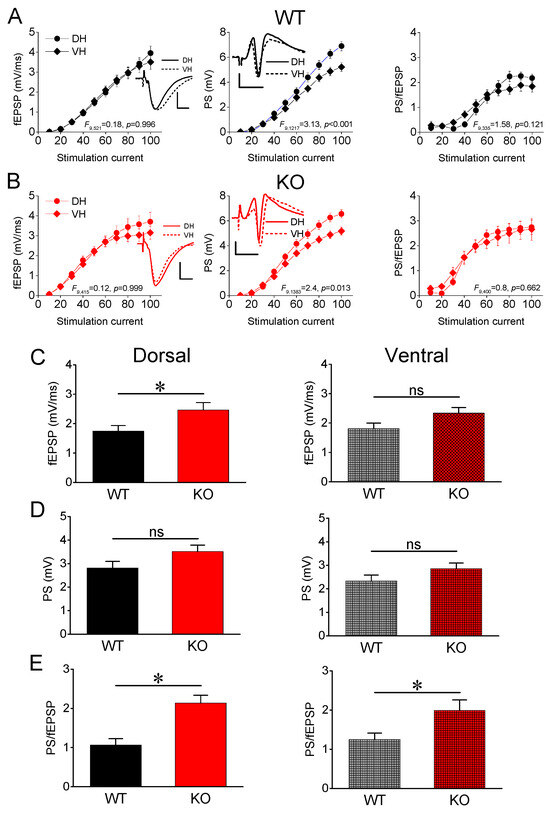
Figure 1
