


Cancers 2023, 15(17), 4337; https://0-doi-org.brum.beds.ac.uk/10.3390/cancers15174337 - 30 Aug 2023
Viewed by 1026
Abstract
►
Show Figures
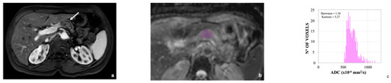
Dynamic biomarkers that permit the real-time monitoring of the tumor microenvironment response to therapy are an unmet need in breast cancer. Breast magnetic resonance imaging (MRI) has demonstrated value as a predictor of pathologic complete response and may reflect immune cell changes in
[...] Read more.
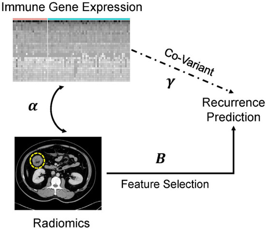
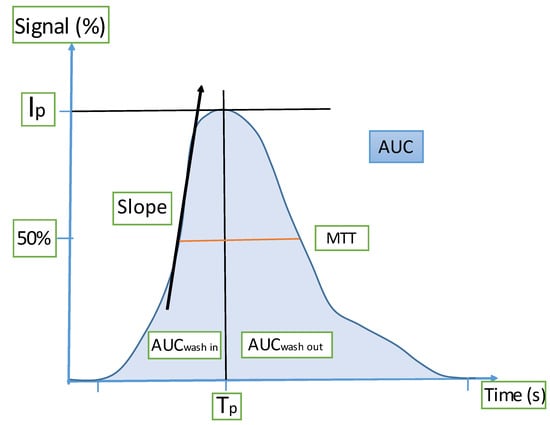
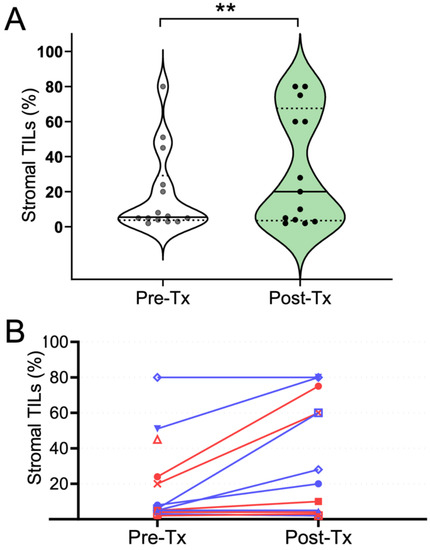
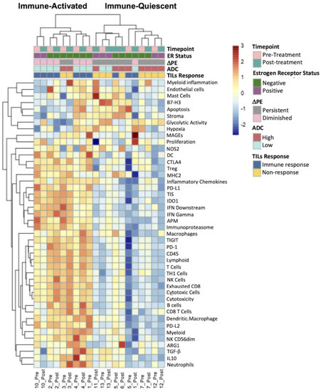
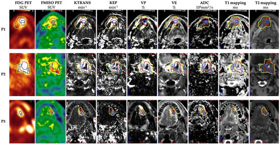
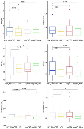
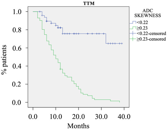
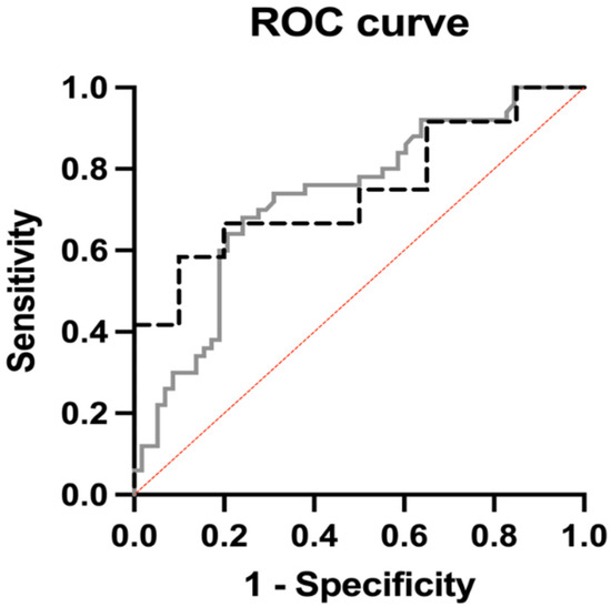
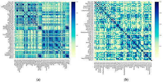
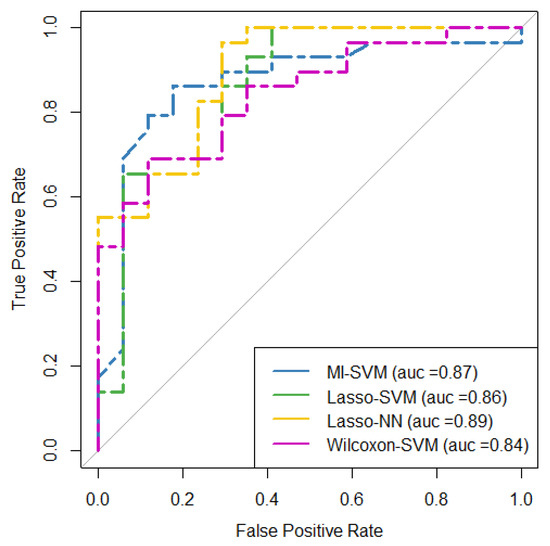
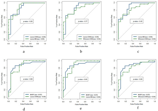
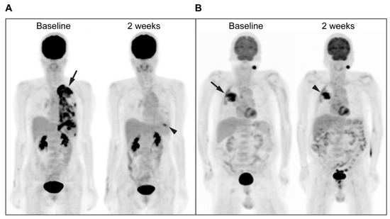
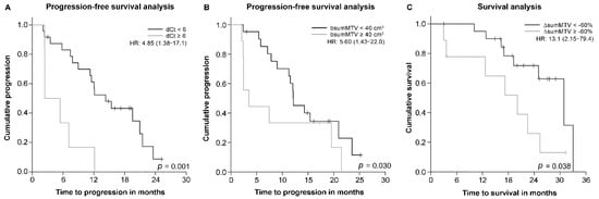
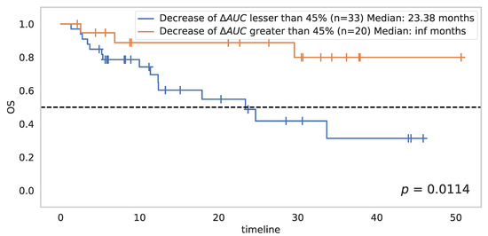
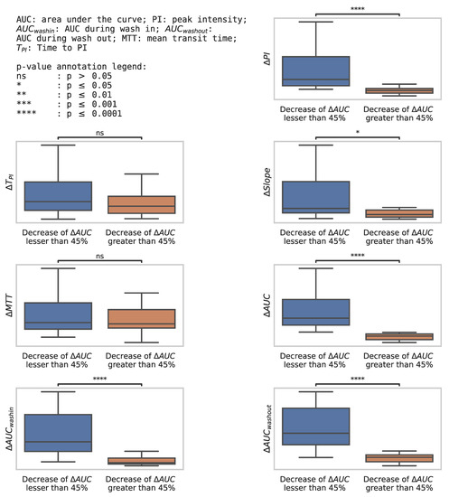
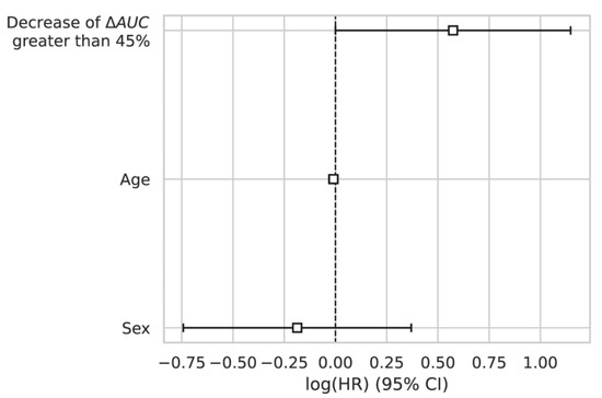
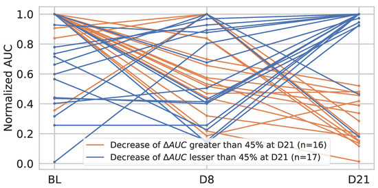
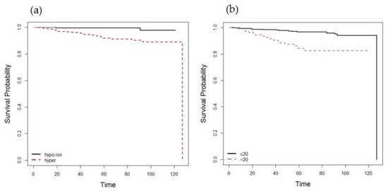
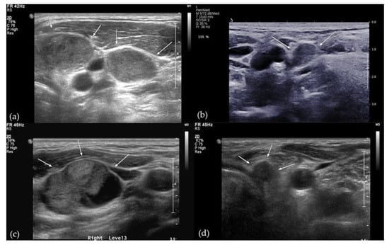
Dynamic biomarkers that permit the real-time monitoring of the tumor microenvironment response to therapy are an unmet need in breast cancer. Breast magnetic resonance imaging (MRI) has demonstrated value as a predictor of pathologic complete response and may reflect immune cell changes in the tumor microenvironment. The purpose of this pilot study was to investigate the value of breast MRI features as early markers of treatment-induced immune response. Fourteen patients with early HER2+ breast cancer were enrolled in a window-of-opportunity study where a single dose of trastuzumab was administered and both tissue and MRIs were obtained at the pre- and post-treatment stages. Functional diffusion-weighted and dynamic contrast-enhanced MRI tumor measures were compared with tumor-infiltrating lymphocytes (TILs) and RNA immune signature scores. Both the pre-treatment apparent diffusion coefficient (ADC) and the change in peak percent enhancement (DPE) were associated with increased tumor-infiltrating lymphocytes with trastuzumab therapy (r = −0.67 and -0.69, p < 0.01 and p < 0.01, respectively). Low pre-treatment ADC and a greater decrease in PE in response to treatment were also associated with immune-activated tumor microenvironments as defined by RNA immune signatures. Breast MRI features hold promise as biomarkers of early immune response to treatment in HER2+ breast cancer.
Full article
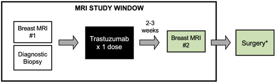
Figure 1


