


Cancers 2023, 15(24), 5727; https://0-doi-org.brum.beds.ac.uk/10.3390/cancers15245727 - 06 Dec 2023
Viewed by 821
Abstract
►
Show Figures
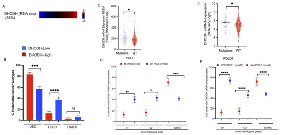
Endometrial carcinoma (EC) is the most common gynecological malignancy in the United States. De novo pyrimidine synthesis pathways generate nucleotides that are required for DNA synthesis. Approximately 38% of human endometrial tumors present with an overexpression of human dihydroorotate dehydrogenase (DHODH). However, the
[...] Read more.
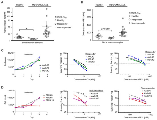
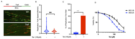
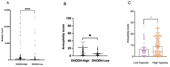
Endometrial carcinoma (EC) is the most common gynecological malignancy in the United States. De novo pyrimidine synthesis pathways generate nucleotides that are required for DNA synthesis. Approximately 38% of human endometrial tumors present with an overexpression of human dihydroorotate dehydrogenase (DHODH). However, the role of DHODH in cancer cell DNA replication and its impact on modulating a treatment response is currently unknown. Here, we report that endometrial tumors with overexpression of DHODH are associated with a high mutation count and chromosomal instability. Furthermore, tumors with an overexpression of DHODH show significant co-occurrence with mutations in DNA replication polymerases, which result in a histologically high-grade endometrial tumor. An in vitro experiment demonstrated that the inhibition of DHODH in endometrial cancer cell lines significantly induced replication-associated DNA damage and hindered replication fork progression. Furthermore, endometrial cancer cells were sensitive to the DHODH inhibitor either alone or in combination with the Poly (ADP-ribose) polymerase 1 inhibitor. Our findings may have important clinical implications for utilizing DHODH as a potential target to enhance cytotoxicity in high-grade endometrial tumors.
Full article
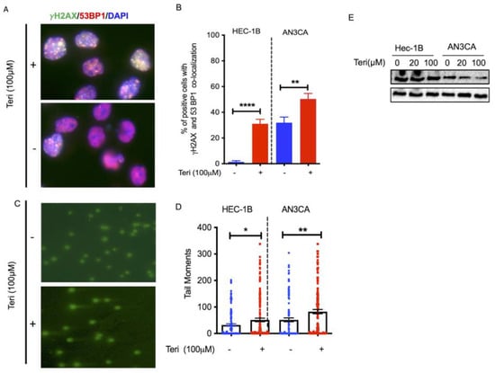
Figure 1

