


Cells 2023, 12(9), 1314; https://0-doi-org.brum.beds.ac.uk/10.3390/cells12091314 - 04 May 2023
Cited by 3 | Viewed by 2934
Abstract
►
Show Figures
According to the 2020 global cancer data released by the World Cancer Research Fund (WCRF) International, gastric cancer (GC) is the fifth most common cancer worldwide, with yearly increasing incidence and the second-highest fatality rate in malignancies. Despite the contemporary ambiguous molecular mechanisms
[...] Read more.
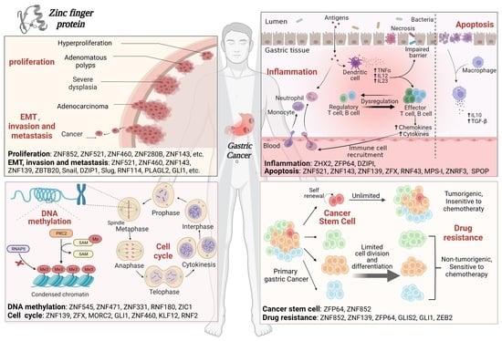
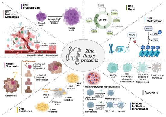
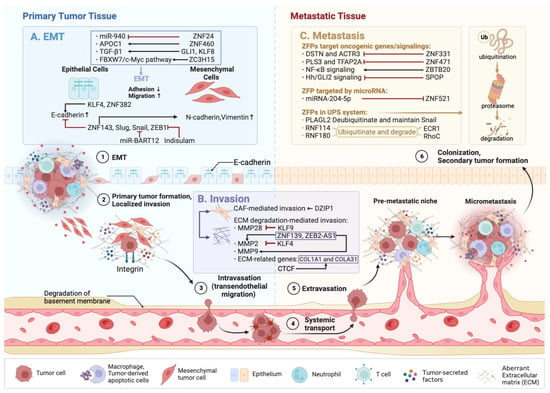
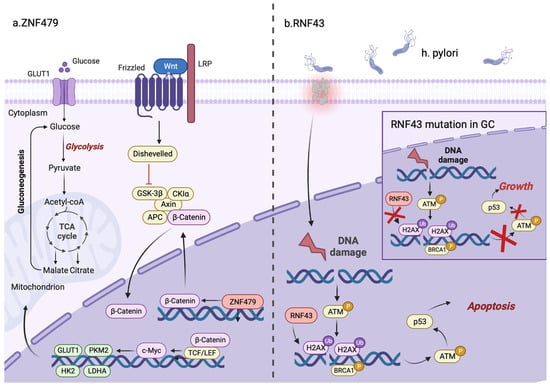
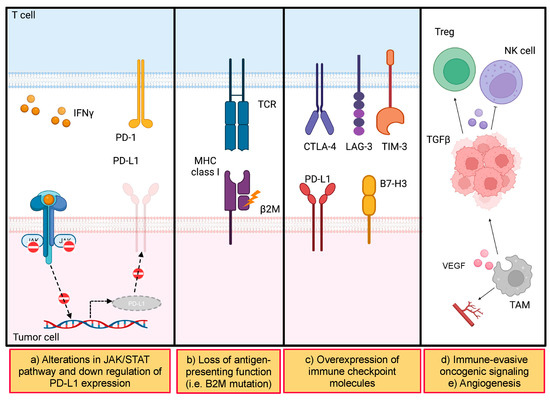
According to the 2020 global cancer data released by the World Cancer Research Fund (WCRF) International, gastric cancer (GC) is the fifth most common cancer worldwide, with yearly increasing incidence and the second-highest fatality rate in malignancies. Despite the contemporary ambiguous molecular mechanisms in GC pathogenesis, numerous in-depth studies have demonstrated that zinc finger proteins (ZFPs) are essential for the development and progression of GC. ZFPs are a class of transcription factors with finger-like domains that bind to Zn2+ extensively and participate in gene replication, cell differentiation and tumor development. In this review, we briefly outline the roles, molecular mechanisms and the latest advances in ZFPs in GC, including eight principal aspects, such as cell proliferation, epithelial–mesenchymal transition (EMT), invasion and metastasis, inflammation and immune infiltration, apoptosis, cell cycle, DNA methylation, cancer stem cells (CSCs) and drug resistance. Intriguingly, the myeloid zinc finger 1 (MZF1) possesses reversely dual roles in GC by promoting tumor proliferation or impeding cancer progression via apoptosis. Therefore, a thorough understanding of the molecular mechanism of ZFPs on GC progression will pave the solid way for screening the potentially effective diagnostic indicators, prognostic biomarkers and therapeutic targets of GC.
Full article
Graphical abstract


