


Cells 2023, 12(12), 1561; https://0-doi-org.brum.beds.ac.uk/10.3390/cells12121561 - 06 Jun 2023
Cited by 1 | Viewed by 1484
Abstract
►
Show Figures
Despite the progress made in treatments, melanoma is one of the cancers for which its incidence and mortality have increased during recent decades. In the research of new therapeutic strategies, natural polyphenols such as chrysin could be good candidates owing to their capacities
[...] Read more.
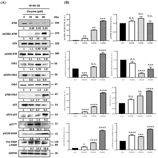
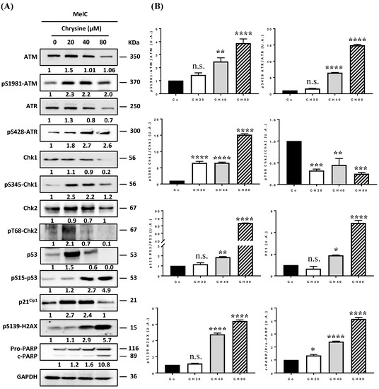
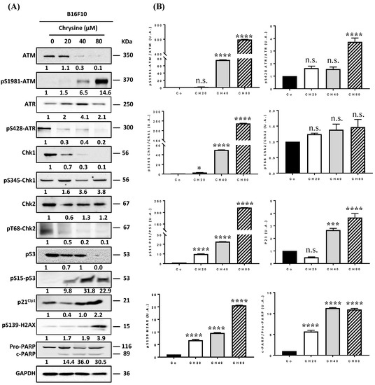
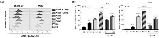
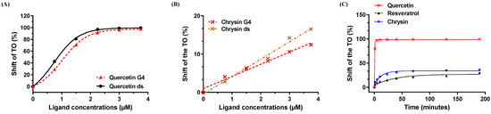
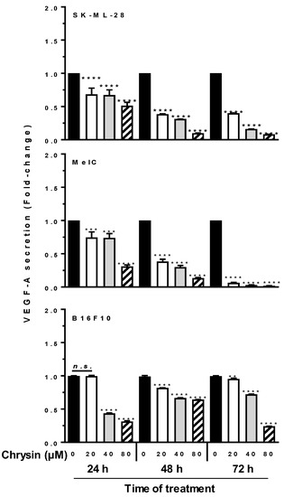
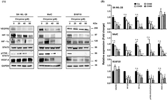
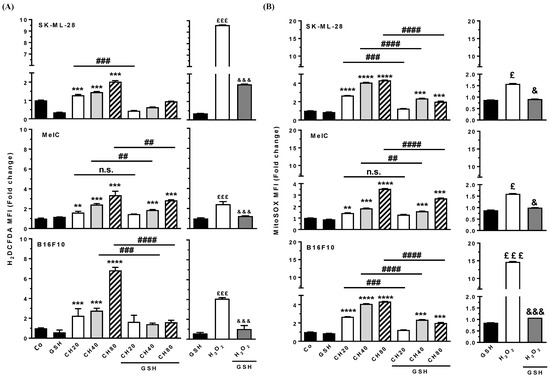
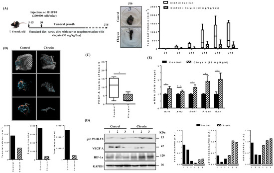
Despite the progress made in treatments, melanoma is one of the cancers for which its incidence and mortality have increased during recent decades. In the research of new therapeutic strategies, natural polyphenols such as chrysin could be good candidates owing to their capacities to modulate the different fundamental aspects of tumorigenesis and resistance mechanisms, such as oxidative stress and neoangiogenesis. In the present study, we sought to determine whether chrysin could exert antitumoral effects via the modulation of angiogenesis by acting on oxidative stress and associated DNA damage. For the first time, we show a link between chrysin-induced antiproliferative effects, the activation of the DNA damage pathway, and its ability to limit angiogenesis. More specifically, herein, we show that chrysin induces single- and double-stranded DNA breaks via the activation of the DNA damage response pathway: ATM (ataxia-telangiectasia-mutated)/Chk2 (checkpoint kinase 2) and ATR (ataxia telangiectasia and Rad3-related)/Chk1 (checkpoint kinase 1) pathways. Strong activation of this DNA damage response was found to be partly involved in the ability of chrysin to limit angiogenesis and may partly involve a direct interaction between the polyphenol and DNA G-quadruplex structures responsible for the replication fork collapse. Moreover, these events were associated with a marked reduction in melanoma cells’ capacity to secrete proangiogenic factor VEGF-A. The disruption of these key protein actors in tumor growth by chrysin was also confirmed in a syngeneic model of B16 melanoma. This last point is of importance to further consider the use of chrysin as a new therapeutic strategy in melanoma treatment.
Full article
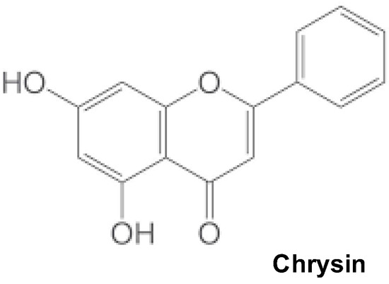
Figure 1