


Dent. J. 2022, 10(12), 238; https://0-doi-org.brum.beds.ac.uk/10.3390/dj10120238 - 13 Dec 2022
Cited by 2 | Viewed by 3088
Abstract
►
Show Figures
Background: Smile aesthetics has a vital role to play in an individual’s life and one of the factors affecting the beauty of the smile is gingival color. A gingival color change or gingival hyperpigmentation causes an unesthetic smile line, especially in patients with
[...] Read more.
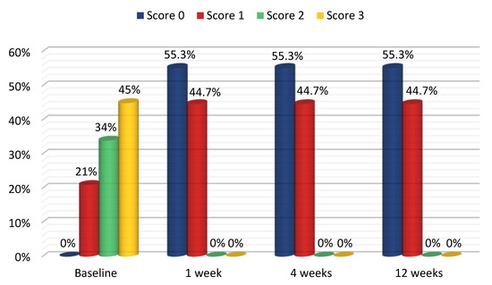
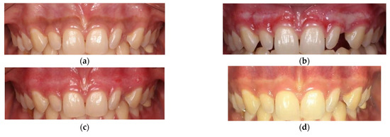
Background: Smile aesthetics has a vital role to play in an individual’s life and one of the factors affecting the beauty of the smile is gingival color. A gingival color change or gingival hyperpigmentation causes an unesthetic smile line, especially in patients with a gummy smile, which is also known as a black gummy smile. Numerous gingival depigmentation methods have been performed successfully for ablating gingival melanin pigmented epithelium. Thus, the aim of this study is to evaluate the treatment efficacy of gingival hyperpigmentation by using a carbon dioxide (CO2) laser. Methods: A cross-sectional descriptive study was carried out with 38 patients at a hospital in Vietnam. Ponnaiyan classification and the Hedin melanin index were used to assess the distribution and extent of gingival pigmentation in the study. Pain assessment was performed using the Visual Analog Scale (VAS) to evaluate the intensity of pain during the laser treatment. In addition, clinical evaluation (i.e., wound healing) of each treatment procedure was conducted using the three level Dummett–Gupta Oral Pigmentation Index (DOPI) assessment. Results: This study showed that less pain was experienced by patients treated by CO2 laser; the rates of no pain, mild pain and moderate pain after treatment were, respectively, 21%, 76% and 2.6%; there was 100% complete epithelization after 1 week. The DOPI rates for turning from a DOPI score of 1, 2 or 3 to a DOPI score of 0 after a 12-week treatment were 87.5%, 76.9% and 24%, respectively. Conclusions: Using a CO2 laser for gingival melanin pigmentation treatment is a safe and effective procedure.
Full article
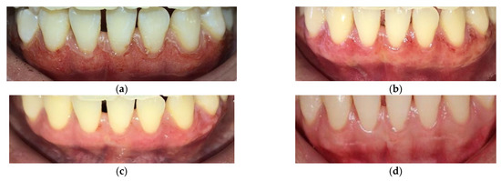
Figure 1
