


Int. J. Mol. Sci. 2023, 24(23), 17032; https://0-doi-org.brum.beds.ac.uk/10.3390/ijms242317032 - 01 Dec 2023
Viewed by 824
Abstract
►
Show Figures
This review summarizes the currently known biochemical neuroadaptive mechanisms of remote ischemic conditioning. In particular, it focuses on the significance of the pro-adaptive effects of remote ischemic conditioning which allow for the prevention of the neurological and cognitive impairments associated with hippocampal dysregulation
[...] Read more.
This review summarizes the currently known biochemical neuroadaptive mechanisms of remote ischemic conditioning. In particular, it focuses on the significance of the pro-adaptive effects of remote ischemic conditioning which allow for the prevention of the neurological and cognitive impairments associated with hippocampal dysregulation after brain damage. The neuroimmunohumoral pathway transmitting a conditioning stimulus, as well as the molecular basis of the early and delayed phases of neuroprotection, including anti-apoptotic, anti-oxidant, and anti-inflammatory components, are also outlined. Based on the close interplay between the effects of ischemia, especially those mediated by interaction of hypoxia-inducible factors (HIFs) and steroid hormones, the involvement of the hypothalamic–pituitary–adrenocortical system in remote ischemic conditioning is also discussed.
Full article
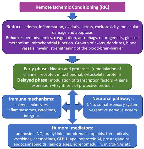
Figure 1


