


Int. J. Mol. Sci. 2024, 25(3), 1707; https://0-doi-org.brum.beds.ac.uk/10.3390/ijms25031707 - 30 Jan 2024
Viewed by 1091
Abstract
►
Show Figures
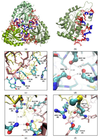
Control of schistosomiasis japonica, endemic in Asia, including the Philippines, China, and Indonesia, is extremely challenging. Schistosoma japonicum is a highly pathogenic helminth parasite, with disease arising predominantly from an immune reaction to entrapped parasite eggs in tissues. Females of this species can
[...] Read more.
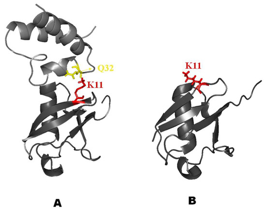
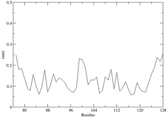
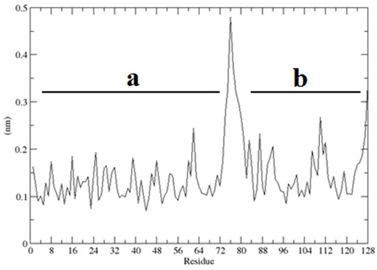
Control of schistosomiasis japonica, endemic in Asia, including the Philippines, China, and Indonesia, is extremely challenging. Schistosoma japonicum is a highly pathogenic helminth parasite, with disease arising predominantly from an immune reaction to entrapped parasite eggs in tissues. Females of this species can generate 1000–2200 eggs per day, which is about 3- to 15-fold greater than the egg output of other schistosome species. Bovines (water buffalo and cattle) are the predominant definitive hosts and are estimated to generate up to 90% of parasite eggs released into the environment in rural endemic areas where these hosts and humans are present. Here, we highlight the necessity of developing veterinary transmission-blocking vaccines for bovines to better control the disease and review potential vaccine candidates. We also point out that the approach to producing efficacious transmission-blocking animal-based vaccines before moving on to human vaccines is crucial. This will result in effective and feasible public health outcomes in agreement with the One Health concept to achieve optimum health for people, animals, and the environment. Indeed, incorporating a veterinary-based transmission vaccine, coupled with interventions such as human mass drug administration, improved sanitation and hygiene, health education, and snail control, would be invaluable to eliminating zoonotic schistosomiasis.
Full article
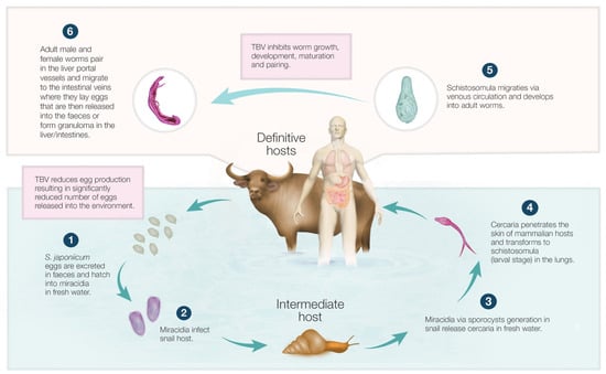
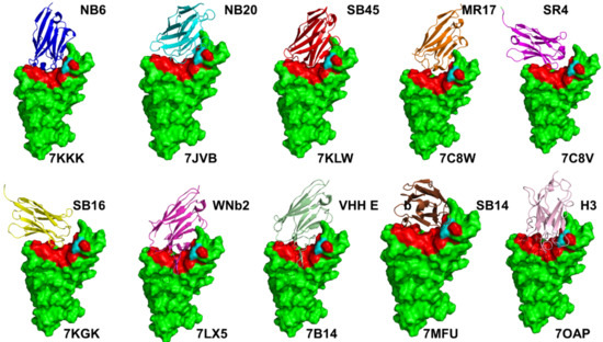
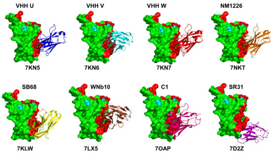
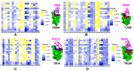
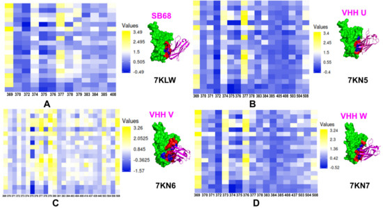
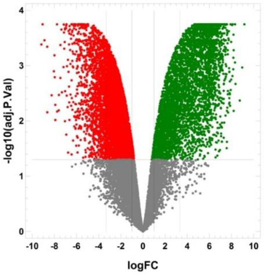
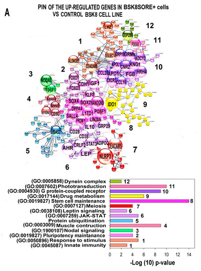
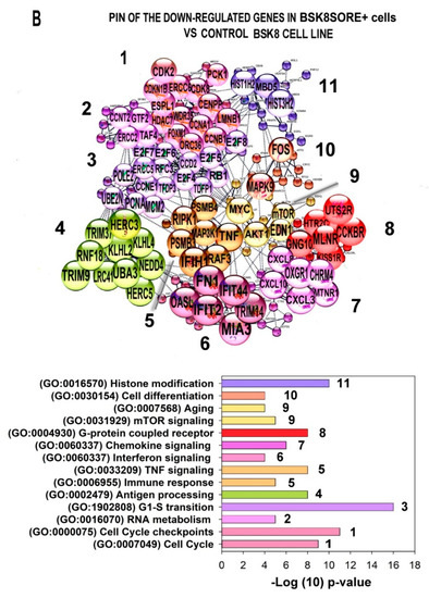
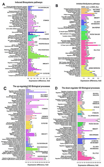
Figure 1


