


J. Funct. Biomater. 2024, 15(5), 114; https://0-doi-org.brum.beds.ac.uk/10.3390/jfb15050114 (registering DOI) - 24 Apr 2024
Abstract
Titanium with apatite-forming ability as well as antibacterial activity is useful as a component of antibacterial dental implants. When Ti was subjected to hydrogen peroxide (H2O2), copper acetate (Cu(OAc)2), and heat (H2O2-Cu(OAc)2
[...] Read more.
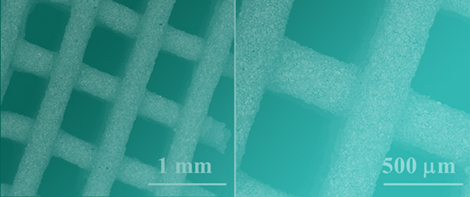
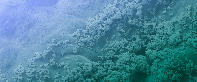
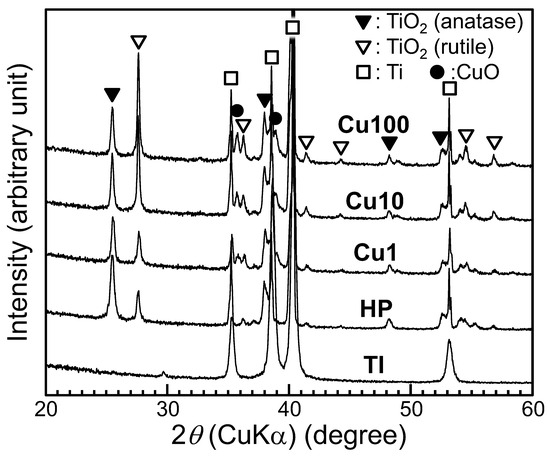
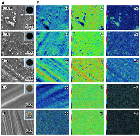
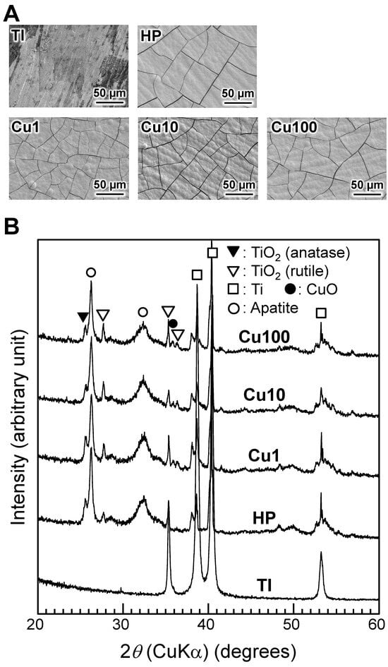
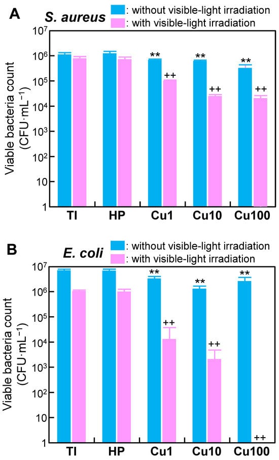
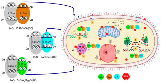
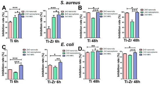
Titanium with apatite-forming ability as well as antibacterial activity is useful as a component of antibacterial dental implants. When Ti was subjected to hydrogen peroxide (H2O2), copper acetate (Cu(OAc)2), and heat (H2O2-Cu(OAc)2-heat) treatments, a network structure of anatase and rutile titanium dioxide (TiO2) and fine copper oxide (CuO) particles was formed on the Ti surface. The resulting samples accumulated a dense and uniform apatite layer on the surface when incubated in simulated body fluid and showed enhanced antibacterial activity against Escherichia coli and Staphylococcus aureus under visible-light irradiation. Electron spin resonance spectra of H2O2-Cu(OAc)2-heat-treated samples showed that hydroxyl radicals (·OH) were generated from the samples, and the concentration of ·OH increased with increasing Cu concentration of the Cu(OAc)2 solution. The enhanced antibacterial activity of these samples under visible-light irradiation may be attributable to the generation of ·OH from samples. These results suggest that Ti implants obtained using H2O2-Cu(OAc)2-heat treatments and subjected to regular or on-demand visible-light irradiation may provide a decreased risk of peri-implantitis.
Full article
(This article belongs to the Special Issue Active Biomedical Materials and Their Applications)
►
Show Figures
Figure 1


