


J. Funct. Biomater. 2012, 3(3), 688-705; https://0-doi-org.brum.beds.ac.uk/10.3390/jfb3030688 - 20 Sep 2012
Cited by 35 | Viewed by 7710
Abstract
►
Show Figures
The osseointegration rate of implants is related to their composition and surface roughness. Implant roughness favors both bone anchoring and biomechanical stability. Osteoconductive calcium phosphate (Ca-P) coatings promote bone healing and apposition, leading to the rapid biological fixation of implants. It has been
[...] Read more.
The osseointegration rate of implants is related to their composition and surface roughness. Implant roughness favors both bone anchoring and biomechanical stability. Osteoconductive calcium phosphate (Ca-P) coatings promote bone healing and apposition, leading to the rapid biological fixation of implants. It has been clearly shown in many publications that Ca-P coating accelerates bone formation around the implant. This review discusses two main routes for the manufacturing of polymer-based osteoconductive scaffolds for tissue engineering, namely the incorporation of bioceramic particles in the scaffold and the coating of a scaffold with a thin layer of apatite through a biomimetic process.
Full article
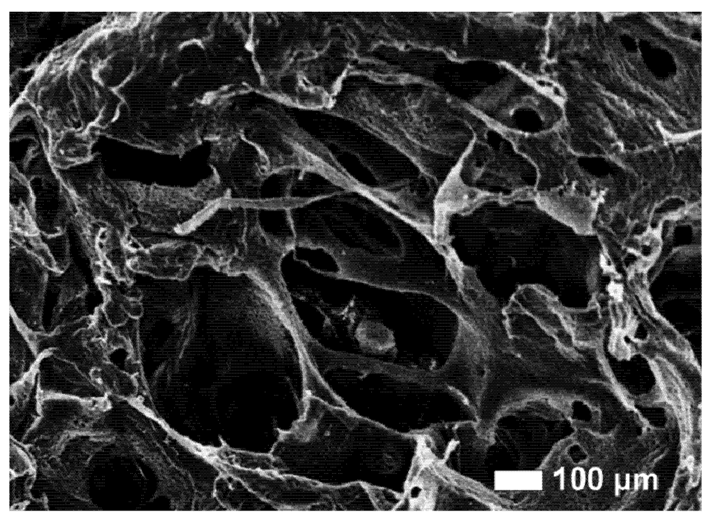
Figure 1