


Life 2022, 12(10), 1481; https://0-doi-org.brum.beds.ac.uk/10.3390/life12101481 - 23 Sep 2022
Cited by 3 | Viewed by 1817
Abstract
►
Show Figures
The role of nutraceuticals in the treatment of glaucoma remains controversial. The aim of this study was to evaluate the effect of citicoline, vitamin C, and docosahexaenoic acid (DHA) in patients with glaucoma. Methods: This was a prospective, randomized study. Patients with glaucoma
[...] Read more.
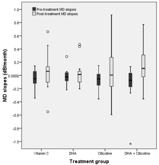
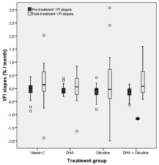
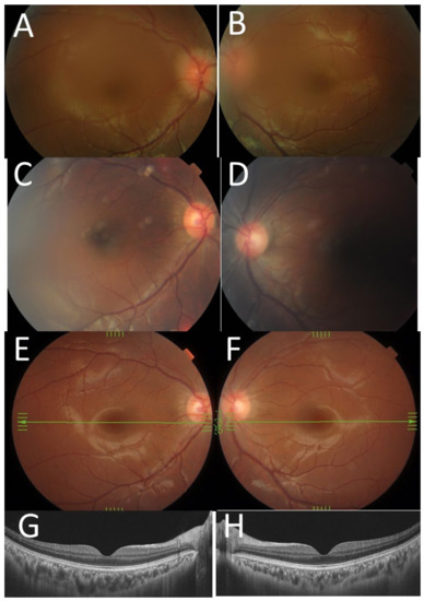
The role of nutraceuticals in the treatment of glaucoma remains controversial. The aim of this study was to evaluate the effect of citicoline, vitamin C, and docosahexaenoic acid (DHA) in patients with glaucoma. Methods: This was a prospective, randomized study. Patients with glaucoma were randomized to one of four groups and treated for 3 months with vitamin C, DHA, citicoline, or a combination of DHA and citicoline. We conducted a complete ophthalmic examination and visual fields each month and calculated the slopes of field indices. Changes in visual field indices (VFIs) and their slopes were assessed in each group and compared. Results: Seventy-three persons were included in the study. Mean defect (MD) significantly improved (p = 0.001) from −9.52 ± 4.36 to −7.85 ± 4.36 dB during the study period in persons taking DHA + citicoline. Similarly, the mean VFI significantly improved (p = 0.001) in this group. The only treatment group showing a statistically significant improvement (p = 0.006) in the MD (from −0.1041 ± 0.2471 to 0.1383 ± 0.2544 dB/month) and VFI slope was the group treated with DHA+citicoline. Conclusions: The combination of oral treatment with DHA + citicoline significantly improved VF indices and their slopes in patients with glaucoma after 3 months of treatment.
Full article
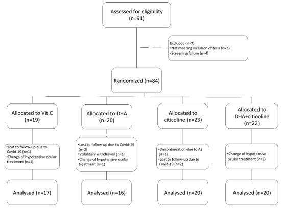
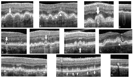
Figure 1

