Modern Approaches and Innovations on Methods and Imaging Protocols of the Maxillofacial District

A special issue of Methods and Protocols (ISSN 2409-9279). This special issue belongs to the section "Biomedical Sciences and Physiology".

Deadline for manuscript submissions: closed (1 November 2022) | Viewed by 12718

Special Issue Editors


E-Mail Website
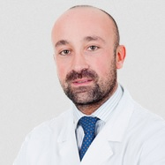
Guest Editor
Department of Oral and Maxillofacial Sciences, Sapienza University, University of Rome, 00161 Rome, Italy
Interests: endodontics; prosthodontics; CBCT; dental imaging; endodontic surgery; MRI
Special Issues, Collections and Topics in MDPI journals

Special Issue Information

Dear Colleagues,

In current imaging diagnostics, it is necessary to implement modern protocols and approaches, which minimize the patient's exposure to ionizing radiation and subsequent biological damage deriving from these. Future evolutions and radiographic diagnostic exams, always guaranteeing an adequate resolution quality, will have to limit the biological damage caused by ionizing radiations. MRI and Ultrasound are getting better and better, and more attention is being paid to these newly applied exams in the literature. The various diagnostic exams, although recently described in the literature in their recent applications, will make it possible to achieve the same accuracy and precision as radiographic examinations, minimizing the side effects of the latter.

Magnetic resonance imaging, of which there is evidence that allows it to be superimposed on CBCT or CT for dental use, has the only negative sides of its large size, its reduced availability, and the problems related to the data acquisition sequences and to the patient.

Ultrasounds, on the other hand, allow a more specific use for the oral mucosa, and for the most superficial area of the bone, for the evaluation of its density and to discriminate in the differential diagnosis of the various bone lesions. The main limitations are represented by the dependence of the exam on the operator who practices it, by the reduced choice of probes for dental use.

I expect to receive from Clinicians and researchers, original research articles, review articles, and any possible contribution that can improve the knowledge of the various specialists in the head and neck area for the prescription of the most appropriate and least invasive diagnostic exam.

Prof. Dr. Luca Testarelli
Dr. Dario Di Nardo
Dr. Rodolfo Reda
Guest Editors

Manuscript Submission Information

Manuscripts should be submitted online at www.mdpi.com by registering and logging in to this website. Once you are registered, click here to go to the submission form. Manuscripts can be submitted until the deadline. All submissions that pass pre-check are peer-reviewed. Accepted papers will be published continuously in the journal (as soon as accepted) and will be listed together on the special issue website. Research articles, review articles as well as short communications are invited. For planned papers, a title and short abstract (about 100 words) can be sent to the Editorial Office for announcement on this website.

Submitted manuscripts should not have been published previously, nor be under consideration for publication elsewhere (except conference proceedings papers). All manuscripts are thoroughly refereed through a single-blind peer-review process. A guide for authors and other relevant information for submission of manuscripts is available on the Instructions for Authors page. Methods and Protocols is an international peer-reviewed open access semimonthly journal published by MDPI.

Please visit the Instructions for Authors page before submitting a manuscript. The Article Processing Charge (APC) for publication in this open access journal is 1800 CHF (Swiss Francs). Submitted papers should be well formatted and use good English. Authors may use MDPI's English editing service prior to publication or during author revisions.

Keywords

  • 3D imaging
  • CBCT
  • MRI
  • ultrasound
  • tomography
  • Micro-CT
  • dental imaging
  • CranioFacial imaging
  • radiography

Published Papers (5 papers)

Order results
Result details
Select all
Export citation of selected articles as:

Editorial

Jump to: Research, Other

3 pages, 204 KiB  
Editorial
Modern Approaches and Innovations on Methods and Imaging Protocols of the Maxillofacial District
by Rodolfo Reda, Maurilio D’Angelo, Alessio Zanza, Dario Di Nardo and Luca Testarelli
Methods Protoc. 2022, 5(1), 2; https://0-doi-org.brum.beds.ac.uk/10.3390/mps5010002 - 27 Dec 2021
Viewed by 2261
Abstract
In recent years, improvements in imaging techniques have profoundly changed the diagnosis of pathologies of the maxillofacial district [...] Full article

Research

Jump to: Editorial, Other

9 pages, 460 KiB  
Article
Relationship between Posterior Permanent Dentition Pattern and Radiographic Changes of the Mandibular Condyle
by Mahsa Esfehani, Marayam Tofangchiha, Neda Poorsayyah, Haniyeh Rahimi, Sarvin Kholafazadeh, Sina Radfar, Francesco Pagnoni, Rodolfo Reda and Luca Testarelli
Methods Protoc. 2022, 5(6), 97; https://0-doi-org.brum.beds.ac.uk/10.3390/mps5060097 - 04 Dec 2022
Cited by 2 | Viewed by 1672
Abstract
This study assessed the relationship between posterior permanent dentition and radiographic changes of the mandibular condyle. This descriptive, cross-sectional study was conducted on 300 panoramic radiographs of patients over 40 years of age (188 females and 112 males). Panoramic radiographs were evaluated for [...] Read more.
This study assessed the relationship between posterior permanent dentition and radiographic changes of the mandibular condyle. This descriptive, cross-sectional study was conducted on 300 panoramic radiographs of patients over 40 years of age (188 females and 112 males). Panoramic radiographs were evaluated for condylar changes such as flattening, subcortical sclerosis, subcortical cyst, erosion, osteophytes, and generalized sclerosis. Presence of muscle pain and temporomandibular joint (TMJ) pain and sounds, and history of TMJ trauma were also assessed. The occlusal scheme of posterior teeth was analyzed according to the Eichner’s index. The frequency of condylar changes was calculated in the right and left sides, and their association with posterior permanent dentition was analyzed by the Chi-square test (alpha = 0.05). The frequency of flattening, muscle pain, TMJ sounds, and erosion was 11.7%, 9.7%, 5.7%, and 3.7% in the right side, respectively. The frequency of flattening, muscle pain, erosion, and subcortical cyst was 12%, 9.3%, 5%, and 5% in the left side, respectively. The frequency of bilateral muscle pain, flattening, TMJ sounds, and TMJ pain was 18%, 16.7%, 11.7%, and 9.3%, respectively. Cases with TMJ trauma, generalized sclerosis, and osteophytes were few. According to the Eichner’s index, most patients with condylar changes had classes A and B, and a smaller percentage had class C. No significant difference was noted between healthy individuals and those with condylar changes regarding dentition patterns. No relationship existed between condylar changes and posterior permanent dentition pattern. Full article
Show Figures

Figure 1

14 pages, 1542 KiB  
Article
Predictive Analysis of Maxillary Canine Impaction through Sella Turcica Bridging, Ponticulus Posticus Calcification, and Lateral Incisor Anomalies: A Retrospective Observational Study
by Rosanna Guarnieri, Francesca Germanò, Federica Altieri, Michele Cassetta, Camilla Grenga, Gabriella Padalino, Roberto Di Giorgio and Ersilia Barbato
Methods Protoc. 2022, 5(6), 91; https://0-doi-org.brum.beds.ac.uk/10.3390/mps5060091 - 21 Nov 2022
Cited by 4 | Viewed by 1646
Abstract
Maxillary canine impaction is an increasing dental anomaly and is often related to other dento-skeletal anomalies. The aim of this work is to support the clinician in evaluating the relationship between a displaced maxillary canine and clinical (the features of lateral incisors)/skeletal ( [...] Read more.
Maxillary canine impaction is an increasing dental anomaly and is often related to other dento-skeletal anomalies. The aim of this work is to support the clinician in evaluating the relationship between a displaced maxillary canine and clinical (the features of lateral incisors)/skeletal (ponticulus posticus and sella turcica bridging) anomalies through orthopanoramic radiographs, lateral cephalograms, and plaster casts to identify the parameters that best predict maxillary canine impaction. A retrospective observational study was carried out on the analysis of the medical records, radiographic findings (panoramic radiographs and lateral cephalograms), and plaster casts of 203 orthodontic patients divided into a case group, with at least one impacted maxillary canine, and a control group, without an impaction. A chi-square test and logistic regression analysis were used to analyze the data. A statistically significant association was found between the impaction of the maxillary canine and the female sex, the bridging of the sella turcica, the ponticulus posticus calcification, and the anomaly of the lateral incisor; a logistic regression revealed that these significant variables were found to be positive predictors of impacted maxillary canines, particularly in reference to the impaction in the palatal area. Finding one of these clinical and radiographic elements can represent a predictive sign of the possible impaction of the maxillary canine. Full article
Show Figures

Figure 1

Other

Jump to: Editorial, Research

12 pages, 644 KiB  
Study Protocol
Effectiveness of 38% Silver Diamine Fluoride Application along with Atraumatic Restorative Treatment for Arresting Caries in Permanent Teeth When Compared to Atraumatic Restorative Treatment in Adults—Study Protocol for a Randomized Controlled Trial
by Anju Varughese, Chandrashekar Janakiram, Balagopal Varma, Anil Mathew, Shankar Rengasamy Venugopalan, Venkitachalam Ramanarayanan and Prabath Singh
Methods Protoc. 2022, 5(6), 87; https://0-doi-org.brum.beds.ac.uk/10.3390/mps5060087 - 26 Oct 2022
Viewed by 2025
Abstract
Introduction: Dental caries in the adult population that require preventive and therapeutic treatment are generally neglected in rural communities. The determination of the effectiveness of the application of 38% silver diamine fluoride (SDF) in arresting caries lesions when combined with atraumatic restorative treatment [...] Read more.
Introduction: Dental caries in the adult population that require preventive and therapeutic treatment are generally neglected in rural communities. The determination of the effectiveness of the application of 38% silver diamine fluoride (SDF) in arresting caries lesions when combined with atraumatic restorative treatment (ART) is very important, as it serves as a preventive and restorative procedure to regain the function of the permanent dentition. The assessment of optimal SDF application with ART, in comparison with ART alone, in managing cavitated carious lesions in a pragmatic setting, is the need of the hour to recommend optimal dental care, especially in rural settings which have minimal access to comprehensive dental care. Methods and Analysis: The clinical trial will enrol 220 adults (18–65 years) with cavitated carious lesions attending the Amrita School of Dentistry in the Ernakulam district, India. This study is a randomized, controlled trial with a 1:1 allocation ratio in two parallel groups. Study arm 1 will receive 38% SDF application and ART, and study arm 2 will receive ART only. A digital radiograph will be taken immediately after restoration (baseline) as well as at the end of the 6th month for evaluation of caries arrest. The assessment of the survival of the restoration will be done on the 7th day, 30th day, and at the end of the 6th month. The final analysis would include both the tooth and person levels. Ethics and Dissemination: This trial adheres to the principles of the Declaration of Helsinki and the guidelines of the Indian Council of Medical Research (ICMR). This study protocol has been approved by the Institutional Review Board. This trial has been registered prospectively with the Clinical Trial Registry of India (Registration No: CTRI/2021/12/038816). Full article
Show Figures

Figure 1

11 pages, 1236 KiB  
Protocol
Assessment of Common Risk Factors of Non-Communicable Diseases (NCDs) and Periodontal Disease in Indian Adults: An Analytical Cross-Sectional Study
by Lakshmi Puzhankara and Chandrashekar Janakiram
Methods Protoc. 2022, 5(2), 22; https://0-doi-org.brum.beds.ac.uk/10.3390/mps5020022 - 03 Mar 2022
Cited by 1 | Viewed by 4252
Abstract
Risk factors that predispose individuals towards major non-communicable diseases (NCDs) and periodontal disease (PD) often co-occur in the same individual. The common risk factor approach (CRFA) for controlling the risk factors associated with NCDs and PD ensures that modifying a few risk factors [...] Read more.
Risk factors that predispose individuals towards major non-communicable diseases (NCDs) and periodontal disease (PD) often co-occur in the same individual. The common risk factor approach (CRFA) for controlling the risk factors associated with NCDs and PD ensures that modifying a few risk factors has an incredible impact on regulating many chronic conditions. To apply CRFA to NCDs and PD, it is essential to quantify the common risk factors of these conditions. The proposed hospital-based analytical cross-sectional study aims to assess the proportion overlap of risk factors that are common or shared between NCDs (cardiovascular diseases (CVD) and diabetes mellitus (DM) type 2) and PD. The risk factors for PD and NCDs will be estimated in subjects aged 18 years and above, diagnosed with NCDs (DM type 2, CVD) or PD. This will be a non-directional study. The dependent variables analyzed will be PD and NCDs (DM type 2, CVD). The explanatory variables that are assessed will be: age, gender, address, occupation, access and affordability of care, familial pattern, family size, insurance, socioeconomic status, obesity, tobacco usage, physical activity, alcohol consumption, food frequency, stress, and oral hygiene. The research is expected to provide data which will aid in the development of advocacy initiatives to implement CRFA for PD and NCDs. Full article
Show Figures

Figure 1

Back to TopTop