


Prosthesis 2024, 6(2), 401-412; https://0-doi-org.brum.beds.ac.uk/10.3390/prosthesis6020030 - 22 Apr 2024
Abstract
(1) Background: Since new intraoral scanner (IOS) versions are introduced to the market and software continues to advance, there is an ongoing need to assess the accuracy of newer IOS models. (2) Methods: Four types of IOSs and one laboratory scanner (used as
[...] Read more.
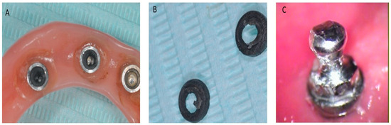
(1) Background: Since new intraoral scanner (IOS) versions are introduced to the market and software continues to advance, there is an ongoing need to assess the accuracy of newer IOS models. (2) Methods: Four types of IOSs and one laboratory scanner (used as a reference) were used to scan an edentulous model with six parallel implants and their respective scan bodies, which were connected to each other. Using dedicated software, the distances between all scan bodies were calculated, generating a total of 540 measurements. Trueness (comparisons to the reference model) and precision (intragroup comparisons) were statistically compared with ANOVA and Tukey tests. (3) Results: When considering trueness values, statistically significant differences were observed between the tested scanner for all subgroups considered (p < 0.05). By contrast, no statistically significant differences were reported for precision values. (4) Conclusions: Within the limitations of the present in vitro study, it can be concluded that all tested IOSs were similar in terms of precision, while Trios and i700W yielded the worst trueness values. Nevertheless, increasing the measuring distance leads to a decrease in both trueness and precision.
Full article


