Topic Editors

ARDEC Academy, Viale Giovanni Pascoli 67, 47923 Rimini, Italy
School of Dentistry, Saint Camillus International University for Health Sciences (Unicamillus), Rome, Italy
Department of Oral & Maxillofacial Surgery, Rabin Medical Center, Beilinson Campus, Petah Tikva 49100, Israel

New Frontiers and Boundaries in the Use of Biomaterials in Dentistry

Abstract submission deadline
closed (10 April 2022)
Manuscript submission deadline
closed (10 June 2022)
Viewed by
42769

Topic Information

Dear Colleagues,

It is a pleasure to invite you to submit manuscripts to the forthcoming Topic “New Frontiers and Boundaries in the Use of Biomaterials in Dentistry”.

Biomaterials play an important role in modern dentistry and, among them, implantology has led to a paradigm shift that has decisively influenced dental therapy and broadened the fields of dental research. However, the placement of implants requires the presence of a sufficient amount of alveolar bone. When the bone volume is not adequate, regenerative procedures are needed, which are performed using different techniques and mostly include the use of biomaterials. Among them, autogenous or heterologous bone and a large variety of synthetic or animal-derived biomaterials have been used. These biomaterials have been proposed in different conformations, such as granules, gel, blocks, membrane, novel structures, and 3D printing devices.

Unfortunately, the clinical results are not always optimal, and the research behind them is often inadequate for guiding the clinician in the correct use of the biomaterial.

This Topic aims to serve as a platform for the publication of papers detailing strong evidence in reporting on the characteristics of various biomaterials and their clinical use in dentistry.

In vitro, experimental, and clinical studies on the use of biomaterials of any type, including oral implants, may be suitable.

We look forward to receiving your submissions.

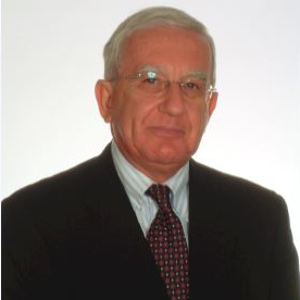
Dr. Daniele Botticelli
Prof. Dr. Adriano Piattelli
Prof. Dr. Gabi Chaushu
Topic Editors

Keywords

  • allograft
  • biomaterial
  • bone regeneration
  • bone substitute
  • tissue engineering
  • xenograft

Participating Journals

Journal Name Impact Factor CiteScore Launched Year First Decision (median) APC
Materials
materials
3.4 5.2 2008 13.9 Days CHF 2600
Dentistry Journal
dentistry
2.6 4.0 2013 27.8 Days CHF 2000
Journal of Clinical Medicine
jcm
3.9 5.4 2012 17.9 Days CHF 2600
Journal of Functional Biomaterials
jfb
4.8 5.0 2010 13.3 Days CHF 2700

Preprints.org is a multidiscipline platform providing preprint service that is dedicated to sharing your research from the start and empowering your research journey.

MDPI Topics is cooperating with Preprints.org and has built a direct connection between MDPI journals and Preprints.org. Authors are encouraged to enjoy the benefits by posting a preprint at Preprints.org prior to publication:

  1. Immediately share your ideas ahead of publication and establish your research priority;
  2. Protect your idea from being stolen with this time-stamped preprint article;
  3. Enhance the exposure and impact of your research;
  4. Receive feedback from your peers in advance;
  5. Have it indexed in Web of Science (Preprint Citation Index), Google Scholar, Crossref, SHARE, PrePubMed, Scilit and Europe PMC.

Published Papers (18 papers)

Order results
Result details
Journals
Select all
Export citation of selected articles as:
11 pages, 851 KiB  
Article
Evaluation of the Shear Bond Strength of Four Bioceramic Materials with Different Restorative Materials and Timings
by Abeer S. Alqahtani, Ayman M. Sulimany, Abdullah S. Alayad, Abdulaziz S. Alqahtani and Omar A. Bawazir
Materials 2022, 15(13), 4668; https://0-doi-org.brum.beds.ac.uk/10.3390/ma15134668 - 03 Jul 2022
Cited by 10 | Viewed by 2066
Abstract
The objectives of this study were to evaluate the shear bond strength (SBS) of a resin composite (RC) and a resin-modified glass ionomer (RMGI) to four different bioceramic materials and to compare the effects of the immediate vs. delayed placement of restoration on [...] Read more.
The objectives of this study were to evaluate the shear bond strength (SBS) of a resin composite (RC) and a resin-modified glass ionomer (RMGI) to four different bioceramic materials and to compare the effects of the immediate vs. delayed placement of restoration on the SBS. A total of 160 Teflon blocks and 40 blocks/material, were randomly filled with one of the bioceramic materials (NeoPUTTY®, NeoMTA2®, TotalFill® BC RRM™ Fast Set Putty, and ProRoot® MTA). The restoration was performed immediately or in a delayed time frame (after 7 days) using a Filtek™ Z350 XT Flowable composite (bonded to the bioceramic materials using Single bond universal 3M) or GC Fuji II LC® RMGI. The SBS test was performed at a crosshead speed of 1 mm/min, and the failure mode was evaluated under a digital microscope by one blinded examiner. One-way analysis of variance (ANOVA) with the Games–Howell post hoc test was used to compare the mean SBS between the groups. The mean SBS of the bioceramic materials to RC was significantly higher than to RMGI except for ProRoot MTA (p-value 0.65). The SBS values to RC were as follows: ProRoot MTA (7.64 MPa); NeoMTA2 (8.57 MPa) which was significantly higher than both NeoPUTTY (4.04 MPa) and TotalFill® BC RRM™ Fast Set Putty (4.38 MPa). For RMGI groups, ProRoot MTA showed the highest SBS (7.18 MPa), followed by NeoMTA2 (4.15 MPa), NeoPUTTY (1.62 MPa), and TotalFill® BC RRM™ Fast Set Putty (1.54 MPa). The delayed timing restoration showed a significantly higher SBS than the immediate, except for the immediate RMGI restoration with MTA. To conclude, the SBS of RC to the bioceramic materials was significantly higher than RMGI, except for ProRoot MTA. Both restorative materials had a significantly higher SBS to the MTA groups in comparison to premixed bioceramics. Delayed RC restoration had a higher SBS than immediate restoration. Similarly, delayed RMGI restoration had a higher SBS than immediate restoration with premixed bioceramic but not with MTA. Full article
Show Figures

Figure 1

18 pages, 27863 KiB  
Article
Fluorinated Agents Effects on Orthodontic Alloys: A Descriptive In Vitro Study
by Roberta Condò, Elisabetta Carli, Alessandro Cioffi, Maria Elena Cataldi, Vincenzo Quinzi, Adriano Casaglia, Aldo Giancotti, Paola Pirelli, Ivano Lucarini, Francesco Maita, Luca Maiolo and Gianluca Mampieri
Materials 2022, 15(13), 4612; https://0-doi-org.brum.beds.ac.uk/10.3390/ma15134612 - 30 Jun 2022
Cited by 4 | Viewed by 1584
Abstract
Fluoride-based mouthwashes and gels are preventive measures in countering demineralization and caries but, modifying environmental acidity, can reduce the wet corrosion resistance of orthodontic alloys. To evaluate chemical stability, in vitro experiments were conducted on stainless steel and nickel–titanium wires, weighed before and [...] Read more.
Fluoride-based mouthwashes and gels are preventive measures in countering demineralization and caries but, modifying environmental acidity, can reduce the wet corrosion resistance of orthodontic alloys. To evaluate chemical stability, in vitro experiments were conducted on stainless steel and nickel–titanium wires, weighed before and after immersion in household fluorinated mouthwashes and gels, measuring weight variations and elution of metal ions from acid corrosion phenomena. Elution samples were analyzed by inductively coupled plasma mass spectrometry, detecting residual ion concentration, while surface changes were analyzed under scanning electron microscopy. Results showed stainless steel wires do not undergo significant erosion when exposed to most fluorinated mouthwashes but, at prolonged exposure, alloys elute gradually greater amounts of metals and Ni–Ti wires become more sensitive to some mouthwashes. Ions’ elution varies considerably, especially for Ni–Ti wires, if exposed to household fluorinated gels, for which significant negative values were obtained. Changes, affecting wires’ outer layer, negatively act on shiny appearance and luster, reducing corrosion resistance. Although examined orthodontic wires showed good chemical stability and low toxicity, surface corrosion from exposure to fluorinated agents was observed. Home use must be accompanied by clinician prescription and, for household dental gels, must follow manufacturers’ recommendations, ensuring prophylactic action without damaging alloys surfaces. Full article
Show Figures

Graphical abstract

9 pages, 516 KiB  
Article
Salivary Markers of Oxidative Stress in Patients Undergoing Orthodontic Treatment with Clear Aligners versus Self-Ligating Brackets: A Non-Randomized Clinical Trial
by Cristina Menéndez López-Mateos, María Luisa Menéndez López-Mateos, Antonio Aguilar-Salvatierra, Gerardo Gómez-Moreno, Javier Carreño Carreño, Hoda Khaldy and Mario Menéndez-Núñez
J. Clin. Med. 2022, 11(12), 3531; https://0-doi-org.brum.beds.ac.uk/10.3390/jcm11123531 - 20 Jun 2022
Cited by 4 | Viewed by 1686
Abstract
The aim of this work was to determine advanced the oxidative protein products (AOPPs), total antioxidant capacity (TAC), and myeloperoxidase activity (MPO) in the saliva of patients undergoing orthodontic treatment with clear removable aligners in comparison with another group in treatment with fixed [...] Read more.
The aim of this work was to determine advanced the oxidative protein products (AOPPs), total antioxidant capacity (TAC), and myeloperoxidase activity (MPO) in the saliva of patients undergoing orthodontic treatment with clear removable aligners in comparison with another group in treatment with fixed passive self-ligating brackets applying light forces, before treatment, after 30 days, and after 90 days of treatment. This non-randomized clinical trial recruited patients consecutively, all of which were over 18 years of age and due to undergo orthodontic treatment. They were divided into two groups according to treatment type: Group A, 48 patients treated with clear aligners (Invisalign®); and Group B, 19 patients treated with Damon System® 0.22″ self-ligating brackets applying light forces. Saliva samples were collected by a single clinician following the same protocol and underwent three analyses—AOPPs, TAC, and MPO levels–at baseline before placing the apparatus, after 30 days, and after 90 days treatment. Orthodontic treatment, whether with clear aligners or fixed self-ligating brackets and light forces, increased AOPPs after the first 30 days of treatment. During the initial phases of orthodontic treatment, neither clear aligners nor fixed self-ligating brackets applying light forces showed changes in TAC and MPO. Orthodontic treatment with both clear aligners and fixed apparatus self-ligating brackets applying light forces increases oxidative stress (AOPPs) after the first 30 days of treatment. There are no differences in AOPP levels between treatment with clear aligners and self-ligating brackets during the first 90 days of treatment. The antioxidative capacity of saliva during the initial phases of orthodontic treatment, whether with self-ligating brackets or clear aligners, does not undergo significant changes. With either orthodontic technique, the patients’ salivary antioxidant capacity is similar. Full article
Show Figures

Figure 1

10 pages, 391 KiB  
Article
Oral and Dental Considerations of Combat-Induced Post Traumatic Stress Disorder (PTSD)—A Cross-Sectional Study
by Nirit Tagger-Green, Carlos Nemcovsky, Naama Fridenberg, Orr Green, Liat Chaushu and Roni Kolerman
J. Clin. Med. 2022, 11(11), 3249; https://0-doi-org.brum.beds.ac.uk/10.3390/jcm11113249 - 06 Jun 2022
Viewed by 2234
Abstract
Objective: This study compared dental, periodontal, oral, and joint/muscle tenderness among Israeli combat-induced post-traumatic stress disorder (Ci-PTSD) war veterans to non-PTSD patients. Study design: This retrospective three-arm study compared oral and facial manifestations between 100 Israeli veterans with Ci- PTSD (study group) and [...] Read more.
Objective: This study compared dental, periodontal, oral, and joint/muscle tenderness among Israeli combat-induced post-traumatic stress disorder (Ci-PTSD) war veterans to non-PTSD patients. Study design: This retrospective three-arm study compared oral and facial manifestations between 100 Israeli veterans with Ci- PTSD (study group) and 103 non-PTSD periodontal patients (Control group). The study group was further divided into two subgroups of individuals who received psychiatric medications (40 patients) or did not (60 patients). All patients underwent complete dental, oral, and periodontal examinations, including assessing signs of parafunction. Results: All PTSD patients had poor oral hygiene. The plaque index (PI) was higher in the PTSD group compared to the control group (0.72 ± 0.28 vs. 0.45 ± 0.29, respectively, p < 0.001). The decayed, missing, and filled teeth score (DMFt) was higher in the PTSD population than in the controls (19.97 ± 8.07 vs. 13.05 ± 6.23 p < 0.05). Severe periodontal disease was more common among the PTSD subgroup taking medications (med -group) (62.5%) compared to the nonmedicated group (non-med group) (30.0%) and the controls (27.2%) (p = 0.001). Heavy smoking was more prevalent in the medicated PTSD patients than in other groups. Conclusions: The present study shows higher morbidities in combat-induced PTSD patients, including oral, dental, and periodontal manifestations, especially in medicated patients. Full article
Show Figures

Figure 1

9 pages, 600 KiB  
Article
Ozone in Patients with Periodontitis: A Clinical and Microbiological Study
by Ana Maria Ramirez-Peña, Arturo Sánchez-Pérez, Matilde Campos-Aranda and Francisco Javier Hidalgo-Tallón
J. Clin. Med. 2022, 11(10), 2946; https://0-doi-org.brum.beds.ac.uk/10.3390/jcm11102946 - 23 May 2022
Cited by 6 | Viewed by 2352
Abstract
The purpose of this article was to assess the effectiveness of ozone therapy as an adjunct to mechanical therapy in periodontitis patients. Thirty-two patients diagnosed with generalized periodontitis were selected, with a total of 655 teeth examined. Each patient’s mouth was divided into [...] Read more.
The purpose of this article was to assess the effectiveness of ozone therapy as an adjunct to mechanical therapy in periodontitis patients. Thirty-two patients diagnosed with generalized periodontitis were selected, with a total of 655 teeth examined. Each patient’s mouth was divided into four quadrants (the split-mouth model) to be randomly treated with four sessions of gaseous ozone or air. The following clinical variables were recorded: the gingival index, the periodontal clinical attachment loss, the Miller’s mobility index and the clinical improvements, as assessed through the visual analog scale (VAS). In addition, the microorganisms were qualitatively compared. After four weeks of treatment, the teeth of the ozone-treated quadrants showed statistically significant reductions in the gingival index and an improvement in the clinical attachment (p < 0.0001). The same treatment also significantly improved mobility by between 70% and 86% compared to the control group (p < 0.0001). Statistically significant differences were also recorded for the VAS (p < 0.0001). In the qualitative study of the subgingival flora, significant differences were observed (p < 0.0001). The overall results of this trial support the view that ozone treatment is effective and well tolerated in cases of generalized chronic periodontitis. Full article
Show Figures

Figure 1

18 pages, 4494 KiB  
Article
Immediate Maxillary Full-Arch Rehabilitation of Periodontal Patients with Terminal Dentition Using Tilted Implants and Bone Augmentation: A 5-Year Retrospective Cohort Study
by Gil S. Slutzkey, Omer Cohen, Liat Chaushu, Arkadi Rahmanov, Eitan Mijiritsky, Ilan Beitlitum and Roni Kolerman
J. Clin. Med. 2022, 11(10), 2902; https://0-doi-org.brum.beds.ac.uk/10.3390/jcm11102902 - 20 May 2022
Cited by 8 | Viewed by 2674
Abstract
Background: All-on-four protocols with tilted implants in the maxilla are used to rehabilitate the terminal dentition of the severe generalized periodontitis patients. Data on long-term biological complications are scarce. Methods: Eighty-four axial and forty-six tilted immediate implants have been placed in the extraction [...] Read more.
Background: All-on-four protocols with tilted implants in the maxilla are used to rehabilitate the terminal dentition of the severe generalized periodontitis patients. Data on long-term biological complications are scarce. Methods: Eighty-four axial and forty-six tilted immediate implants have been placed in the extraction sockets of 23 patients according to a four–six implants protocol combined with ridge augmentation. Within 72 h, a provisional prosthesis was cemented to the implants; after 6 months, a cemented ceramic–metallic prosthesis was delivered. The patients were followed for up to 5 years. Results: The 5-year survival rate of the straight and tilted implants was 100% and 97.8, and the prosthetic one was 100%. Marginal bone loss (MBL) of the straight implants was 0.42 ± 0.67 and 0.59 ±1.01 mm on the mesial and distal sides; for the tilted, it was 0.37 ± 0.68 and 0.34 ±0.62 mm, and the differences were not statistically significant. Implant position, smoking, keratinized mucosal width, and cantilever did not affect MBL. Peri-implant mucositis involved 29.4% and 22.2% of the straight and tilted implants, respectively; peri-implantitis involved 5.8% and 4.4% of the straight and tilted implants, respectively, without statistical significance. Conclusions: This immediate loading protocol’s 5-year survival and success rates were high. No difference between the straight and tilted implants was found regarding survival, success rates, and MBL. Full article
Show Figures

Figure 1

12 pages, 1295 KiB  
Article
Cytotoxicity of Methacrylate Dental Resins to Human Gingival Fibroblasts
by Jolanta Sulek, Elzbieta Luczaj-Cepowicz, Grazyna Marczuk-Kolada, Maciej Rosłan and Adam Holownia
J. Funct. Biomater. 2022, 13(2), 56; https://0-doi-org.brum.beds.ac.uk/10.3390/jfb13020056 - 11 May 2022
Cited by 1 | Viewed by 2205
Abstract
This study aimed to assess the acute and delayed cytotoxicity of three, popular light-cured methacrylate-based restorative resins (MRs): Charisma (C), Estelite (E), and Filtek (F), to human gingival fibroblasts in culture. Cells were grown for up to 24 h with light-cured (or pre-cured) [...] Read more.
This study aimed to assess the acute and delayed cytotoxicity of three, popular light-cured methacrylate-based restorative resins (MRs): Charisma (C), Estelite (E), and Filtek (F), to human gingival fibroblasts in culture. Cells were grown for up to 24 h with light-cured (or pre-cured) resins. We evaluated resin cytotoxicity, redox imbalance, necrosis/apoptosis, miR-9, and heat shock protein 70 (HSP70). The role of resin-induced oxidative stress (damage) in HSP70-response (repair) was assessed using binary fluorescence labeling. All MRs decreased viable cell numbers and cell proliferation and damaged cell membranes, and their 24 h-delayed toxicity was lower (C), higher (F), or similar (E) to that induced by freshly-cured resins. Cell membrane damage induced by C and E decreased with time, while F produced a linear increase. All resins generated intracellular oxidative stress with the predominant necrotic outcome, and produced heterogeneous responses in miR-9 and HSP70. The double fluorescence (damage/repair) experiments pointed to common features of E and F but not C. In the subset of cells, the binary response induced by E and F was different from C, similar to each other, and positively interrelated. Experimental data show that selective MR cytotoxicity should be taken into account when considering repetitive use or massive reconstruction. Full article
Show Figures

Graphical abstract

8 pages, 1646 KiB  
Article
Effectiveness of Different Irrigant Activation Systems on Smear Layer Removal: A Scanning Electron Microscopic Study
by Ramón Miguéns-Vila, Benjamín Martín-Biedma, Saleta Aboy-Pazos, David Uroz-Torres, Pablo Álvarez-Nóvoa, Ana Belén Dablanca-Blanco, Iván Varela-Aneiros and Pablo Castelo-Baz
J. Clin. Med. 2022, 11(4), 1003; https://0-doi-org.brum.beds.ac.uk/10.3390/jcm11041003 - 15 Feb 2022
Cited by 3 | Viewed by 1888
Abstract
The aim of this study was to evaluate the effectiveness of smear layer removal after the use of different irrigation methods (passive ultrasonic irrigation (PUI), continuous ultrasonic irrigation (CUI), apical negative pressure irrigation and conventional irrigation) using scanning electron microscopy (SEM) as an [...] Read more.
The aim of this study was to evaluate the effectiveness of smear layer removal after the use of different irrigation methods (passive ultrasonic irrigation (PUI), continuous ultrasonic irrigation (CUI), apical negative pressure irrigation and conventional irrigation) using scanning electron microscopy (SEM) as an analytical tool. A total of 100 single-canal teeth were decoronated and randomly divided into five groups (n = 20) according to the irrigation method used: conventional irrigation with front outlet syringe, conventional irrigation with lateral outlet syringe, apical negative pressure irrigation (EndoVac), PUI with Irrisafe and CUI with ProUltra PiezoFlow ultrasonic irrigation needle. Root canal preparation was performed with the ProTaper Gold system up to the F4 instrument, and 5.25% NaOCl was used as an irrigant. After chemical-mechanical preparation, the roots were split longitudinally, and the coronal, middle and apical thirds were examined. SEM digital photomicrographs were taken at ×1000 magnification to evaluate the amount of smear layer in each root canal third; CUI significantly removed more smear layer than any other irrigant activation protocol (p < 0.05); CUI was more effective in removing the smear layer than the other irrigation protocols. However, none of the irrigation protocols were able to produce root canals completely free from smear layer. Full article
Show Figures

Figure 1

10 pages, 1069 KiB  
Article
Microtensile Bond Strength of Bioactive Pit and Fissure Sealants Bonded to Primary and Permanent Teeth
by Abdulelah AlQahtani, Yousef Al-Dlaigan and Ahmed Almahdy
Materials 2022, 15(4), 1369; https://0-doi-org.brum.beds.ac.uk/10.3390/ma15041369 - 12 Feb 2022
Cited by 3 | Viewed by 2493
Abstract
Background: Sealing occlusal pits and fissures is an effective preventive measure against dental caries. Pit and fissure sealants (PFS) should be strongly bonded to the teeth to prevent partial or complete loss of the sealant, which may limit its preventive effect. Objective: The [...] Read more.
Background: Sealing occlusal pits and fissures is an effective preventive measure against dental caries. Pit and fissure sealants (PFS) should be strongly bonded to the teeth to prevent partial or complete loss of the sealant, which may limit its preventive effect. Objective: The objective of the study was to compare the microtensile bond strength (μTBS) of bioactive resin-based sealants (Bio-RBS) and resin-based sealants (RBS), with and without the use of a bonding agent, to the enamel of primary and permanent teeth. Methods: One hundred and twenty caries-free primary molar specimens and 120 permanent molar specimens were divided to eight groups (30 specimens per group), both primary and permanent teeth were sealed with a Bio-RBS BioCoatTM (Premier®, Plymouth Meeting, PA, USA) or with a RBS ClinproTM (3M ESPE, Saint Paul, MN, USA), with or without the use of a bonding agent (Prime & Bond NT; Dentsply, Inc., Charlotte, NC, USA). Half the specimens were aged with 5000 thermal cycles, and all specimens were tested for the μTBS and failure mode. Results: The mean μTBS of aged Bio-RBS was higher in permanent teeth than primary teeth, and the aging process reduced the μTBS of RBS more than that of Bio-RBS. Moreover, the addition of a bonding agent improved the μTBS of aged RBS in permanent teeth. Conclusion: We concluded that Bio-RBS exhibit superior μTBS than RBS when applied to permanent teeth. Full article
Show Figures

Figure 1

12 pages, 19309 KiB  
Article
Guided Bone Regeneration in the Edentulous Atrophic Maxilla Using Deproteinized Bovine Bone Mineral (DBBM) Combined with Platelet-Rich Fibrin (PRF)—A Prospective Study
by João Manuel Mendez Caramês, Filipe Araújo Vieira, Gonçalo Bártolo Caramês, Ana Catarina Pinto, Helena Cristina Oliveira Francisco and Duarte Nuno da Silva Marques
J. Clin. Med. 2022, 11(3), 894; https://0-doi-org.brum.beds.ac.uk/10.3390/jcm11030894 - 08 Feb 2022
Cited by 6 | Viewed by 3035
Abstract
Background: Bone regeneration procedures represent a major challenge in oral surgery. This study aimed to evaluate a composite PRF/particulate xenograft in guided bone regeneration. Methods: Edentulous patients with horizontal ridge deficiencies in the anterior maxilla and candidates to an immediate-loading full-arch rehabilitation were [...] Read more.
Background: Bone regeneration procedures represent a major challenge in oral surgery. This study aimed to evaluate a composite PRF/particulate xenograft in guided bone regeneration. Methods: Edentulous patients with horizontal ridge deficiencies in the anterior maxilla and candidates to an immediate-loading full-arch rehabilitation were included. Horizontal linear measurements indicating bone gain were assessed from computer beam computer tomography (CBCT) scans obtained at pre-surgery, post-surgery, and the 12-month follow-up. Mean bone values were presented as mean ± 95% CI. Non-parametric tests were used as appropriate, and the effect size was calculated with Cohen’s d repeated measures. Results: Eighteen patients were rehabilitated with 72 implants. The mean horizontal bone width was 4.47 [4.13–4.80] mm pre-surgically, 9.25 [8.76–9.75] mm post-surgically, and 7.71 [7.28–8.14] mm 12 months after. Conclusions: PRF associated with a xenograft seems to promote an effective horizontal bone gain. Randomized clinical trials are needed to confirm the benefits of this surgical approach. Full article
Show Figures

Figure 1

18 pages, 11636 KiB  
Systematic Review
Physico-Chemical Properties of Calcium-Silicate vs. Resin Based Sealers—A Systematic Review and Meta-Analysis of Laboratory-Based Studies
by Viresh Chopra, Graham Davis and Aylin Baysan
Materials 2022, 15(1), 229; https://0-doi-org.brum.beds.ac.uk/10.3390/ma15010229 - 29 Dec 2021
Cited by 11 | Viewed by 2361
Abstract
Introduction: The aim of this systematic review is to analyse the effect of physico-chemical properties of calcium silicate-based sealers in comparison to epoxy resin sealers in permanent teeth using a single-cone obturation technique. Methods: The study was conducted according to the guidelines of [...] Read more.
Introduction: The aim of this systematic review is to analyse the effect of physico-chemical properties of calcium silicate-based sealers in comparison to epoxy resin sealers in permanent teeth using a single-cone obturation technique. Methods: The study was conducted according to the guidelines of Cochrane Handbook for Systematic Reviews of Interventions and Preferred Reporting Items for Systematic Review and Meta-Analyses (PRISMA) statement. Literature search was performed using the PubMed/MEDLINE, Cochrane Central Register of Controlled Trials, Web of Science, DOAJ, Open Gray with no language restrictions until October 2020. Two reviewers assessed the studies for eligibility. Grading of Recommendations, Assessment, Development, and Evaluations (GRADE) was carried out to assess the evidence. Meta-analysis of the pooled data with subgroups was performed using the RevMan software (p < 0.05). Results: Results from the 28 included studies showed that the mean difference in adaptation to root canal walls (marginal adaptation, interfacial gaps and void volume) for both sealers were non-significant. However, void volume values showed a significant mean difference (p < 0.00001) favouring the calcium silicate-based sealers. The pooled meta-analysis reported statistically significant differences for apical microleakage (p = 0.0007) whilst there were non-significant mean differences for fracture resistance (p = 0.09) and push-out bond strength (p = 0.63). The heterogeneity among the included studies was 97% (I2). Conclusions: Within the limitations of this review, calcium silicate-based sealers demonstrated a similar or superior performance in comparison to resin-based sealers in terms of the physico-chemical properties. Full article
Show Figures

Figure 1

18 pages, 2936 KiB  
Article
Effect of Topical PTH 1-34 Functionalized to Biogran® in the Process of Alveolar Repair in Rats Submitted to Orchiectomy
by Paula Buzo Frigério, Pedro Henrique Silva Gomes-Ferreira, Fábio Roberto de Souza Batista, Juliana Moura, Idelmo Rangel Garcia Júnior, Daniele Botticelli, Paulo Noronha Lisboa-Filho and Roberta Okamoto
Materials 2022, 15(1), 207; https://0-doi-org.brum.beds.ac.uk/10.3390/ma15010207 - 28 Dec 2021
Cited by 6 | Viewed by 1770
Abstract
(1) Background: There are many therapies for osteoporosis control and bone maintenance; anabolic drugs such as teriparatide and bone grafts help in the repair process and stimulate bone formation. Thus, the aim of the present study was to evaluate the behavior of repaired [...] Read more.
(1) Background: There are many therapies for osteoporosis control and bone maintenance; anabolic drugs such as teriparatide and bone grafts help in the repair process and stimulate bone formation. Thus, the aim of the present study was to evaluate the behavior of repaired bone in the presence of PTH (teriparatide) associated with Biogran® (biomaterial) through a sonochemical procedure after extraction in rats. (2) Methods: The insertion of Biogran® with PTH in the alveolus was performed 30 days after incisor extraction. Euthanasia occurred after 60 days. (3) Results: The use of local treatment of PTH loaded with Biogran® in healthy rats promoted good results for micro-CT, with an increase in percentage and bone volume, number and trabecular separation and less total porosity. Greater immunostaining for Wnt, β-Catenin and osteocalcin proteins and lower expression for Thrombospondin-Related Adhesive Protein (TRAP), which shows an increase in the number of osteoblasts and inhibition of osteoclast action. However, the treated orchiectomized groups did not obtain such expressive results. (4) Conclusion: The use of Biogran® with PTH improved alveolar repair in rats. However, new researches with more efficient doses must be studied to collaborate effectively with the formation of a quality bone after the orchiectomy. Full article
Show Figures

Figure 1

10 pages, 1927 KiB  
Article
Comparison of Silane Heat Treatment by Laser and Various Surface Treatments on Microtensile Bond Strength of Composite Resin/Lithium Disilicate
by Goknil Ergun-Kunt, Rafat Sasany, Mehmet Faruk Koca and Mutlu Özcan
Materials 2021, 14(24), 7808; https://0-doi-org.brum.beds.ac.uk/10.3390/ma14247808 - 16 Dec 2021
Cited by 3 | Viewed by 2254
Abstract
In the current study, we evaluated the effects of heat treatment (by Er:YAG or furnace) and various surface treatments on the microtensile bond strength (μTBS) of silanized lithium disilicate ceramic. Seventy lithium disilicate (IPS e. max Press; Ivoclar Vivadent) and composite resin (Tetric [...] Read more.
In the current study, we evaluated the effects of heat treatment (by Er:YAG or furnace) and various surface treatments on the microtensile bond strength (μTBS) of silanized lithium disilicate ceramic. Seventy lithium disilicate (IPS e. max Press; Ivoclar Vivadent) and composite resin (Tetric N-Ceram; Ivoclar Vivadent) blocks were made and distributed into seven groups (n = 10) at random: S: silanization alone; ALS: airborne particle abrasion (APA) and silanization; SC: APA modified with silica and silanization; SHT1: silanization and heat treatment by Er:YAG; SHT2: silanization and heat treatment performed in the furnace (100 °C, 1 min); HF: etching with HF; and HFS: etching with HF and silanization. Every ceramic specimen was cemented to a composite resin block after surface treatment. Cemented specimens were embedded into acrylic resin and were tested with the μTBS test. Data were analyzed using one-way ANOVA and Tamhane T2 tests (α = 0.05). The SHT1 group had the highest bond of strength compared to the other groups (27.46 MPa). The ALS group had the lowest strength of the groups (15.56 MPa). Between SHT2 and HFS (p = 1), the comparison of the mean µTBS values showed no significant differences. It was concluded that silane heat treatment increased the resin composite–ceramic bond strength; however, within the terms of μTBS, the Er:YAG laser treatment was more successful than other surface treatment applications. Full article
Show Figures

Figure 1

11 pages, 1554 KiB  
Article
Evaluation of Newly Introduced Bioactive Materials in Terms of Cavity Floor Adaptation: OCT Study
by Heba B. Abdel-Maksoud, Aziza W. Bahanan, Lujain J. Alkhattabi and Turki A. Bakhsh
Materials 2021, 14(24), 7668; https://0-doi-org.brum.beds.ac.uk/10.3390/ma14247668 - 12 Dec 2021
Cited by 1 | Viewed by 2662
Abstract
Objective. The aim of the present study was to evaluate the adaptation of newly introduced bioactive restorative materials to the cavity floor using cross-polarization optical coherence tomography (CP-OCT). Materials and Methods. Round class V cavities were prepared on the proximal surfaces of sixty [...] Read more.
Objective. The aim of the present study was to evaluate the adaptation of newly introduced bioactive restorative materials to the cavity floor using cross-polarization optical coherence tomography (CP-OCT). Materials and Methods. Round class V cavities were prepared on the proximal surfaces of sixty non-carious human anterior teeth (0.5 mm depth × 4 mm diameter), which were divided into groups according to the restorative material (n = 15). In the VF group, Vertise flow composite (Kerr, Orange, CA, USA) was used, in the BF group, Beautifil II composite (Shofu, Koyoto, Japan) was used, and in the AB group, ACTIVA BioACTIVE composite (Pulpdent, Watertown, NY, USA) was used. Cavities were restored using the bulk filling technique and cured according to the manufacturers’ instructions. Then, the specimens were immersed in a contrasting agent, and image acquisitions were taken by CP-OCT to calculate the adaptation percentage by using an image analysis software. Results. B-scans showed a diffuse bright band of white pixels at the tooth-resin interface that was interpreted as a micro-gap present between the cavity floor and restorative material. The Kruskal-Wallis test showed a statistically significant difference between all tested groups with the AB group representing the least gap formation, followed by the BF group, and then the VF group, which demonstrated the highest gap formation. Conclusions. In class V cavities, better adaptation to the cavity floor can be obtained when using ACTIVA BioACTIVE more than Vertise flow and Beautifil II composites. In addition, CP-OCT is considered a non-destructive imaging tool that helps in evaluating the quality of the tooth-restoration interface when bioactive composites are used. Full article
Show Figures

Figure 1

15 pages, 4774 KiB  
Article
Effect of Two Brands of Glaze Material on the Flexural Strength and Probability of Failure of High Translucent Monolithic Zirconia
by Raj Gaurav Singh, Kai Chun Li, Karl Michael Lyons and John Neil Waddell
Materials 2021, 14(22), 7022; https://0-doi-org.brum.beds.ac.uk/10.3390/ma14227022 - 19 Nov 2021
Cited by 5 | Viewed by 1844
Abstract
(1) Background: The effect of glazing on the mechanical properties of monolithic high translucent zirconia is not well reported. Therefore, the purpose of this study was to evaluate the effect of glazing on the flexural strength of high translucent zirconia; (2) Methods: Ninety [...] Read more.
(1) Background: The effect of glazing on the mechanical properties of monolithic high translucent zirconia is not well reported. Therefore, the purpose of this study was to evaluate the effect of glazing on the flexural strength of high translucent zirconia; (2) Methods: Ninety specimens were prepared from second-generation 3Y-TZP high translucent blocks and divided into three groups. Glaze materials were applied on one surface of the specimen and subjected to a four-point bending test and flexural stress and flexural displacement values were derived. Descriptive fractographic analysis of surfaces was conducted to observe the point of failure and fracture pattern.; (3) Results: Control-nonglazed (647.17, 1σ = 74.71 MPa) presented higher flexural strength values compared to glaze I (541.20, 1σ = 82.91 MPa) and glaze II (581.10, 1σ = 59.41 MPa). Characteristic strength (σƟ) from Weibull analysis also observed higher (660.67 MPa) values for the control specimens. Confocal microscopy revealed that glazed surfaces were much rougher than control surfaces. Descriptive fractographic analysis revealed that there was no correlation between the point of failure initiation and flexural strength; (4) Conclusions: The test results demonstrated that glazing significantly decreased the flexural strength and flexural displacement of the zirconia specimens. Full article
Show Figures

Figure 1

7 pages, 791 KiB  
Article
Siloranes–Suitability of a Novel Adhesive for Orthodontic Bracket Bonding
by Lorenz Brauchli and Markus Steineck
Dent. J. 2021, 9(11), 135; https://0-doi-org.brum.beds.ac.uk/10.3390/dj9110135 - 17 Nov 2021
Cited by 1 | Viewed by 2237
Abstract
Recently, an epoxy-based resin-Filtek Silorane-has been proposed for restorative fillings. It was the aim of the investigation to evaluate the suitability of this novel resin for orthodontic bracket bonding on unground enamel. Shear bond strength was measured for two adhesives-Filtek Silorane, Transbond XT-in [...] Read more.
Recently, an epoxy-based resin-Filtek Silorane-has been proposed for restorative fillings. It was the aim of the investigation to evaluate the suitability of this novel resin for orthodontic bracket bonding on unground enamel. Shear bond strength was measured for two adhesives-Filtek Silorane, Transbond XT-in combination with steel, ceramic and polymer brackets. For Filtek Silorane etching was performed with the Silorane self-etching primer, as well as phosphoric acid. The Transbond XT samples were etched with phosphoric acid only and served as the control group. All samples were thermo-cycled (1000×, 5–55 °C). Shear testing was carried out with an Instron 3344. In addition, ARI scores were evaluated. The Shear bond strength showed a weak adhesion of Filtek Silorane to unprepared enamel, either with the self-etching primer or the conventional etching (0.87–4.28 MPa). The Shear bond strength of the control group was significantly higher (7.6–16.5 MPa). The ARI scores showed a clear failure at the enamel-adhesive interface for all Filtek Silorane samples. For the combination of Transbond XT and different brackets the failure was found at the adhesive–bracket interface. The novel epoxy-based resin Filtek Silorane is not appropriate for bracket bonding to unprepared enamel. Full article
Show Figures

Figure 1

7 pages, 2004 KiB  
Article
Push-Out Bond Strength of Resin-Modified Glass Ionomer Cement and Flowable Composite Luting Systems on Glass Fiber Post of Root Canal
by Jalison Jacob Cheruvathoor, Lincy Rachel Thomas, Lirin Ann Thomas, Madhuniranjanswamy Mahalakshmamma Shivanna, Pramod Machani, Sachin Naik and Abdulaziz Abdullah Al Kheraif
Materials 2021, 14(22), 6908; https://0-doi-org.brum.beds.ac.uk/10.3390/ma14226908 - 16 Nov 2021
Cited by 2 | Viewed by 1834
Abstract
Background: Posts that have been properly fitted can withstand torsion forces and so provide better retention. The push-out bonding strength of glass fiber posts to the root canal was evaluated using resin-modified glass ionomer cement (RMGIC) and flowable composite (FC). Method: Forty single-rooted [...] Read more.
Background: Posts that have been properly fitted can withstand torsion forces and so provide better retention. The push-out bonding strength of glass fiber posts to the root canal was evaluated using resin-modified glass ionomer cement (RMGIC) and flowable composite (FC). Method: Forty single-rooted maxillary central incisors were used in the study. The samples were randomly divided into two groups of 20 teeth each. The crown-down procedure was used to clean and shape the pulp area. A Tenax fiber trans Coltene whaletene post was used by both groups. The first group utilized FC (Filtek Z 350 3M ESPE) to coat the post, whereas the second group used RMGIC (Rely X 3M ESPE). The specimens were cross-sectioned after 24 h. Specimens were cross-sectioned four millimeters thick into coronal and middle parts using a sectioning machine, yielding 40 specimens per group. The strength of the bond between the luting cement and the posts was measured using push-out bond strength testing. We loaded the components at a cross speed of 0.5 mm/min on a universal testing machine until the bond failed. Results: The FC group had a 4.80 N push-out bond strength, whereas the RMGIC group had a 7.11 N push-out bond strength. Conclusion: FC’s mean push-out bond strength score is lower than RMGIC’s. Full article
Show Figures

Figure 1

20 pages, 3841 KiB  
Article
Evaluation of Marginal/Internal Fit and Fracture Load of Monolithic Zirconia and Zirconia Lithium Silicate (ZLS) CAD/CAM Crown Systems
by Haneen A. Sadeqi, Mirza Rustum Baig and Mohammad Al-Shammari
Materials 2021, 14(21), 6346; https://0-doi-org.brum.beds.ac.uk/10.3390/ma14216346 - 23 Oct 2021
Cited by 12 | Viewed by 3327
Abstract
Fit accuracy and fracture strength of milled monolithic zirconia (Zi) and zirconia-reinforced lithium silicate (ZLS) crowns are important parameters determining the success of these restorations. This study aimed to evaluate and compare the marginal and internal fit of monolithic Zi and ZLS crowns, [...] Read more.
Fit accuracy and fracture strength of milled monolithic zirconia (Zi) and zirconia-reinforced lithium silicate (ZLS) crowns are important parameters determining the success of these restorations. This study aimed to evaluate and compare the marginal and internal fit of monolithic Zi and ZLS crowns, along with the fracture load, with and without mechanical aging. Thirty-two stone dies acquired from a customized master metal molar die were scanned, and ceramic crowns (16 Zi Ceramill Zolid HT+ and 16 ZLS Vita Suprinity) were designed and milled. Absolute marginal discrepancies (AMD), marginal gaps (MG), and internal gaps (IG) of the crowns, in relation to the master metal die, were evaluated using x-ray nanotomography (n = 16). Next, thirty-two metal dies were fabricated based on the master metal die, and crowns (16 Zi; 16 ZLS) cemented and divided into four groups of eight each; eight Zi with mechanical aging (MA), eight Zi without mechanical aging (WMA), eight ZLS (MA), and eight ZLS (WMA). Two groups of crowns (Zi-MA; ZLS-MA) were subjected to 500,000 mechanical cycles (200 ± 50 N, 10 Hz) followed by axial compressive strength testing of all crowns, until failure, and the values were recorded. Independent sample t tests (α = 0.05) revealed no significant differences between Zi and ZLS crowns (p > 0.05); for both internal and marginal gaps, however, there were significant differences in AMD (p < 0.005). Independent samples Mann–Whitney U and Kruskal–Wallis tests revealed significant differences between the two materials, Zi and ZLS, regardless of fatigue loading, and for the individual material groups based on aging (α = 0.05). Multiple comparisons using Bonferroni post-hoc analysis showed significant differences between Zi and ZLS material groups, with or without aging. Within the limitations of this study, the ZLS crown fit was found to be on par with Zi, except for the AMD parameter. As regards fracture resistance, both materials survived the normal range of masticatory forces, but the Zi crowns demonstrated greater resistance to fracture. The monolithic Zi and ZLS crowns seem suitable for clinical application, based on the fit and fracture strength values obtained. Full article
Show Figures

Figure 1

Back to TopTop