Article

Biological Activity of Carbazole Alkaloids and Essential Oil of Murraya koenigii Against Antibiotic Resistant Microbes and Cancer Cell Lines

by
Thilahgavani Nagappan
1,
Perumal Ramasamy
2,
Mohd Effendy Abdul Wahid
3,
Thirukanthan Chandra Segaran
3 and
Charles S. Vairappan
1,*
1
Laboratory of Natural Products Chemistry, Institute for Tropical Biology and Conservation, Universiti Malaysia Sabah, 88999 Kota Kinabalu, Sabah, Malaysia
2
School of Medicine, Universiti Malaysia Sabah, 88999 Kota Kinabalu, Sabah, Malaysia
3
Institute of Marine Biotechnology, University Malaysia Terengganu, 21030 Kuala Terengganu, Terengganu, Malaysia
*
Author to whom correspondence should be addressed.
Submission received: 22 September 2011 / Revised: 9 November 2011 / Accepted: 11 November 2011 / Published: 21 November 2011
(This article belongs to the Section Natural Products Chemistry)

Abstract

:
A total of three carbazole alkaloids and essential oil from the leaves of Murraya koenigii (Rutaceae) were obtained and examined for their effects on the growth of five antibiotic resistant pathogenic bacteria and three tumor cell lines (MCF-7, P 388 and Hela). The structures of these carbazoles were elucidated based on spectroscopy data and compared with literature data, hence, were identified as mahanine (1), mahanimbicine (2) and mahanimbine (3). The chemical constituents of the essential oil were identified using Gas Chromatography-Mass Spectroscopy (GCMS). These compounds exhibited potent inhibition against antibiotic resistant bacteria such as Staphylococcus aureus (210P JTU), Psedomonas aeruginosa (ATCC 25619), Klebsiella pneumonia (SR1-TU), Escherchia coli (NI23 JTU) and Streptococcus pneumoniae (SR16677-PRSP) with significant minimum inhibition concentration (MIC) values (25.0–175.0 mg/mL) and minimum bacteriacidal concentrations (MBC) (100.0–500.0 μg/mL). The isolated compounds showed significant antitumor activity against MCF-7, Hela and P388 cell lines. Mahanimbine (3) and essential oil in particular showed potent antibacteria and cytotoxic effect with dose dependent trends (≤5.0 μg/mL). The findings from this investigation are the first report of carbazole alkaloids’ potential against antibiotic resistant clinical bacteria, MCF-7 and P388 cell lines.

1. Introduction

Two major medical issues in the 21st century are the sudden rise in antibiotic resistant bacteria and the high incidence of cancer [1,2,3]. Emergence of antibiotic-resistant bacteria is often regarded as an inevitable phenomenon due to the immediate availability of good healthcare system and antibiotics. The surge of antibiotic resistant microbes cannot be taken lightly, as they have started to show resistance against vancomycin, which was regarded as the last resort in clinical practice [4]. This has become a strong catalyst in reigniting a frantic search for novel metabolites with potent antimicrobial potential [5].
In the last few decades, reports on carcinoma cases have shown a drastic increase, although there have been significant advancements in cancer treatment. Changes in lifestyle and environment are regarded as the leading causes, resulting in over 10 million reported cases globally in year 2001 [6]. Two of the most common type of cancer in females are cervical and breast cancer, with the mortality rate for cervical cancer in the United States in 2006 being 37%. This surge in mortality is also attributed to resistance against treatment with cisplatin [5]. Breast cancer on the other hand, has become the most commonly diagnosed malignant tumor in women, accounting for 24% of all female cancer and being the second most lethal cancer among women [7,8]. On the other hand, leukemia is a common hemato-oncological disorder that affects different age groups with the chronic phase of leukemia affecting mostly adults [9]. The common correlation between these three malignancies is that they tend to relapse and develop drug resistance towards cancer treatments. As such, there is a strong need for the establishment of new chemopreventives and the discovery of new drugs to combat these carcinomas [6,10].
The quest for new antimicrobial and chemopreventive drugs based on traditional herbs is of great interest [1]. The carbazole alkaloids of M. koenigii from the family Rutaceae have been reported to show potent cytotoxic activity against human leukemia cells, prostate cancer cell lines, viral and clinical pathogens [11,12,13,14,15]. However, no information is available pertaining to their activity against clinical antibiotic resistant bacteria and breast cancer cell lines (MCF-7). As stated above, these two medical problems are on the rise and there is an urgent need to find sources of alternative lead drugs in an effort to supplement the ones available. Therefore, we embarked on an investigation of the possible usage of M. koengii to address these issues. This report presents the pioneering findings on the potent bioactive activity of M. koenigii’s chemicals, particularly against clinical antibiotic resistant bacteria strains and the MCF7, P388 cell lines.

2. Results and Discussion

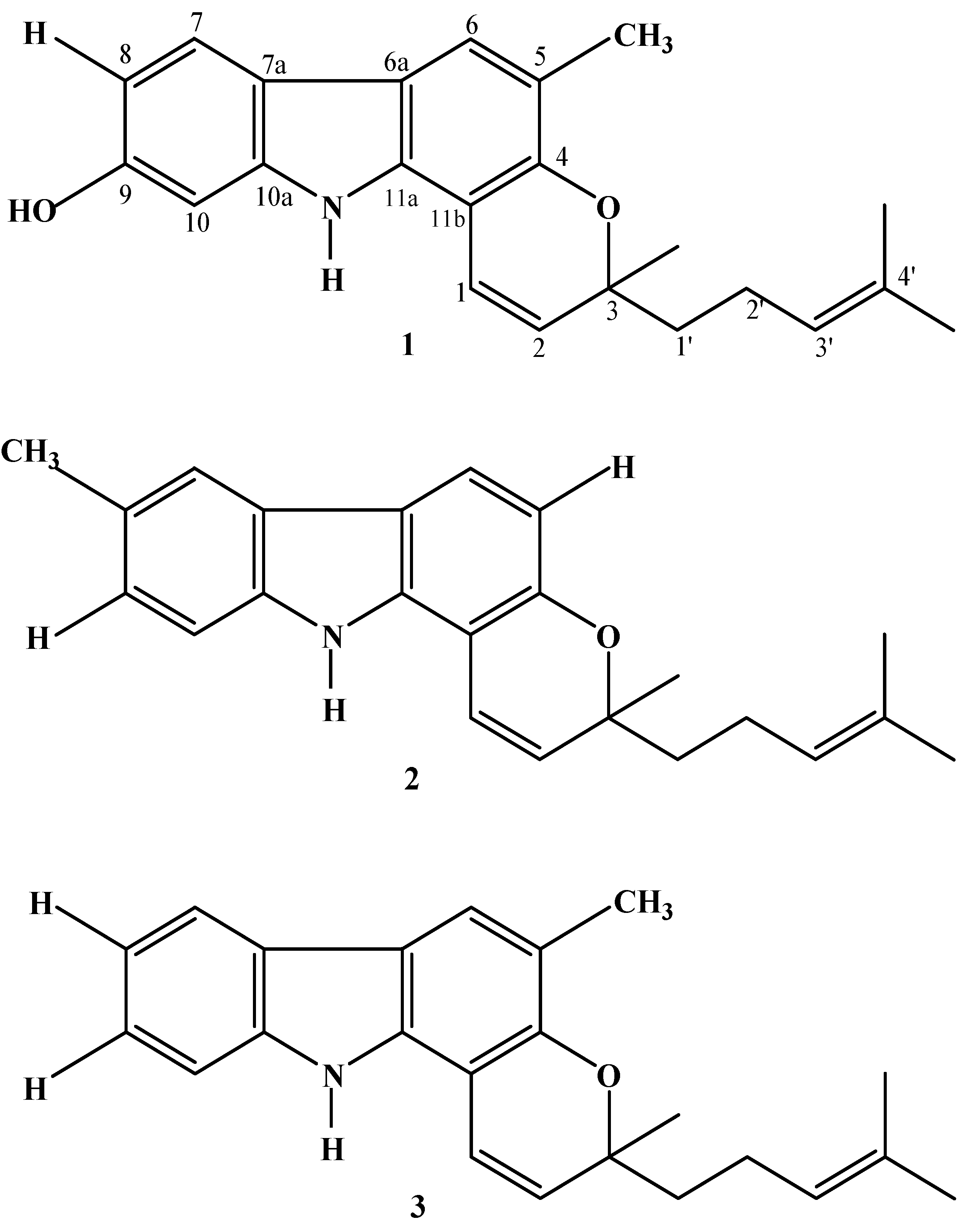
2.1. Structure Elucidation of Alkaloids from the Leaves of Murraya koenigii

A dark green ethanol extract was obtained from 270 g of leaves and an aliquot (5 g) was subjected to silica gel flush column chromatography. Six fractions were obtained by gradient elution using a hexane-ethyl acetate solvent system. Fractions were spotted on silica gel normal phase Thin Layer Chromatography plates (Merck, Germany), and visualized under UV light at 254 nm and using molybdo-phosphoric acid spray. Fractions with secondary metabolites were subjected to semi-preparative HPLC to isolate the alkaloids. A total of three major alkaloids were isolated from M. koenigii, namely compound 1 (0.40%) from fraction 3, while compounds 2 (0.24%) and 3 (0.66%) were isolated from fraction 2. The isolated compounds were subjected to spectroscopic measurements and the resulting data are given in Table 1.
Table 1. 1H-NMR and 13C-NMR spectral data of the isolated compounds from leaves of Murraya koenigii (recorded at 600/150 MHz in CD3OD; δ in ppm).
PositionCompound 1Compound 2Compound 3
δCδHδCδHδCδH
1117.56.60118.86.57118.36.84
2129.25.64126.75.67128.95.63
379.3-79.6-79.3-
4149.9-152.9-150.9-
5118.3-109.76.84119.3-
6120.67.48120.17.71121.87.61
6a116.7-118.9-117.9-
7119.67.64119.37.69119.77.86
7a118.1-125.3-124.8-
8109.16.62129.3-119.97.06
9156.5-125.67.09125.57.23
1097.96.81111.57.27111.47.36
10a143.3-140.2-141.6-
11a136.9-138.7-136.9-
11b105.8-106.0-105.5-
3-CH326.0 1.41 26.6 1.41 26.4 1.41
5-CH316.4 2.27 --16.2 2.29
8-CH3--21.7 2.46--
9-OH-5.01----
1′-CH242.21.72–1.78 42.31.70–1.74 42.11.71–1.73
2′-CH224.12.17–2.20 24.02.14–2.18 23.92.15–2.19
3′125.75.12122.45.12124.95.10
4′132.2-132.6-132.3-
4′-CH317.8 1.64 17.8 1.64 17.61.63
4′-CH326.0 1.56 26.01.57 25.8 1.55
NH-7.65-7.78-7.85
Based on independent structure elucidation the structures of these three compounds were determined as mahanine (1) with the molecular formula, C23H25NO2, electron impact mass spectra (EIMS) (70 eV m/z) mass value of 347 [M]+ (100), 332 (19), 304 (8), 278 (15), 264 (75) and optical rotation, [α]D25 = + 7.6° (CHCl3; c 5.38); mahanimbicine (2) with the molecular formula, C23H25NO, electron impact mass spectra (EIMS)(70 eV m/z) mass value of 331[M]+ (23), 316 (6), 248 (100), 210 (3) and optical rotation, [α]D25 = + 58.1° (CHCl3; c 0.26); mahanimbine (3) with the molecular formula C23H25NO, electron impact mass spectra (EIMS)(70 eV m/z) mass value of 331[M]+ (24), 248 (100), 210 (5) and optical rotation, [α]D25 = + 30.0° (CHCl3; c 0.69). Further, data comparison with published data confirmed them to be known carbazole alkaloids, whose structures are shown in Figure 1.
Figure 1. Chemical structures of carbazole alkaloids [mahanine (1), mahanimbicine (2), mahanimbine (3)] isolated from leaves of Murraya koenigii (L) Spreng.
Molecules 16 09651 g001

2.2. Profiling of Essential Oil from Leaves of Murraya koenigii

The volatile aromatic hydrocarbons were identified based on their Retention Indices (AART) and mass fragment pattern with reference to the NIST 08 and FFNSC version 1.2 databases. A total of 34 aromatic volatile constituents were identified from the oil of M. koenigii, where two sesquiterpene hydrocarbons, β-caryophyllene (19.50%) and α-humulene (15.24%) were present as the major volatile metabolites. Detailed analysis revealed that the 34 volatile constituents could be further grouped into 12 oxygenated monoterpenes, 12 sesquiterpene hydrocarbons, nine oxygenated sesquiterpenes and one oxygenated diterpene. The identified volatile constituents from oils of M. koenigii investigated are presented in Table 2.
Table 2. Composition (%) of volatile compounds in essential oil of Murraya koenigii from Sabah, Malaysia.
RT (min)Ref RIRIVolatile compoundConcentration (%)
15.731082 a1079Linalol0.56
15.911099 b1095trans-Sabinene hydrate 0.53
17.011109 a1112trans-2-Cyclohexen-1-ol 0.48
17.881110 a1113cis-2-Cyclohexen-1-ol 0.54
19.701189 b1185para-Cymen-8-ol 10.31
20.421143 b1139β-Terpineol2.52
21.031175 a1170trans-Piperitol0.40
21.741276 a1273Chrysanthenyl acetate0.39
24.161284 b1279Lavandulyl acetate1.67
24.371285 b1285Bornyl acetate1.68
28.311375 b1370α-Copaene 0.82
28.911390 b1385β-Elemene 0.35
29.391394 a1390( Z)-Jasmone0.11
30.291494 a1489β-Caryophyllene19.50
31.091438 b1436Aromadendrene0.72
31.841454 b1448α-Humulene 15.24
32.701420 a1425Butanedioic acid2.18
33.291487 b1480β-Selinene 3.81
33.301470 a1472Naphthalene1.90
33.551474 a1478α-Selinene6.10
34.371518 b1512δ-Cadinene 2.03
36.031562 b1566Nerolidol2.64
36.051564 b1569trans-Nerolidol1.32
36.281475 a1481Cycloheptane0.13
36.921576 b1580Spathulenol1.98
37.131587 b1591Caryophyllene oxide2.14
37.261594 b1590Viridiflorol1.51
38.131598 a15922-Naphthalenemethanol0.66
38.261079 b1074Trivertal0.35
38.551696 b1694Juniper camphor1.57
38.831581 b1579Cubenol0.57
39.441472 a1476β-Cadina-1(6),4-diene0.50
40.161593 a1596Selina-6-en-4-ol4.78
54.952106 b2105Phytol10.07
Composition of grouped volatile compounds (%)
Monoterpenes (oxygenated)35.29
Sesquiterpenes (hydrocarbon)35.29
Sesquiterpenes (oxygenated)26.47
Diterpenes (oxygenated)2.94
* Identification of volatile components is based on mass spectra value in reference to NIST 08 (a) and FFNSC Ver.1.2 (b) standard libraries.

2.3. Antibacterial Activity

Results on antibacterial activities involving the evaluation of inhibition zones, minimum inhibition concentrations (MICs) and minimum bacteriacidal concentrations (MBCs) of the isolated carbazole alkaloids and essential oil are shown in Table 3.
Table 3. Antibacterial properties of mahanine (1), mahanimbicine (2), mahanimbine (3) and essential oil tested on five strains of clinical human pathogens.
Antibacterial PropertiesCompoundsTested Bacteria
SaPaKpEcSp
DIZ (mm) (mean ± SD)118.5 ± 0.518.5 ± 0.514.5 ± 1.012.5 ± 0.518.0 ± 1.0
216.0 ± 0.512.5 ± 0.518.5 ± 0.514.0 ± 0.511.0 ± 0.5
38.5 ± 0.5NT10.5 ± 0.510.5 ± 0.58.0 ± 0.5
EO12.5 ± 0.516.5 ± 0.518.5 ± 1.014.5 ± 0.510.0 ± 1.0
MIC (mg/mL)125.025.050.075.012.5
225.050.050.025.025.0
375.0NT125.0150.0175.0
EO50.025.025.050.075.0
MBC (μg/mL)1300.0300.0325.0250.0100.0
2>500325.0250.0200.0150.0
3>500NT325.0>500250.0
EO>500250.0>500325.0200.0
Note: Sa-Staphylococcus aureus (210P JTU); Pa-Pseudomonas aeruginosa (ATCC25619); Kp-Klebsiella pneumoniae (SR1-TU); Ec-Escherichia coli (NI23 JTU); Sp-Streptococcus pneumoniae (SR16677-PRSP). The DIZ value of negative control for each bacterium was 4.6 mm (bored well diameter in the agar plates). The concentration of DIZ test were 30 mg/mL. P1: mahanine, P2: mahanimbicine, P3: mahanimbine, EO: essential oil.
Selective antibacterial activities were exhibited by the carbazole alkaloids although they have similar chemical skeletons. Overall, the diameter of inhibition zone for all the tested compounds fell in the range of 8.0 mm to 18.0 mm. When examined against S. aureus, P. aeruginosa and S. pneumoniae, mahanine (1) exhibits the highest susceptibility with inhibition zone of 18.5 ± 0.5 mm, 18.5 ± 0.5 mm and 18.0 ± 0.5 mm respectively while mahanimbicine (2) specifically exhibits the highest susceptibility against K. pneumonia, with an inhibition zone of 18.5 ± 0.5 mm. Mahanimbine (3) appear to be less susceptible to antibiotic resistance bacteria strains as its inhibition zones were in the range of 8.0 mm to 10.0 mm. The minimum inhibition concentration (MIC) of these carbazole alkaloids ranges between 12.5 mg/mL to 175.0 mg/mL. A minimum of 12.5 mg/mL of mahanine (1) was observed as the MIC against S. pneumoniae whereas mahanimbicine (2) exhibits a moderate MIC of 25.0 mg/mL against S. aureus, E. coli and S. pneumoniae, respectively. Meanwhile, the minimum bacteriacidal concentration (MBC) (μg/mL) values of these carbazole alkaloids ranged between 125.0 μg/mL to 500.0 μg/mL. The MBCs of mahanine (1), mahanimbicine (2) and mahanimbine (3) against S. pneumoniae were 100.0 μg/mL, 150.0 μg/mL, 250.0 μg/mL, respectively. Based on this finding, it can be concluded that mahanine (1) was most effective in combating antibiotic resistant clinical bacteria followed by mahanimbicine (2) and mahanimbine (3).
As the essential oil of M. koenigii is a mixture of volatile bioactive terpene classes, positive inhibition were observed according to bacterial strains studied. Diameter of inhibition of M. koenigii essential oil range between 10.00 mm to 18.50 mm as it was most effective in inhibiting S. pneumoniae. The MIC of the essential oil was moderate compared to the MIC values of the individual carbazole alkaloids. The minimal value of inhibition (MIC) was 25.00 μg/mL against P. aeruginosa and K. pneumoniae while 200.00 μg/mL was the minimal bactericidal concentration (MBC) against S. pneumoniae.

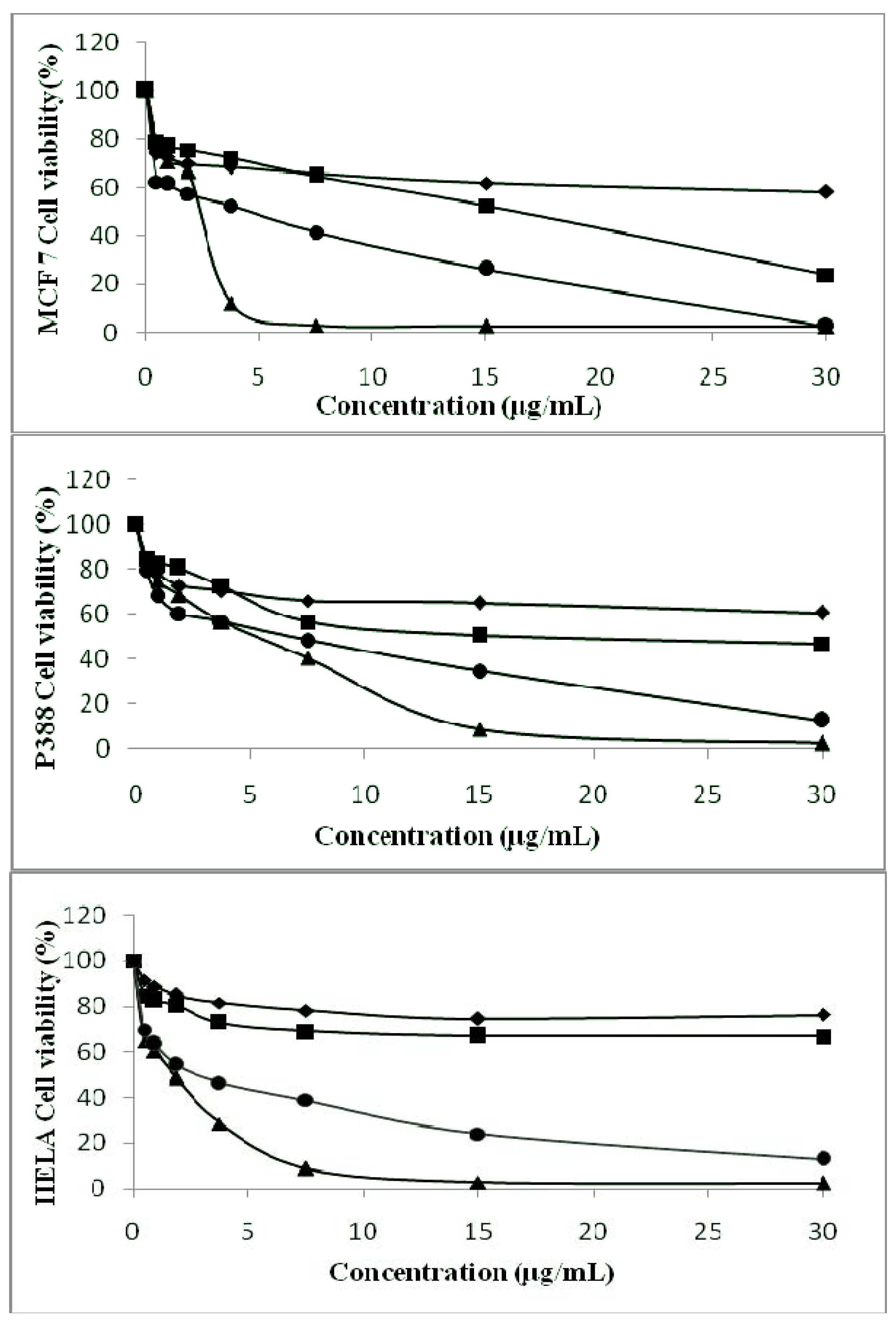
2.4. Anti-Tumor Properties of Isolated Carbazole Alkaloids and Essential Oil

The MTT assay is a simple and reliable technique to measure cell viability used for screening of anti-proliferative agents. Figure 2 presents the summarized plots of cell viability (%) versus concentrations of carbazole alkaloids and essential oil analyzed.
Figure 2. Effect of mahanine (1) (♦), mahanimbicine (2) ( ■), essential oil (•) and mahanimbine (3) (▲) on the growth of MCF-7 cells, P388 cells and HELA cells according to concentration tested.
Molecules 16 09651 g002
The results showed that all the carbazole alkaloids and essential oil exhibit anti-proliferative effects against the three cell lines in dose-dependent manner although taking into account that the growth rate of cancer cells differs. It was found that higher the concentration of compound tested, the lower the cell viability percentages were. Overall, mahanimbine (3) and essential oil of M. koenigii were found to inhibit the proliferation of the cell lines. Mahanimbine (3) was found to exhibit significant cytotoxic activity against all the cancer cell lines studied, with half inhibition concentration (IC50) values of 2.12 μg/mL, 5.00 μg/mL, 1.98 μg/mL in the MCF-7, P388 and HeLa cell lines, respectively. Meanwhile, essential oil of M. koenigii displays IC50 values of 6.00 μg/mL, 7.01 μg/mL, 2.83 μg/mL in the MCF-7, P388 and HeLa cell lines, respectively. Mahanimbicine (2) exhibits a moderate level of cytotoxicity with the IC50 values of 17.00 μg/mL and 18.31 μg/mL against the MCF-7 cell line and P388 cell line. In comparison, mahanine (1) were found to be less cytotoxic against all the cell line studied as the IC50 values fall beyond 30.00 μg/mL.
Carbazole alkaloids from the genus Murraya are also well known for their phytomedicine properties as astringents, analgesic, antidysentric, treatment for snake bites, influenza, rheumatism and traumatic injury [12,19,20]. In addition, this investigation has shown that carbazole alkaloids and essential oil from Malaysian M. koenigii have antibacterial potential against antibiotic resistant bacteria strains. Despite having the similar chemical skeletons, these carbazole alkaloids displayed selective antibacterial and antitumor activity due to their different side chain moieties. Results of antibacterial activity revealed mahanine (1) is the most effective in inhibiting antibiotic resistant bacteria while mahanimbine (3) was found to be the most cytotoxic against the tested cancer cell lines. Positioning of functional groups (-H, -OH and -CH3) in the chemical skeleton could be responsible for these selective bioactive attributes [21]. Generally, the bioactivity of carbazoles were reported to decrease with the introduction of an oxygen atom on the carbazole nucleus, however this effect can be reversed by the replacement of the hydrogen atom of the hydroxyl group by an alkyl or alkoxy group, especially acetyl and methyl rest [22,23,24]. This might explain why the positioning of -CH3 in the mahanimbine (3) skeleton displayed a vital role in its selective antitumor activity [25,26]. To the best of our knowledge, this is the first report on the bioactive potential of mahanimbine (3) against the MCF-7 cell line. This result could be used as a basis for the development of a structure-orientated drug discovery program. In addition, it has provided an important indication that mahanimbine (3) should be further explored as a possible chemotherapeutic agent in human breast cancer studies.
Essential oils of plants in the family Rutaceae are often composed of mono- and sesquiterpenes. Monoterpenes and sesquiterpenes derivatives such as linalool, terpineol, camphor, selinene are reported to possess antibacterial, insect-repelling and toxic activities [27]. As such, the presence of high concentrations of β-caryophyllene (19.50%) along with caryophyllene oxide (2.16%) in the oil of M. koenigii clearly contributes to its bacteria susceptibility properties [28,29] as the activity exhibited could also be explained as a synergistic effect of the dominant volatile constituents [30]. Previous investigations [28,29,30,31] have explained that the antimicrobial activity of the essential oils derived from plants are found to be more pronounced against Gram-positive than against Gram-negative bacteria. This action is often correlated to the presence of terpene constituents although the mechanism of action of compounds from this class against Gram-negative bacteria is not fully understood, but the absence of an outer phospholipidic membrane in Gram-positive bacteria allows the penetration of essential oil’s bioactive hydrocarbons that can cause leakage of vital intracellular constituents and impairment of the bacterial enzyme systems [32].
Classes of terpenes are also known for their antitumor attributes. Studies report that the volatile sesquiterpene hydrocarbons α-humulene, β-caryophyllene and α-caryophyllene isolated from the family Rutaceae are active against human alveolar basal epithelial cells (A-549), colon carcinoma cells (DLD-1) and human prostate adenocarcinoma (LNCaP) cell lines as well as possessing anti-proliferative ability towards myeloid leukemia (K562) cells. Ironically, constituents of monoterpene groups, such as β-pinene, γ-terpinene and ρ-cymene were found to be inactive against DLD-1 cancer cell lines [28]. This could explain the synergism of active hydrocarbon sesquiterpenes and oxygenated sesquiterpenes, which constitute 61.76% of the volatile chemical found in oil of M. koenigii, responsible for the anti-proliferation of HeLa, P388 and MCF-7 cell lines. Reports have suggested [29] that oils that have IC50 values less than 0.13 mg/mL could be possible candidates for further development as cancer therapeutic agent. As such, essential oil of M. koenigii could be such a possible candidate given its IC50 value ranging between 2.83 μg/mL to 7.01 μg/mL.

3. Experimental

3.1. Plant Material

Pest free leaves of Murraya koenigii (L.) Spreng (Rutaceae) were collected from Kg. Bobot, Kota Belud, Sabah in August 2010. Voucher specimens were deposited at the BORNEENSIS, Herbarium of the Institute for Tropical Biology and Conservation, Universiti Malaysia Sabah (BORH 37581).

3.2. Extraction and Identification

Air-dried M. koenigii leaves (270 g) were extracted with ethanol (3 liter) (Merck, Germany) for 7 days using a soxhlet apparatus. Extract were concentrated under reduced pressure at 40 °C to yield a dark green extract (14.75 g). A portion (5 g) was then subjected to successive silica gel (Merck, Kiesegel 60, 70–230 mesh) column chromatography with a stepwise gradient of hexane-ethyl acetate (hex-EtOAc; 9:1, 8:2, 7:3, 6:4, 1:1) before the column was washed with chloroform-methanol-water (65:25:4) to obtain six fractions of different polarity. Fractions were then profiled using a high performance liquid chromatography (HPLC) apparatus equipped with a UV-Vis detector monitored at 254 nm (Prominence, Shimadzu, Japan) coupled with a reverse phase Phenomenex C-18 ODS (10 mm × 250 mm × 5 μm) column separation. Mobile phase in gradient elution was: A: 50% MeCN: 50% H2O, B: 100% MeCN with the profile of: 0 min–30 min: A: 30% B: 70%, 30.01 min–60.00 min A: 0% B: 100% and flow rate was set at 2.00 mL/min under 40 °C. A manual injection of 50 μL of pre-filtered solution (0.2 μm nylon membrane syringe filters, Whatman) was analyzed.

3.3. Essential Oil Extraction and Analysis

Selected pest-free leaves of M. koenigii (50 g) were chopped and subjected to hydro-distillation using a Clevenger-type apparatus for a duration of 8 h. Yield of oil based on fresh weight was 0.12%. Distilled oil was collected in pentane, dried over anhydrous Na2SO4, concentrated in vacuo, stored in air-tight glass vials flushed with nitrogen (N2) gas and kept at −20 °C prior to analysis. Analysis of the essential oil was performed using a Shimadzu QP-2010 gas chromatograph coupled with a Shimadzu GCMSQP-2010Plus detector (Shimadzu, Japan) using a SGE BPX-5 (30.0 m × 0.25 μm i.d., film thickness 0.25 μm) fused silica capillary column. High purity helium was used as the carrier gas at a constant flow rate of 0.8 mL/min. A total of 1 μL sample was injected (split ratio 100:1) into GCMS using AOC5000 auto injector for analysis. The initial temperature was set at 50 °C, heated at a rate of 3 °C/min to 280 °C and held isothermally for 5 min. Ion source temperature for these analysis was set at 200 °C, while the interface temperature was set at 280 °C and the mass spectrometer was set to operate in electron ionization mode with an ionizing energy of 70 eV as acquisition mass range from 40 a.m.u to 450 a.m.u. at 0.25 scan/s. Identification of volatile organic constituents was confirmed using published electron impact-mass spectra (EI-MS) in the National Institute for Standard and Technology (NIST) 1998 and Shimadzu’s Flavours and Fragrance of Natural and Synthetic Compounds (FFNSC) version 1.2 computerized mass spectral libraries. The retention indices were determined based on a homologous series of n-alkanes (C8–C40; Custom Retention Time Index Standard, Restek Corp, USA) external standard analyzed under the same operating conditions and calibrated based on the Automatic Adjustment of Compound Retention Time (AART) function of the GCMS. Relative concentrations of the essential oil components were calculated based on GC peak area with the AART correction factors.

3.4. Isolation and Structure Elucidation of Alkaloids

Carbazole alkaloids were isolated based on their Thin Layer Chromatography (TLC) and High Performance Liquid Chromatography profile data. Presence of alkaloids was detected on TLC upon spraying with Dragendoff’s reagent and isolation of the respective fractions was done using semi-preparative HPLC using a C-18 ODS column (Inertsil ODS-3, 20 mm × 250 mm × 5 μm) coupled with a UV-Vis detector (Prominence, Shimadzu Corp., Japan) monitored at 254 nm. Gradient elution conditions were; A: 50% MeCN: 50% dH2O and B: 100% MeCN under the profile of; 0 min–30 min: A: 30% B: 70%, 30.01 min–60.00 min A: 0% B: 100% with flow rate of 6.00 mL/min with injection volume of 200 μL at 40 °C. All solutions were pre-filtered using Whatman 13 mm, 0.2 μm nylon membrane syringe filters before injections. A total of three major peaks were isolated from Fraction 2 and Fraction 3.
Isolated peaks were subjected to TLC confirmation with Drangendoff spray and 1H-NMR revealed the purity of the isolate and confirmed the identity of the alkaloids with the presence of aromatic and amine protons. Three compounds were isolated and subjected to 1H-, 13C- and 2D NMR spectroscopic analyses. The structures of compounds 13 were determined based on the comparison of their 1H- and 13C-NMR data with those reported in the literature [28,29,30,31]. All spectral data were obtained on the following instruments; IR on a ThermoNicolet FT-IR spectrometer, optical rotations was measured on an AUTOPOL IV automatic polarimeter (Rudolph Research Analytical), 1H-NMR (600 MHz) and 13C-NMR (150 MHz) were recorded with a JEOL ECA 600 (Japan Electronic Optics Laboratory Co. Ltd., Tokyo, Japan) spectrometer, with TMS as internal standard. HR-ESI-TOFMS data were obtained using LCMS-IT-TOF (Shimadzu).

3.5. Cell Culture and Assay

3.5.1. Antibacterial Activity Assay

Antibacterial activity was assayed with the standard agar well diffusion method (NCCLS, 2000). The bacterial suspension (106 CFU/mL) was “flood-inoculated” onto the surface of PCA medium and wells (4.6 mm) were cut from the agar. Test compounds and standards were dissolved in DMSO, sterilized by filtration through 0.22 mm sterilizing Milipore express filter (Millex-GP, Bedford, OH, USA) and 60 μL of these solutions were delivered into the wells. Gentamicin (600 mg/well) was used as positive reference standards to determine the sensitivity of each microbial species tested and DMSO solutions were applied as negative controls. The inoculated plates were incubated at 37 °C for 24–48 h. The diameter of inhibition zone (DIZ) of the tested bacteria was measured and expressed in millimeters to evaluate the antibacterial activity of the samples. Tests were performed in triplicates.

3.5.2. Minimum Inhibitory Concentration (MIC) Assay

MIC was assayed using two-fold microdilution broth method (NCCLS, 2003). Dilutions were used to dispense 0.1 mL into each of the sterile 96 wells of a standard tray. Each well contained 5 × 10⁵ colony forming units (CFU/mL) of test bacteria, serially diluted samples and respective growth medium. Negative and positive controls were prepared accordingly using DMSO and gentamicin, respectively. After incubated at 37 °C for 24 h, the microdilution trays were checked with unaided eyes to detect the growth inhibition of the bacteria. The growth end points were determined by comparing the amount of growth in the wells containing test samples with that in the control wells. The acceptable growth (≥2 mm button or definite turbidity) must occur in the positive control well. When a single skipped well occurred, the highest MIC was read. Tests were performed in triplicate for each test concentration.

3.5.3. Minimum Bactericidal Concentration (MBC) Assay

A method in ASM Pocket Guide to Clinical Microbiology was slightly modified to determine the MBC values. Briefly, samples (50 μL) were taken from the wells of the MIC assays, where no visible turbidity (growth) was observed, and spread on freshly prepared PCA plates. The plates were incubated at 37 °C for 24 h so as to determine the MBC, which was defined as the lowest concentration of the samples which allowed less than 0.1% of the original inoculums treated with the compounds to survive and grow on the surface of the medium used. Tests were performed in triplicate.

3.6. Anti-Tumor Assay

3.6.1. Cell Lines and Cultivation Conditions

The three cell lines used in this investigation comprised human breast (MCF-7), human cervical (HeLa) and murine leukemia cell lines (P388). The MCF-7, HeLa and P388 cells each were cultured as monolayers in RPMI-1640, supplemented with 10.0% (v/v) heat-inactivated FBS, 100 U/mL penicillin and 100.0 μg/mL streptomycin. All cell cultures were grown in a humidified incubator at 37 °C in 5.0% CO2 and 95% O2.

3.6.2. Measurement of Cell Viability by MTT Assay

The cytotoxic effects of mahanine (1), mahanimbicine (2), mahanimbine (3) and essential oil were determined by measuring conversion of 3-(4,5-dimethylthiazol-2-yl)-2,5-diphenyltetrazolium bromide (MTT) dye. All pure compounds and essential oil were dissolved in DMSO to a final concentration of 30 μg/mL. Briefly, 2.0 × 10⁴ MCF-7 cells were treated in triplicate with each pure compound and the essential oil. The same treatments were applied on 5.0 × 10⁵ HeLa cells and 1.0 × 10⁵ P388 cells. MTT reagent (5.0 mg/mL) was added and cells were incubated in the dark at 37 °C. DMSO was added to dissolve purple formazon crystals and a microtiter plate reader (Tecan, Switzerland) was used to measure the absorbance at 570 nm with 630 nm as the reference wavelength.

3.7. Statistical Analysis

The significance of differences was estimated using Student’s t-test. A p-value of less than 0.05 was considered significant.

4. Conclusions

In conclusion, this study has revealed the bioactive potentials of carbazole alkaloids from M. koenigii where mahanine (1) and mahanimbicine (2) inhibited antibiotic resistant bacteria and mahanimbine (3) was found to significantly suppress the proliferation of MCF-7 cells. The differences in biological activities could be correlated to differences in their functionality since the chemical skeletons was similar. To the best of our knowledge, this report is the first pertaining to the selectivity of these metabolites towards antibiotic resistant bacteria and MCF-7 cells. This information could be of importance in the development of new antibacterial and antitumor lead metabolites.

Acknowledgments

The authors would like to acknowledge the Ministry of Science Technology and Innovation of Malaysia for its financial assistance (Grant No: 02-01-10-SF0131). The corresponding author would like to express his gratitude to the support staff of the Institute for Tropical Biology and Conservation, Universiti Malaysia Sabah, for their assistance during the course of this investigation.

References and Notes

  1. Brandao, G.C.; Kroon, E.G.; Duarte, M.G.; Braga, F.C.; Filho, J.D.S.; Oliveira, A.B. Antimicrobial, antiviral and cytotoxic activity of extracts and constituents from Polygonum spectabile Mart. Phytomedicine 2010, 17, 926–929. [Google Scholar] [CrossRef]
  2. Li, Z.B.; Wang, J.Y.; Jiang, B.; Zhang, X.L.; An, L.J.; Bao, Y.M. Benzobijuglone, a novel cytotoxic compound from Juglans mandshurica induced apoptosis in HeLa cervical cancer cells. Phytomedicine 2007, 14, 846–852. [Google Scholar] [CrossRef]
  3. Chen, C.Y.; Liu, T.Z.; Tseng, W.C.; Lu, F.J.; Hung, R.P.; Chen, C.H.; Chen, C.H. (−)-Anonaine induces apoptosis through Bax- and caspase-dependent pathways in human cervival cancer (HeLa) cells. Food Toxicol. Chem. 2008, 46, 2694–2702. [Google Scholar] [CrossRef]
  4. Zhang, F.F.; Gan, L.L.; Zhou, C.H. Synthesis, antibacterial and antifungal activities of some carbazole derivatives. Bioorg. Med. Chem. Lett. 2010, 20, 1881–1884. [Google Scholar] [CrossRef]
  5. Antilla, A.; Ronco, G. Working group on the registration and monitoring of cervical cancer screening programmes in the European Union within the European Network for Information on Cancer (EUNICE). Description of the national situation of cervical cancer screening in the member states of the European Union. Eur. J. Cancer 2009, 45, 2685–2708. [Google Scholar] [CrossRef]
  6. Parkin, D.M.; Bray, F.I.; Devesa, S.S. Cancer burden in the year 2000: The global picture. Eur. J. Cancer 2001, 37, 4–66. [Google Scholar]
  7. Mao, W.W.; Wang, T.T.; Zeng, H.P.; Wang, Z.H.; Chen, J.P.; Shen, J.G. Synthesis and evaluation of novel substituted 5-hydroxycoumarin and pyranocoumarin derivatives exhibiting significant antiproliferative activity against breast cancer cell lines. Bioorg. Med. Chem. Lett. 2009, 19, 4570–4573. [Google Scholar] [CrossRef]
  8. Nagaiah, K.; Venkatesham, A.; Rao, R.S.; Saddanapu, V.; Yadav, J.S.; Basha, S.J.; Sarma, A.V.S.; Sridhar, B.; Addlagatta, A. Synthesis of new cis-fused tetrahydrochromeno [4,3-b]quinolines and their antiproliferative activity studies against MDA-MB-231 and MCF-7 breast cancer cell lines. Bioorg. Med. Chem. Lett. 2010, 20, 3259–3264. [Google Scholar]
  9. Bhattacharya, K.; Samanta, S.K.; Tripathi, R.; Mallick, A.; Chandra, S.; Pal, C.B.; Shaha, C.; Mandal, C. Apoptotic effects of mahanine on human leukemic cells are mediated through crosstalk between Apo-1/Fas signaling and the Bid protein and via mitochondrial pathways. Biochem. Pharmacol. 2010, 79, 361–372. [Google Scholar]
  10. Roy, K.M.; Thalang, V.N.; Trakoontivakorn, G.; Nakahara, K. Mechanism of mahanine-induced apoptosis in human leukemia cells (HL-60). Biochem. Pharmacol. 2004, 67, 41–51. [Google Scholar]
  11. Delamare, A.P.L.; Pistorello, I.T.M.; Artico, L.; Serafini, L.A.; Echeverrigaray, S. Antibacterial activity of the essential oils of Salvia officinalis L. and Salvia triloba L. cultivated in South Brazil. Food Chem. 2007, 100, 603–608. [Google Scholar] [CrossRef]
  12. Ito, C.; Itoigawa, M.; Onoda, S.; Hosokawa, A.; Ruangrungsi, N.; Okuda, T.; Tokuda, H.; Nishino, H.; Furukawa, H. Chemical constituents of Murraya siamensis: Three coumarins and their anti-tumor promoting effect. Phytochemistry 2005, 66, 567–572. [Google Scholar]
  13. Jagadeesh, S.; Sinha, S.; Pal, B.C.; Bhattacharya, S.; Banerjee, P.P. Mahanine reverses an epigenetically silenced tumor suppressor gene RASSF1A in human prostate cancer cells. Biochem. Biophys. Res. Commun. 2007, 362, 212–217. [Google Scholar] [CrossRef]
  14. Sathaye, S.; Bagal, Y.; Gupta, S.; Kaur, H.; Redkar, R. Hepatoprotective effects of aqueous leaf extract and crude isolates of Murraya koenigii against in vitro ethanol-induced hepatotoxicity model. Exp. Toxicol. Pathol. 2011, 63, 587–591. [Google Scholar] [CrossRef]
  15. Cordell, G.A. The Alkaloids: Chemistry and Biology; Academic Press: Waltham, MA, USA, 2008. [Google Scholar]
  16. Tachibana, Y.; Kikuzaki, H.; Lajis, N.; Nakatani, N. Antioxidative activity of carbazoles from Murraya koenigii leaves. J. Agric. Food Chem. 2001, 45, 5589–5594. [Google Scholar]
  17. Ramsewak, R.S.; Nair, M.G.; Strasburg, G.M.; DeWitt, D.L.; Nitiss, J.L. Biologically active carbazole alkaloids from Murraya koenigii. J. Agric. Food Chem. 1999, 47, 444–447. [Google Scholar]
  18. Rahman, M.M.; Gray, A.I. A benzoisofuranone derivative and carbazole alkaloids from Murraya koenigii and their antimicrobial. Phytochemistry 2005, 66, 1601–1606. [Google Scholar]
  19. Wu, T.S.; Wang, M.L.; Wu, P.L. Seasonal variations of carbazole alkaloids in Murraya euchrestifolia. Phytochemistry 1996, 43, 785–789. [Google Scholar]
  20. Voss, M.E.; Ralph, J.M.; Xie, D.; Manning, D.D.; Chen, X.; Frank, A.J.; Leyhane, A.J.; Liu, L.; Stevens, J.M.; Budde, C.; et al. Synthesis and SAR of vinca alkaloids analogues. Bioorg. Med. Chem. Lett. 2009, 19, 1245–1249. [Google Scholar]
  21. Adebajo, A.C.; Ayoola, O.F.; Iwalewa, E.O.; Akindahunsi, A.A.; Omisore, N.O.A.; Adewunmi, C.O.; Adenowo, T.K. Anti-trichomonal, biochemical and toxicological activities of methanolic extract and some carbazole alkaloids isolated from the leaves of Murraya koenigii growing in Nigeria. Phytomedicine 2006, 13, 246–254. [Google Scholar] [CrossRef]
  22. Caballero, E.; Adeva, M.; Calderon, S.; Sahagun, H.; Tome, F.; Medarde, M.; Fernandez, J.L.; Lopez-Lazaro, M.; Ayuso, M.J. Synthesis and cytotoxic activity of different open indocarbazole alkaloid analogues. Bioorg. Med. Chem. Lett. 2003, 11, 3413–3421. [Google Scholar] [CrossRef]
  23. Knolker, H.J. Transition metal complexes in organic synthesis. Part 70. Synthesis of biologically active carbazole alkaloids using organometallic chemistry. Curr. Org. Synth. 2004, 1, 309–331. [Google Scholar] [CrossRef]
  24. Zhang, F.F.; Gan, L.L.; Zhou, C.H. Synthesis, antibacterial and antifungal activities of some carbazole derivatives. Bioorg. Med. Chem. Lett. 2010, 20, 1881–1884. [Google Scholar] [CrossRef]
  25. Knolker, H.J.; Reddy, K. Isolation and synthesis of biologically active carbazole alkaloids. Chem. Rev. 2002, 102, 4303–4427. [Google Scholar] [CrossRef]
  26. Manosroi, J.; Dhumtanom, P.; Manosroi, A. Anti-proliferative activity of essential oil extracted from Thai medicinal plants on KB and P388 cell lines. Cancer Lett. 2006, 235, 114–120. [Google Scholar] [CrossRef]
  27. Cardile, V.; Russo, A.; Formisano, C.; Rigano, D.; Senatore, F.; Arnold, N.A.; Piozzi, F. Essential oils of Salvia bracteata and Salvia rubifolia from Lebanon: Chemical composition, antimicrobial activity and inhibitory effect on human melanoma cells. J. Ethnopharmacol. 2009, 126, 265–272. [Google Scholar] [CrossRef]
  28. Ulubelen, A.; Topcu, G.; Eris, C.; Sonmez, U.; Kartal, M.; Kurucu, S.; Bozok-Johansson, C. Terpenoids from Salvia sclarea. Phytochemistry 1994, 36, 971–974. [Google Scholar]
  29. Bouaziz, M.; Yangui, T.; Sayadi, S.; Dhouib, A. Disinfect properties of essential oils from Salvia officinalis L. cultivated in Tunisia. Food Chem. Toxicol. 2009, 47, 2755–2760. [Google Scholar] [CrossRef]
  30. Rota, C.M.; Herrera, A.; Martinez, R.M.; Sotomayor, J.A.; Jordan, M.J. Antimicrobial activity and chemical composition of Thymus vulgaris, Thymus zygis and Thymus hyemalis essential oils. Food Control 2008, 19, 681–687. [Google Scholar] [CrossRef]
  31. Sandri, I.G.; Zacaria, J.; Fracaro, F.; Delamare, A.P.L.; Echeverrigaray, S. Antimicrobial activity of the essential oils of Brazilian species of the genus Cunila against foodborne pathogens and spoiling bacteria. Food Chem. 2007, 103, 823–828. [Google Scholar] [CrossRef]
  32. Delamare, A.P.L.; Pistorello, I.T.M.; Artico, L.; Serafini, L.A.; Echeverrigaray, S. Antibacterial activity of the essential oils of Salvia officinalis L. and Salvia triloba L. cultivated in South Brazil. Food Chem. 2007, 100, 603–608. [Google Scholar] [CrossRef]
  • Sample Availability: Not available.

