Article

Two New Phenolic Glucosides from Lagerstroemia speciosa

1
College of Pharmacy and Research Institute of Pharmaceutical Science, Seoul National University, Seoul 151-742, Korea
2
Department of Genetic Engineering, Sungkyunkwan University, Suwon 440-746, Korea
3
College of Pharmacy, The Catholic University of Korea, Bucheon 420-743, Korea
4
Department of Life Science, Hallym University, Chuncheon 200-702, Korea
5
Popa mountain park, Forest Department, Kyaukpadaung Township, Mandalay Division, Myanmar
6
College of Pharmacy and RFIND-BKplus Team, Dongguk University-Seoul, 32 Dongguk-lo, Ilsan dong-gu, Goyang 410-820, Korea
7
Department of Forest Sciences, Seoul National University, Seoul 151-921, Korea
*
Author to whom correspondence should be addressed.
These authors contributed equally to this work.
Submission received: 6 February 2015 / Revised: 4 March 2015 / Accepted: 5 March 2015 / Published: 10 March 2015
(This article belongs to the Section Natural Products Chemistry)

Abstract

:
Two new phenolic glucosides, 1-O-benzyl-6-O-E-caffeoyl-β-d-glucopyranoside and 1-O-(7S,8R)-guaiacylglycerol-(6-O-E-caffeoyl)-β-d-glucopyranoside, were isolated from the aerial parts of Lagerstroemia speciosa, along with ten known compounds. The structures of the isolated compounds were determined based on 1D- and 2D-NMR, Q-TOF MS and optical rotation spectroscopic data. All of the compounds showed moderate inhibitory activities against nitric oxide production in lipopolysaccharide-treated RAW264.7 cells, with IC50 values of 69.5–83.3 μM.

Graphical Abstract

1. Introduction

Lagerstroemia speciosa L. Pers., which belongs to the Lythraceae family, is a tropical plant distributed in areas of Southeast Asia, including the Philippines, Vietnam, Myanmar and southern China. In these regions, L. speciosa is commonly called banaba, and it has been traditionally used for the treatment of diabetes, obesity and kidney malfunction [1,2].
Earlier phytochemical investigations have revealed that L. speciosa possessed triterpenes and sterols [3,4], phenolic compounds, including ellagic and gallic acid [5], ellagitannins and several flavonoids [6,7]. In vivo and in vitro studies indicated that banaba extract shows anti-α-glucosidase activity [3], anti-HIV-1 protease and reverse transcriptase activities [5], anti-human rhinovirus activity [6], hypoglycemic activity [7,8] and free radical scavenging and anti-inflammatory effects [9]. For further phytochemical investigation of L. speciosa, diverse column chromatography methods were performed to isolate two novel phenolic compounds, 1-O-benzyl-6-O-E-caffeoyl-β-D-glucopyranoside 1 and 1-O-(7S,8R)-guaiacylglycerol-(6-O-E-caffeoyl)-β-D-glucopyranoside 2, along with ten known compounds, 312. In addition, the inhibitory activities of these isolated compounds against nitric oxide (NO) production in lipopolysaccharide (LPS)-treated RAW 264.7 cells were evaluated.

2. Results and Discussion

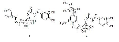
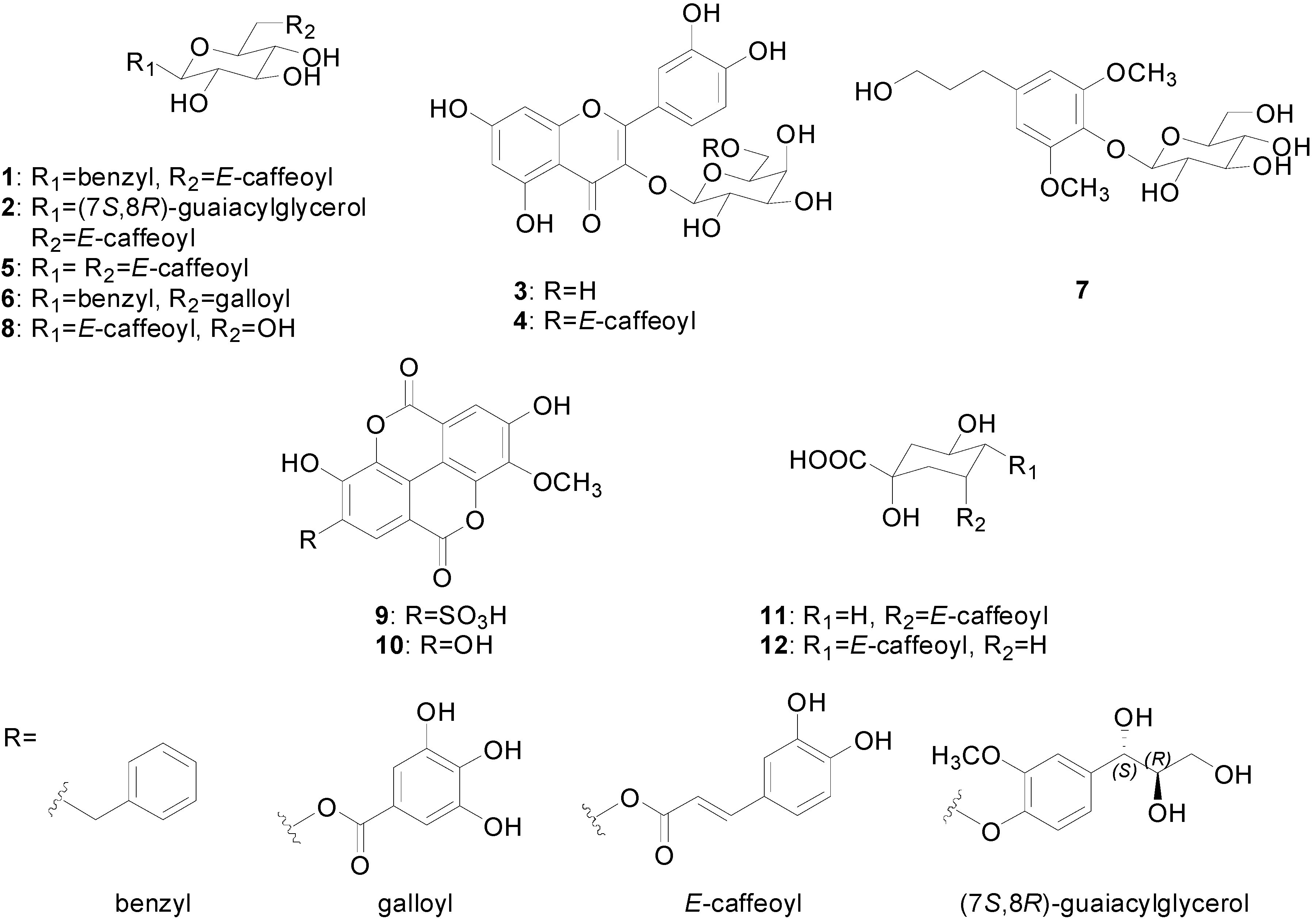
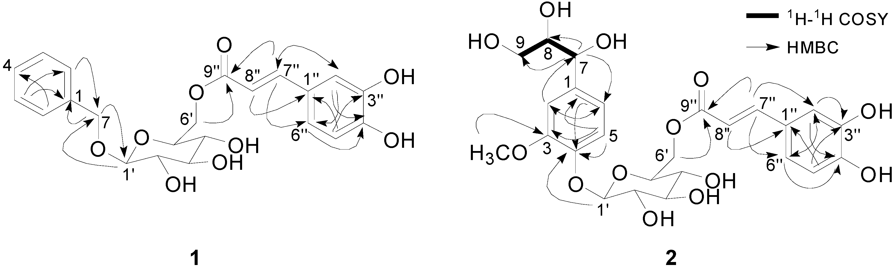
2.1. Structural Elucidation of Isolated Compounds

The structure elucidation of two new compounds (1 and 2) was accomplished by interpretation of the spectroscopic data, including 1D- and 2D-NMR, Q-TOF MS and optical rotation data. The ten known compounds were determined to be quercetin-3-O-β-d-galactopyranoside 3 [10], quercetin-3-O-(6''-O-E-caffeoyl)-β-d-galactopyranoside 4 [11], 1,6-di-O-E-caffeoyl-β-d-glucopyranoside 5 [12], benzyl-6'-O-galloyl-β-d-glucopyranoside 6 [13], dihydrosyringin 7 [14], 1-O-E-caffeoyl-β-d-glucopyranoside 8 [15], 3-O-methylellagic acid 4'-sulfate 9 [7], 3-O-methylellagic acid 10 [7], chlorogenic acid 11 [16] and cryptochlorogenic acid 12 [16] (Figure 1). To the best of our knowledge, the known eight compounds (38 and 1112) were isolated for the first time from L. speciosa.
Compound 1 was isolated as an amorphous brownish powder, and its molecular formula was assigned as C22H24O9 from the [M+H]+ and [M+Na]+ ion peaks at m/z 433.1506 (calcd. for C22H25O9: 433.1499) and 455.1328 (calcd. for C22H24O9Na: 455.1318), respectively, by Q-TOF MS. The 1H-NMR spectrum showed resonances corresponding to a benzyl moiety (δH 7.38 (2H, d, J = 7.3 Hz; H-2, H-6), 7.31 (2H, t, J = 7.3 Hz; H-2, H-6), 7.27 (1H, t, J = 7.3 Hz; H-4), 4.76 (1H, d, J = 12.1 Hz; H-7a), 4.58 (1H, d, J = 12.1 Hz; H-7b)) and an E-caffeoyl residue (δH 7.51 (1H, d, J = 15.9 Hz; H-7''), 7.04 (1H, br s; H-2''), 6.99 (1H, br d, J = 7.2 Hz; H-6''), 6.72 (1H, d, J = 7.8 Hz; H-5''), and 6.30 (1H, d, J = 15.9 Hz; H-8'')), as well as a signal assigned to the anomeric proton of the glucosyl moiety (δH 4.28 (1H, d, J = 7.9 Hz; H-1')) (Table 1). The 13C-NMR spectrum of 1 showed six glucopyranosyl signals at δC 102.0, 76.5, 73.8, 73.4, 70.1 and 63.5, and the configuration was confirmed as the β-form from the coupling constant. HMBC correlations of H-7 (δH 4.76 and 4.58) to C-1' (δC 102.0) and H-6' (δH 4.41 and 4.19) to C-9'' (δC 166.6) indicated that 1 is a caffeic acid ester of 1-O-benzyl-β-d-glucopyranoside (Figure 2). Based on the spectroscopic evidence above, 1 was determined to be 1-O-benzyl-6-O-E-caffeoyl-β-d-glucopyranoside.
Figure 1. Chemical structures of Compounds 112 from L. speciosa.
Molecules 20 04483 g001
Figure 2. Key 1H-1H COSY and HMBC correlations for Compounds 1 and 2.
Molecules 20 04483 g002
The positive Q-TOF MS spectrum of Compound 2 showed a sodiated pseudomolecular ion peak at m/z 561.1595 (calcd. for C25H30O13Na: 561.1584); thus, its molecular formula was determined to be C25H30O13. The 1H- and 13C-NMR spectra of 2 indicated the presence of 6-O-E-caffeoyl-β-d-glucopyranoside, similar to the case of Compound 1. The difference between 1 and 2 is that the guaiacylglycerol moiety is linked to a glucosyl group instead of a benzyl moiety. 1D- and 2D-NMR spectra, including 1H-1H COSY and HMBC spectra, revealed the presence of a guaiacylglycerol skeleton. The 1H-NMR spectrum indicated the presence of a 1,3,4-trisubstituted aromatic ring (δH 7.09 (1H, d, J = 8.3 Hz; H-5), 7.08 (1H, d, J = 1.8 Hz; H-2) and 6.83 (1H, dd, J = 8.3, 1.8 Hz; H-6)), a methene group (δH 3.48 (1H, m; H-9a) and 3.35 (1H, m; H-9b)), two methine protons (δH 4.52 (1H, d, J = 4.2 Hz; H-7) and 3.62 (1H, m; H-8)) and a methoxy group (δH 3.87 (3H, s)) (Table 1). A 1H-1H COSY experiment revealed the partial structure of -CH(7)-CH(8)-CH2(9), and the HMBC correlation of H-1' (δH 4.87) to C-4 (δC 147.0) revealed the C4-O-C1' connectivity between the 6-O-E-caffeoyl-β-d-glucopyranoside and guaiacylglycerol moieties (Figure 2). The small JH7,H8 coupling constant (J = 4.2 Hz) in the 1H-NMR spectrum of 2 suggested that C-7 and C-8 were in the erythro configuration (JH7,H8 ≤ 4.4 Hz); a relatively large coupling constant (JH7,H8 ≥ 6.0 Hz) would be expected for the threo configuration [17]. Enzymatic hydrolysis of 2 afforded 2a, which had a pseudomolecular ion peak at m/z 237.0743, [M+Na]+ (calcd. for C10H14O5Na: 237.0739), in the Q-TOF MS data, and an optical rotation of [ α ] D 22   =   + 11.2 , which demonstrated the presence of the (7S,8R)-guaiacylglycerol unit [17,18]. According to the spectroscopic data above, Compound 2 was assigned as 1-O-(7S,8R)-guaiacylglycerol-(6-O-E-caffeoyl)-β-d-glucopyranoside.
Table 1. 1H- (500 MHz, δ ppm, J in Hz) and 13C-NMR (125 MHz, δ ppm) of Compounds 1 and 2 in CD3OD.
12
PositionδH (J in Hz)δCPositionδH (J in Hz)δC
1137.81138.7
2,67.38, d (7.3)127.827.08, d, (1.8)112.5
3,57.31, t (7.3)128.13150.8
47.27, t (7.3)127.44147.0
7a4.76, d (12.1)69.657.09, d (8.3)117.8
7b4.58, d (12.1)66.83, dd (8.3, 1.8)120.7
1'4.28, d (7.9)102.074.52, d (4.2)75.2
2'3.07, m73.483.62, m77.5
3'3.17, m76.59a3.48, m64.4
4'3.16, m70.19b3.35, m
5'3.40, m73.81'4.87, d (7.6)102.9
6'a4.41, brd (11.7)63.52'3.52, m75.0
6'b4.19, dd (11.7, 6.6)3'3.49, m77.9
1''125.14'3.41, m72.0
2''7.04, brs114.55'3.68, m75.8
3''145.56'a4.53, dd (11.9, 2.1)64.8
4''149.26'b4.32, dd (11.9, 6.9)
5''6.72, d (7.8)115.71''127.9
6''6.99, d (7.2)121.52''7.06, d (2.0)115.4
7''7.51, d (15.9)145.43''147.3
8''6.30, d (15.9)113.44''149.9
9''166.65''6.80, d (8.2)116.8
6''6.96, dd (8.2, 2.0)123.2
7''7.57, d (15.9)147.3
8''6.28, d (15.9)115.1
9''169.1
OCH33.87, s56.8

2.2. Inhibitory Activity against NO Production

The inhibitory effects of 112 against LPS-induced NO production in RAW264.7 cells were evaluated: all isolates showed moderate inhibitory activities, with IC50 values of 69.5–83.3 μM (Table 2).
Table 2. Inhibitory effects of Compounds 112 against lipopolysaccharide-induced nitric oxide production in RAW 264.7 cells.
CompoundCell Viability
IC50 (μM)
NO Inhibition
IC50 (μM)
1>10081.8 ± 1.3
287.7 ± 3.571.1 ± 1.9
3>10081.9 ± 1.9
4>10083.3 ± 0.9
5>10069.5 ± 1.4
699.0 ± 0.473.8 ± 0.5
7>10076.2 ± 0.6
884.8 ± 5.070.7 ± 1.2
9>10079.1 ± 1.3
10>10078.0 ± 2.3
11>10081.2 ± 0.7
12>10081.1 ± 0.5

3. Experimental Section

3.1. General Experimental Procedures

1H- and 13C-NMR spectra were recorded on an AscendTM 500 spectrometer (Bruker, Germany). Mass spectra were obtained with a 6530 ESI-Q-TOF MS instrument (Agilent Technologies, Santa Clara, CA, USA). UV spectra were recorded with a UV-1800 spectrometer (Shimadzu, Japan). Optical rotations were measured with a P-2000 polarimeter (Jasco, Tokyo, Japan). A Gilson preparative HPLC system (Gilson, Middelton, WI, USA), equipped with binary pumps, a UV/Vis-155 detector and a GX-271 liquid handler, was used to isolate the compounds. Semi-preparative high-performance countercurrent chromatography (HPCCC) was performed with a Dynamic Extractions Spectrum HPCCC instrument that contained two experimental bobbins. One bobbin possessed a semi-preparative coil of 70.5 mL, 3.2 mm inner diameter. The other bobbin contained a semi-preparative coil of 70.5 mL. The β-values ranged from 0.64 to 0.81 for the analytical column and from 0.52 to 0.86 for the semi-preparative column. Organic solvents used for chromatography were of analytical grade and obtained from Daejung Chemical and Metals (Gyunggido, Korea). HPLC-grade solvents, including methanol and water, were purchased from Fisher Scientific Korea (Seoul, Korea). Silica gel 60 and RP-C18 silica gel were purchased from Merck (Kenilworth, NJ, USA), and Sephadex LH-20 was obtained from Pharmacia Co. (Stockholm, Sweden). Analytical HPLC was performed with a YMC-Pack ODS-A column (250 × 20 mm ID, 5 μm; YMC, Japan). Nω-nitro-l-arginine methyl ester hydrochloride (l-NAME) and LPS (Escherichia coli 0111:B4) were purchased from Sigma Chemical Co. (St. Louis, MO, USA).

3.2. Plant Material

Leaves and twigs of L. speciosa were collected at Popa Mountain National Park (Mandalay, Myanmar) in August 2011, and identified by Young Dong Kim (Hallym University, Chuncheon, Korea). A voucher specimen (No. MM-0097) was deposited at the herbarium of the National Institute of Biological Research (Incheon, Korea).

3.3. Extraction and Isolation

The dried and ground aerial parts of L. speciosa (534 g) were extracted with methanol in an ultrasonic bath (3 h × 3 times) and evaporated under reduced pressure to give a methanol extract (47 g). The methanol extract was suspended in water and successively partitioned to give n-hexane (11 g), ethyl acetate (8 g) and n-butanol (10 g) soluble extracts. The ethyl acetate fraction was subjected to Diaion HP-20 column chromatography (CC) with gradient elution (20%–100% methanol) to give eight subfractions (Fraction E1-1–Fraction E1-8). Fraction E1-4 (760 mg) was subjected to HPCCC (n-hexane/ethyl acetate/methanol/water, 2:8:2:8 v/v; 1,600 rpm; 3 mL/min) to yield four subfractions (Fraction E1-4-1–Fraction E1-4-4). Compound 3 (14 mg) was obtained from E1-4-3 by HPLC on ODS silica gel (ODS HPLC) (eluent, 47:53 v/v methanol/water mixture). Compounds 4 (2.8 mg) and 8 (1.4 mg) were isolated from Fraction E1-7 by Sephadex LH-20 CC (eluent: methanol), followed by repetitive reversed-phase HPLC (RP-HPLC) with gradient elution (methanol-water, 15:85 → 60:40, v/v). Fraction E1-6 was subjected to Sephadex LH-20 CC (eluent: methanol), followed by repetitive ODS HPLC with gradient elution using a methanol-water mixture (20%–65% methanol) to obtain Compounds 1 (1.7 mg), 2 (2.8 mg), 5 (2.1 mg) and 6 (4.2 mg).
The n-butanol fraction was fractionated into eleven subfractions (Fraction B1–Fraction B11) by gravity-driven column chromatography with gradient elution (chloroform–methanol mixture, 9:1 → 10:0, v/v). Fraction B3 was subjected to Sephadex LH-20 CC, followed by RP-HPLC with gradient elution (methanol-water mixture, 15%–50% methanol) to give Compound 7 (1.1 mg). Fraction B5 was subjected to HPCCC (ethyl acetate/n-butanol/water system, 6:4:10 v/v; 1,600 rpm; 3 mL/min); the lower phase was used as the mobile phase to obtain six subfractions (Fraction B5-1–B5-6). Compound 8 (5.8 mg) was isolated from Fraction B5-3 by RP-HPLC with gradient elution (methanol-water mixture, 15%–60% methanol). Fraction B7 was subjected to normal-phase column chromatography to afford five subfractions (Fraction B7-1–Fraction B7-5); then, Fraction B7-3 was subjected to RP-HPLC to give Compounds 9 (6.0 mg) and 10 (4.1 mg). Compounds 11 (5.7 mg) and 12 (1.7 mg) were obtained from Fraction B7-5 after RP-HPLC with gradient elution (methanol-water mixture, 10:90 → 50:50, v/v). The structures of the isolated compounds were determined by 1D- and 2D-NMR spectroscopy, MS analysis and comparison with the corresponding spectroscopic data in the literature.

3.4. Enzymatic Hydrolysis of Compound 2

Compound 2 (1.5 mg) was dissolved in H2O (2 mL) and hydrolyzed with β-glucosidase (4 mg, from almonds, Sigma-Aldrich) at 37 °C for 72 h. The reaction mixture was evaporated under reduced pressure, and the residue was dissolved in methanol (2 mL). The methanol solution was centrifuged at 10,000 rpm, and then, the supernatant was subjected to RP-HPLC (10:90 MeCN/H2O) to yield 2a (0.24 mg).

3.5. Measurement of NO Production

RAW264.7 cells were plated at 1 × 10⁶ cells/mL and incubated for 18 h. After incubation, the cells were treated with Compounds 112 for 30 min and then continuously incubated with LPS (1.0 μg/mL) for 24 h. The inhibitory effects of the isolated compounds on NO production were evaluated by using the Griess reagent and an EIA kit to determine the NO levels, as described previously [19].

3.6. Cell Viability Test

RAW264.7 cells were plated at 1 × 10⁶ cells/mL and incubated for 18 h followed by the addition of Compounds 112 to the cells (25 to 150 μM), and the cells were incubated for 24 h. The cytotoxicity was evaluated by an MTT (3-(4,5-dimethylthiazol-2-yl)-2,5-diphenyltetrazolium bromide) assay. The 10 μM of MTT solution (10 mg/mL in phosphate buffered saline, pH 7.4) were added to the cells 3 h prior to the end of the culture period. The incubation was terminated by the addition of 15% sodium dodecyl sulfate to each well to solubilize the formazan. The absorbance was measured by a microplate reader at 570 nm (OD570-630).

Supplementary Materials

Supplementary materials can be accessed at: https://0-www-mdpi-com.brum.beds.ac.uk/1420-3049/20/03/4483/s1.

Acknowledgments

This work was supported by a National Institute of Biological Resources (NIBR) grant, a Basic Science Research Program through the National Foundation of Korea (NRF) funded by the Ministry of Education, Science and Technology (2011–0011899) and a research fund of the Catholic University of Korea (2011).

Author Contributions

K.D. Yoon designed the study and wrote the manuscript. J. Choi, J.Y. Cho, S.J. Choi and H. Jeon performed the experiment. K.D. Yoon, J.Y. Cho, Y.W. Chin and J. Kim analyzed the data. Y.D. Kim, K.M. Htwe and W.S. Lee collected and identified L. speciosa.

Conflicts of Interest

The authors declare no conflict of interest.

References

  1. Klein, G.; Kim, J.; Himmeldirk, K.; Cao, Y.; Chen, X. Antidiabetes and anti-obesity activity of Lagerstroemia speciosa. Evid. Based Complement. Altern. Med. 2007, 4, 401–407. [Google Scholar] [CrossRef]
  2. Stohs, S.J.; Miller, H.; Kaats, G.R. A review if the efficacy and safety of banaba (Lagerstroemia speciosa L.) and corosolic acid. Phytother. Res. 2012, 26, 317–324. [Google Scholar] [PubMed]
  3. Hou, W.; Li, Y.; Zhang, Q.; Wei, X.; Peng, A.; Chen, L.; Wei, Y. Triterpene acids isolated from Lagerstroemia speciosa leaves as α-glucosidase inhibitors. Phytother. Res. 2009, 23, 614–618. [Google Scholar] [CrossRef] [PubMed]
  4. Ragasa, C.Y.; Ngo, H.T.; Rideout, J.A. Terpenoids and sterols from Lagerstroemia speciosa. J. Asian Nat. Prod. Res. 2005, 7, 7–12. [Google Scholar] [CrossRef] [PubMed]
  5. Nutan; Modi, M.; Goel, T.; Das, T.; Malik, S.; Suri, S.; Rawat, A.K.S.; Srivastava, S.K.; Tuli, R.; Malhotra, S.; et al. Ellagic acid & gallic acid from Lagerstroemia speciosa L. inhibit HIV-1 infection through inhibition of HIV-1 prorotease & reverstranscriptase activity India. J. Med. Res. 2013, 137, 540–548. [Google Scholar]
  6. Song, J.H.; Park, K.S.; Kwon, D.H.; Choi, H.J. Anti-human rhinovirus 2 activity and mode of action of quercetin-7-glucoside from Lagerstroemia speciosa. J. Med. Food. 2013, 16, 274–279. [Google Scholar] [CrossRef] [PubMed]
  7. Bai, N.; He, K.; Roller, M.; Zheng, B.; Chen, X.; Shao, Z.; Peng, T.; Zheng, Q. Active compounds from Lagerstroemia speciosa, insulin-like glucose uptake-stimulatory/inhibitory and adipocyte differentiation-inhibitory activities in 3T3-L1 cells. J. Agric. Food Chem. 2008, 56, 11668–11674. [Google Scholar] [CrossRef] [PubMed]
  8. Liu, F.; Kim, J.K.; Li, Y.S.; Liu, X.Q.; Li, J.; Chen, X.Z. An extract of Lagerstroemia speciosa L. has insulin-like glucose uptake-stimulatory and adipocyte differentiation-inhibitory activities in 3T3-L1 cells. J. Nutr. 2001, 131, 2242–2247. [Google Scholar] [PubMed]
  9. Priya, T.T.; Sabu, M.C.; Jolly, C.I. Free radical scavenging and anti-inflammatory properties of Lagerstroemia speciosa (L.). Inflammopharmacology 2008, 16, 182–187. [Google Scholar] [CrossRef] [PubMed]
  10. Liu, C.; Chen, J.; Wang, J. A novel kaemfperol triglycoside from flower buds of Panax quinquefolium. Chem. Nat. Compd. 2009, 45, 808–810. [Google Scholar] [CrossRef]
  11. Calzada, F.; Cedillo-Rivera, R.; Mata, R. Antiprotozoal activity of the constituents of Conyza filaginoides. J. Nat. Prod. 2001, 64, 671–673. [Google Scholar] [CrossRef] [PubMed]
  12. Hamerski, L.; Bomm, M.D.; Silva, D.H.; Young, M.C.; Furlan, M.; Eberlin, M.N.; Castro-Gamboa, I.; Cavalheiro, A.J.; da Silva Bolzani, V. Phenylpropanoid glucosides from leaves of Coussarea hydrabgeufolia (Rubiaceae). Phytochemistry 2005, 66, 1927–1932. [Google Scholar] [CrossRef] [PubMed]
  13. Isaza, J.H.; Uto, H.; Yoshida, T.A. Flavonol-lignan ester and accompanying acylated, glucoside from Monochaetum multiflorum. Phytochemistry 2001, 58, 321–327. [Google Scholar] [CrossRef] [PubMed]
  14. Pan, H.; Chen, B.; Fu, L.; Wang, M. Chemical constituents from the stems of Dendrobium denneanum (II). Chin. J. Appl. Environ. Biol. 2013, 19, 952–955. [Google Scholar]
  15. Galland, S.; Mora, N.; Abert-Vian, M.; Rakotomanomana, N.; Dangles, O. Chemical synthesis of hydroxycinnamic acid glucosides and evaluation of their ability to stabilize natural color via anthocyanin copigmentation. J. Agric. Food. Chem. 2007, 55, 7573–7579. [Google Scholar] [CrossRef] [PubMed]
  16. Hyun, S.K.; Jung, H.A.; Min, B.S.; Jung, J.H.; Choi, J.S. Isolation of phenolics, nucleosides, saccharides and an alkaloid from the root of Aralia cordata. Nat. Prod. Sci. 2010, 16, 20–25. [Google Scholar]
  17. Gan, M.; Zhang, Y.; Lin, S.; Liu, M.; Song, W.; Zi, J.; Yang, Y.; Fan, X.; Shi, J.; Hu, J.; et al. Glycosides from the root of Iodes cirrhosa. J. Nat. Prod. 2008, 71, 647–654. [Google Scholar] [CrossRef] [PubMed]
  18. Ishikawa, T.; Fujimatu, E.; Kitajima, J. Water-soluble constituents of anis: New clucosides of Anethol glycol and related compounds. Chem. Pharm. Bull. 2002, 50, 1460–1466. [Google Scholar] [CrossRef] [PubMed]
  19. Kim, D.H.; Chung, J.H.; Yoon, J.S.; Ha, Y.M.; Bae, S.; Lee, E.K.; Jung, K.J.; Kim, M.S.; Kim, Y.J.; Kim, M.K.; et al. Ginsenoside Rd inhibits the expressions of iNOS and COX-2 by suppressing NF-kappaB in LPS-stimulated RAW264.7 cells and mouse liver. J. Ginseng Res. 2013, 37, 54–63. [Google Scholar] [CrossRef] [PubMed]
  • Sample Availability: Samples of the compounds 6, 811 are available from the authors.

