
NO-Releasing Enmein-Type Diterpenoid Derivatives with Selective Antiproliferative Activity and Effects on Apoptosis-Related Proteins

Abstract
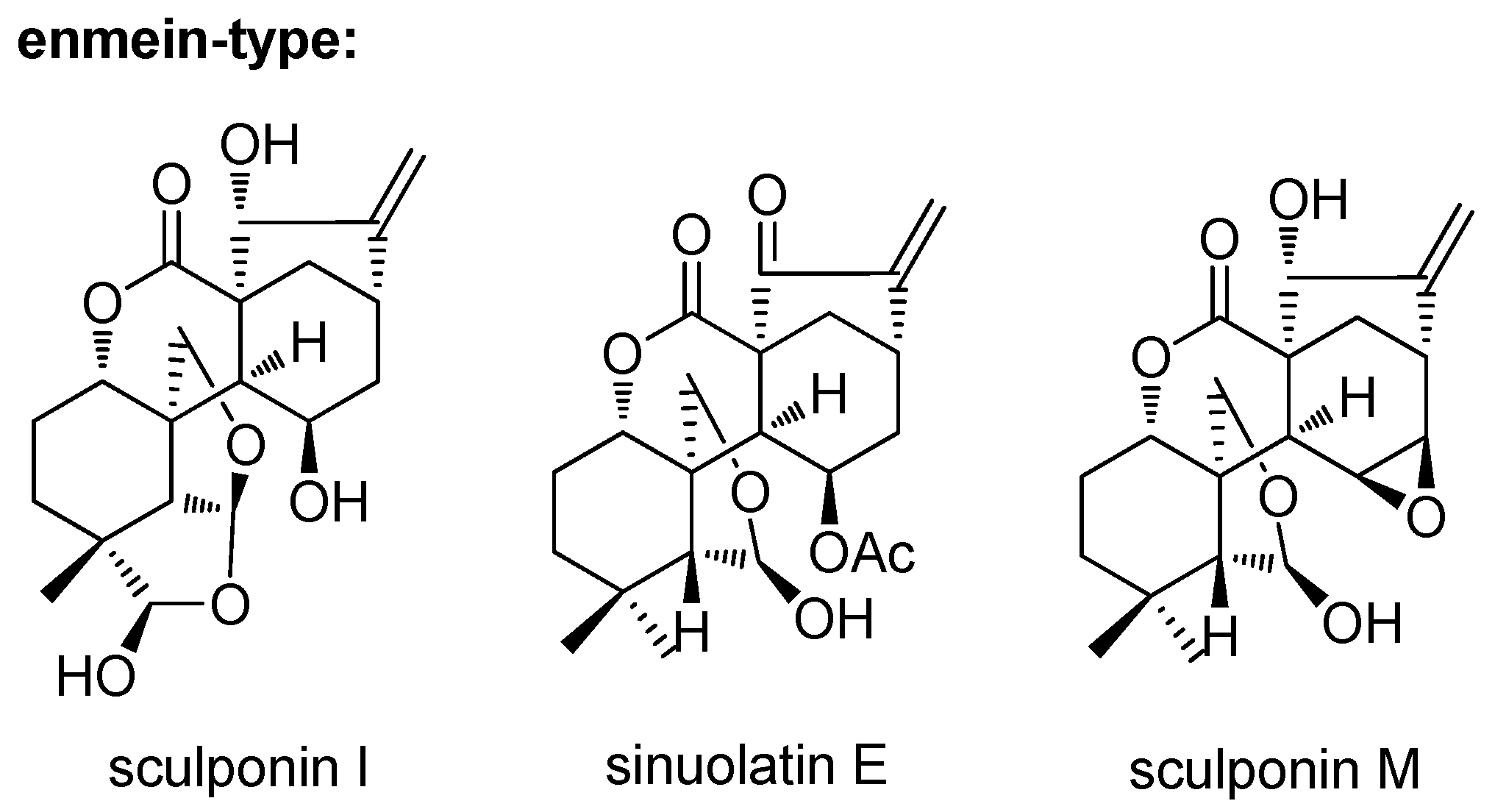
:1. Introduction
2. Introduction
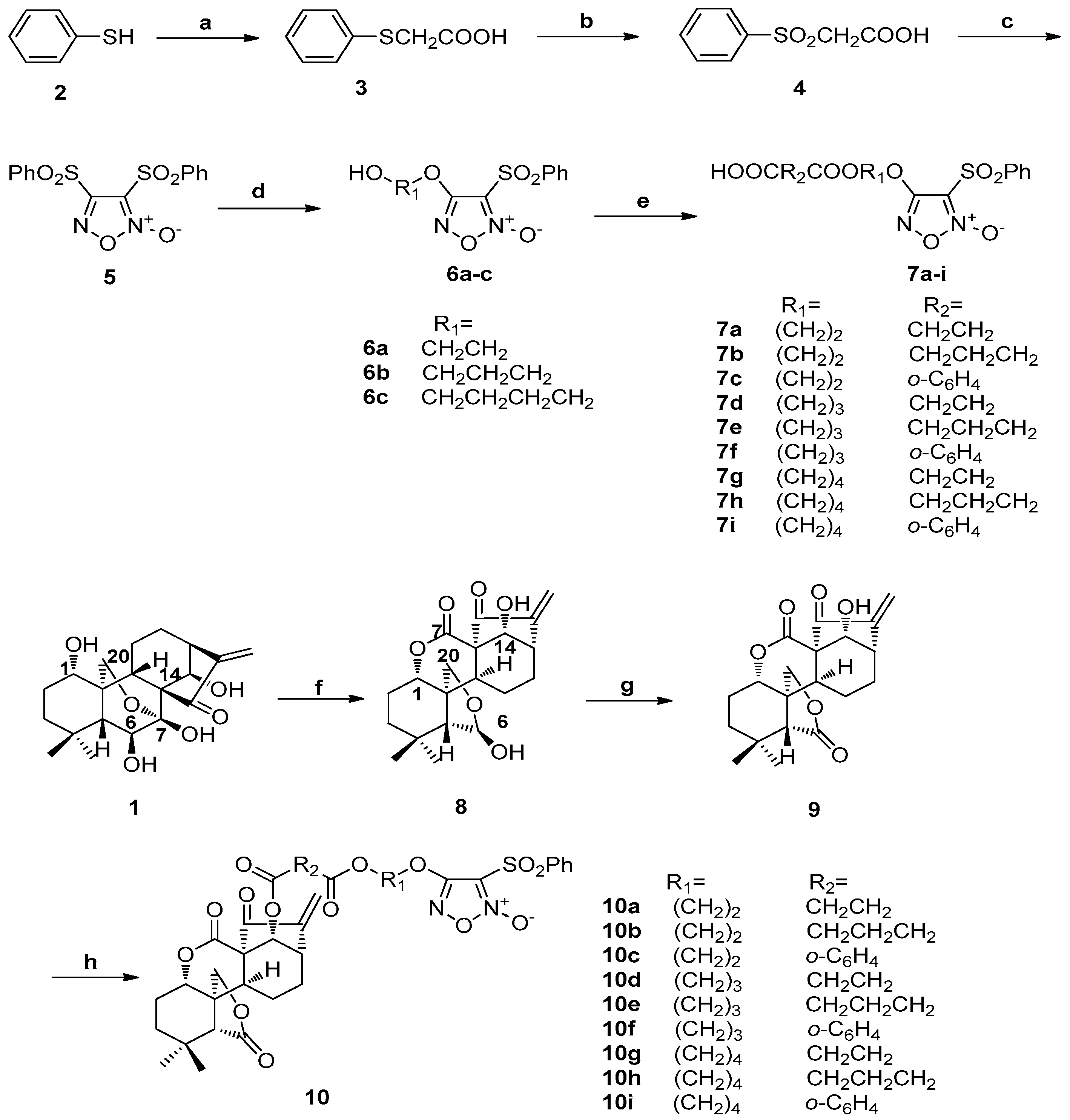
2.1. Synthesis of Compounds 2–9 and 10a–10i
2.2. Antiproliferative Activities in Vitro and SAR
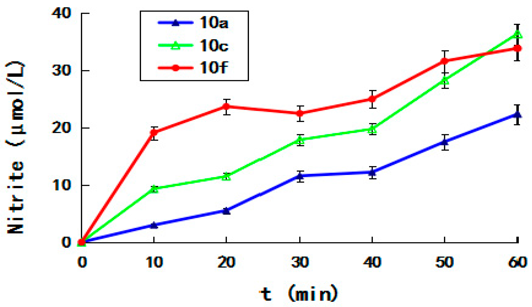
2.3. NO-Releasing Ability
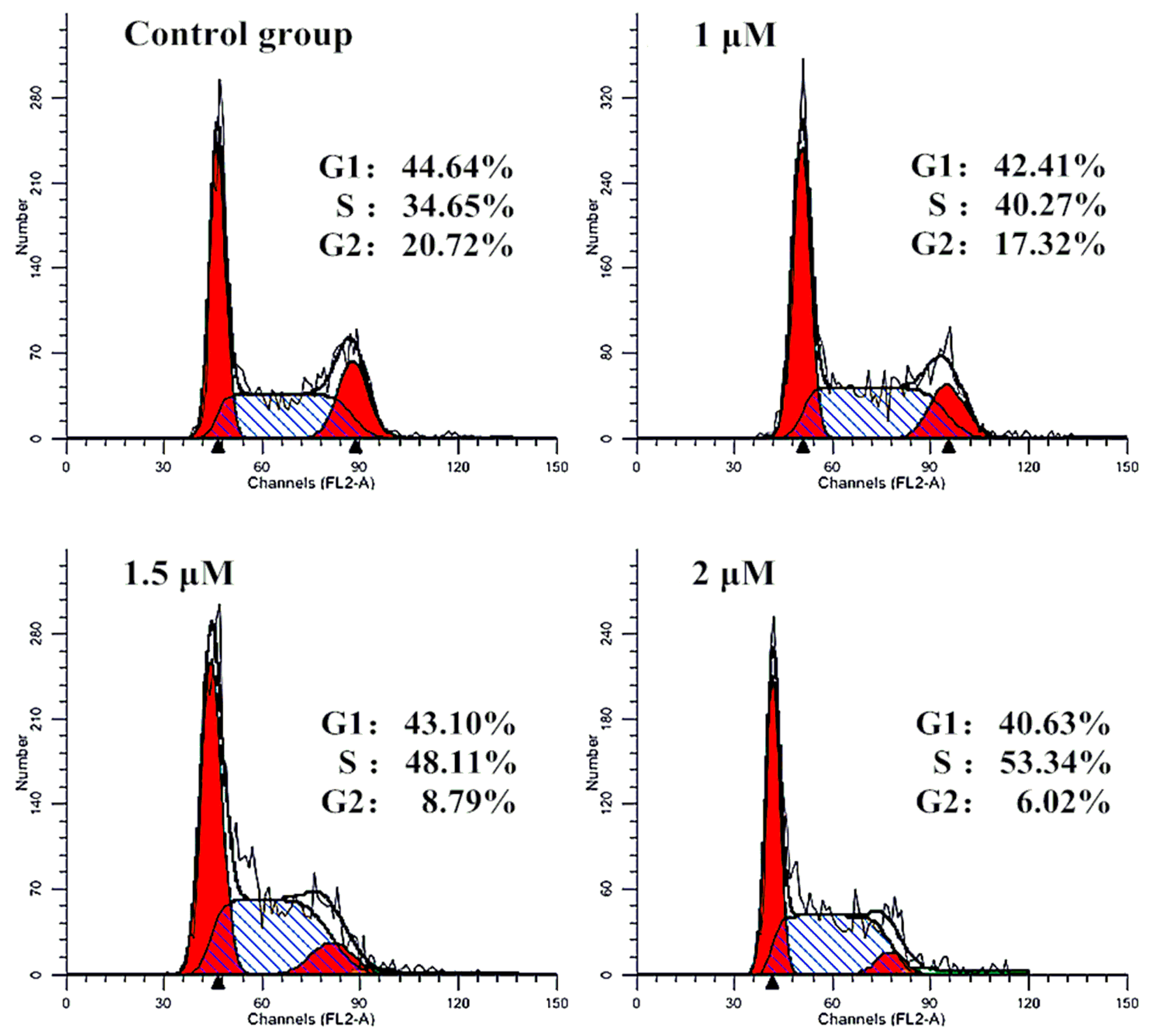
2.4. Effect of Cell Cycle
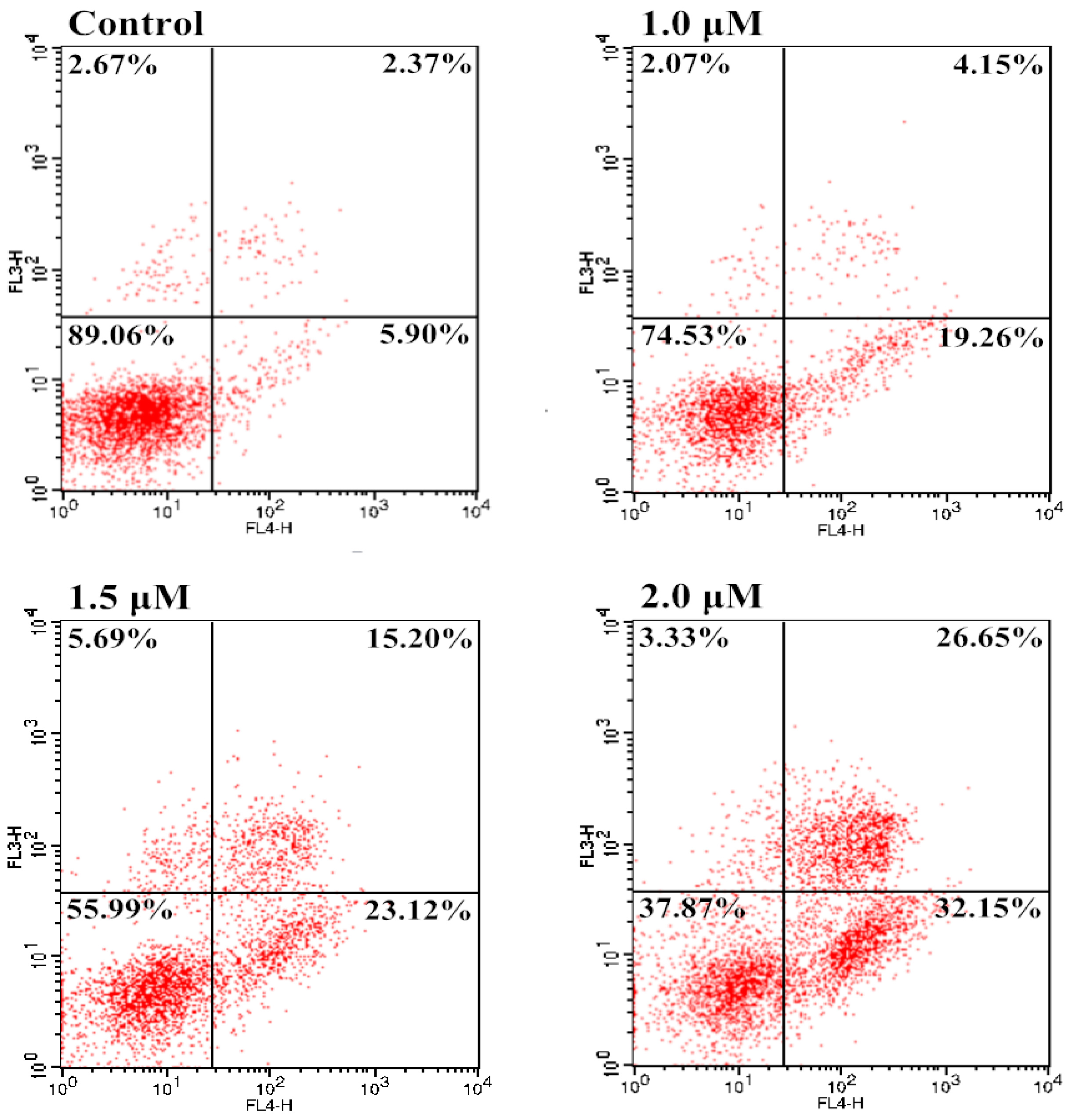
2.5. Induction of Apoptosis
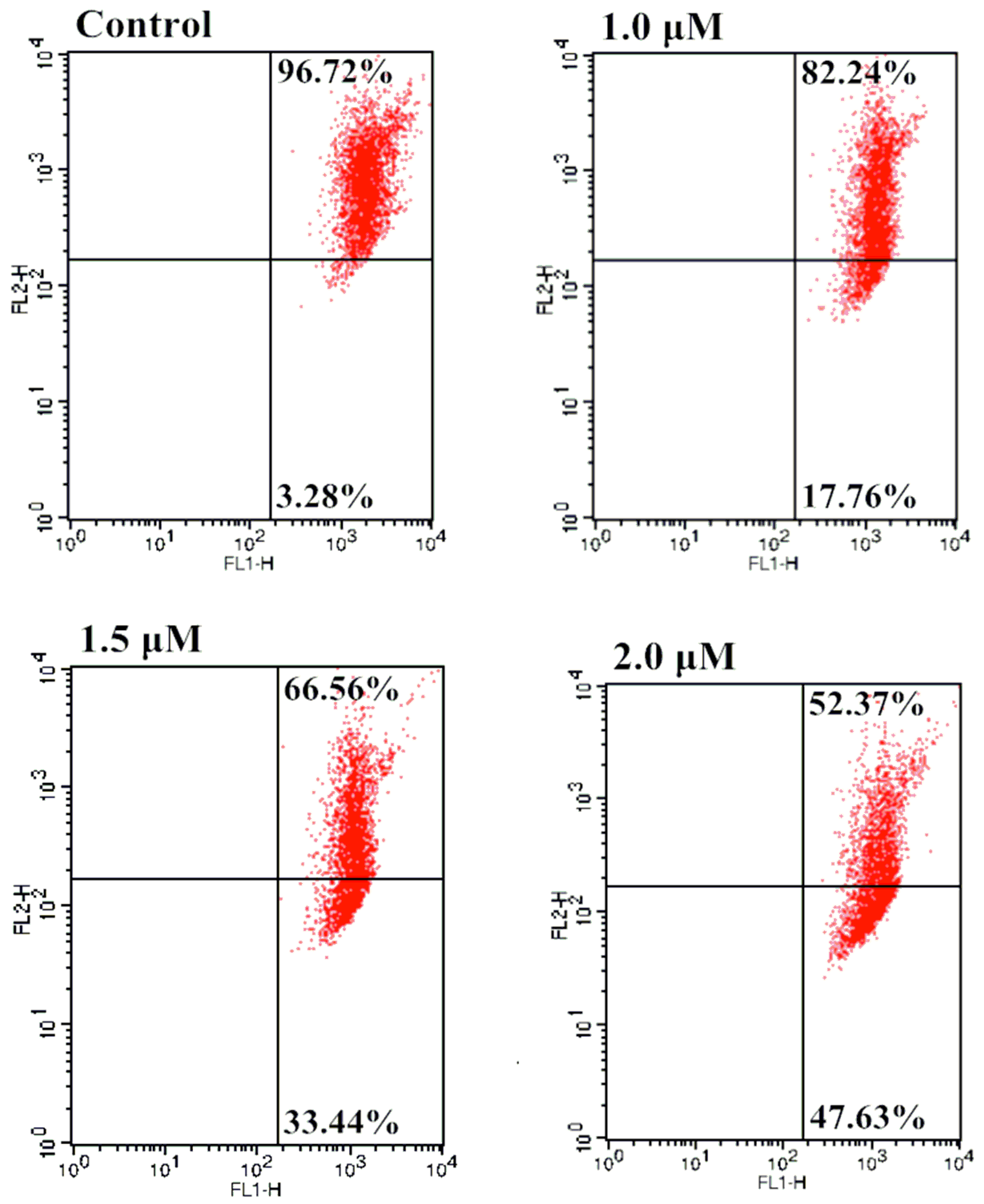
2.6. Effect of Mitochondrial Depolarization
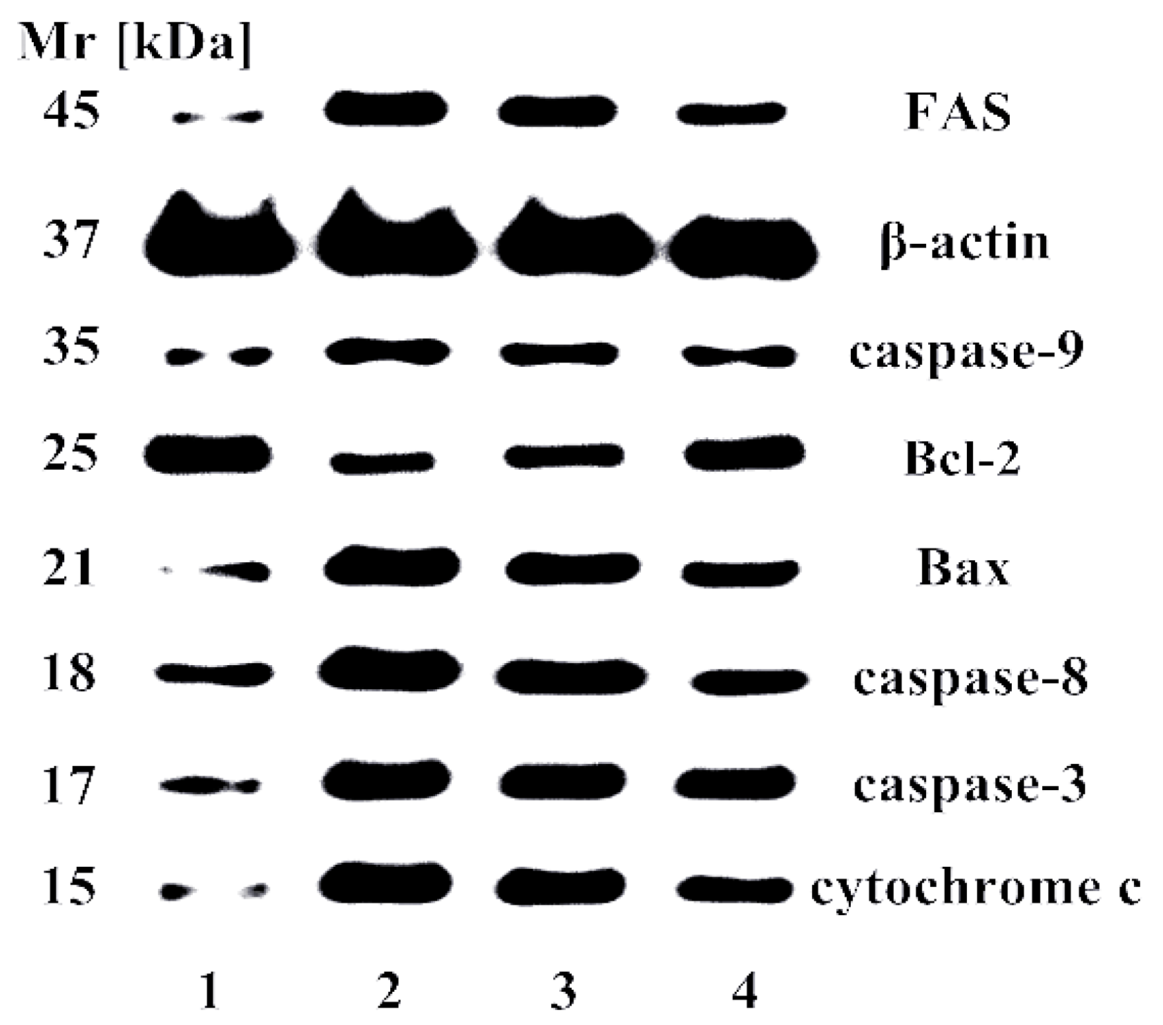
2.7. Effect of Apoptosis-Related Proteins
3. Materials and Methods
3.1. Chemistry
3.1.1. General
3.1.2. General Procedure to Synthesize 10
3.2. Biology
3.2.1. MTT Assay
3.2.2. Griess Assay
3.2.3. Cell Cycle Study
3.2.4. Analysis of Cellular Apoptosis
3.2.5. Mitochondrial Membrane Potential Assay
3.2.6. Western Blot Analysis
4. Conclusions
Supplementary Materials
Acknowledgments
Author Contributions
Conflicts of Interest
Abbreviations
| SAR | Structure Activity Relationship |
| 1H-NMR | Proton Nuclear Magnetic Resonance |
| TMS | Tetramethylsilane |
| MS | Mass Spectrometry |
| ESI | Electrospray Ionization |
| DCM | Dichloromethane |
| MTT | 3-(4,5-Dimethylthiazol-2-yl)-2,5-diphenyltetrazolium bromide |
| FBS | Fetal Bovine Serum |
| NT | Not Test |
| FITC | Fluorescein Isothiocyanate |
References
- Newman, D.J.; Cragg, G.M. Natural Products as Sources of New Drugs from 1981 to 2014. J. Nat. Prod. 2016, 79, 629–661. [Google Scholar] [CrossRef] [PubMed]
- Sun, H.D.; Huang, S.X.; Han, Q.B. Diterpenoids from Isodon species and their biological activities. Nat. Prod. Rep. 2006, 23, 673–698. [Google Scholar] [CrossRef] [PubMed]
- Li, L.M.; Li, G.Y.; Pu, J.X.; Xiao, W.L.; Ding, L.S.; Sun, H.D. ent-Kaurane and cembrane diterpenoids from Isodon sculponeatus and their cytotoxicity. J. Nat. Prod. 2009, 72, 1851–1856. [Google Scholar] [CrossRef] [PubMed]
- He, F.; Xiao, W.L.; Pu, J.X.; Wu, Y.L.; Zhang, H.B.; Li, X.N.; Zhao, Y. Cytotoxic ent-kaurane diterpenoids from Isodon sinuolata. Phytochemistry 2009, 70, 1462–1466. [Google Scholar] [CrossRef] [PubMed]
- Jiang, H.Y.; Wang, W.G.; Zhou, M.; Wu, H.Y.; Zhan, R.; Li, X.N.; Du, X.; Li, Y.; Pu, J.X.; Sun, H.D. Enmein-type 6,7-seco-ent-kauranoids from Isodon sculponeatus. J. Nat. Prod. 2013, 76, 2113–2119. [Google Scholar] [CrossRef] [PubMed]
- Gong, J.X.; Lin, G.A.; Sun, W.B.; Li, C.C.; Yang, Z. Total synthesis of (±) maoecrystal V. J. Am. Chem. Soc. 2010, 132, 16745–16746. [Google Scholar] [CrossRef] [PubMed]
- Moritz, B.J.; Mack, D.J.; Tong, L.; Thomson, R. Total synthesis of the Isodon diterpene sculponeatin N. J. Angew. Chem. Int. Ed. Engl. 2014, 53, 2988–2991. [Google Scholar] [CrossRef] [PubMed]
- Nicolaou, K.C.; Dong, L.; Deng, L.J.; Talbot, A.C.; Chen, D.Y. Synthesis of functionalized maoecrystal V core structures. Chem. Commun. 2010, 46, 70–72. [Google Scholar] [CrossRef] [PubMed]
- Wang, L.; Li, D.H.; Xu, S.T.; Cai, H.; Yao, H.Q.; Zhang, Y.H.; Jiang, J.Y.; Xu, J.Y. The conversion of oridonin to spirolactone-type or enmein-type diterpenoid: Synthesis and biological evaluation of ent-6,7-seco-oridonin derivatives as novel potential anticancer agents. Eur. J. Med. Chem. 2012, 52, 242–250. [Google Scholar] [CrossRef] [PubMed]
- Li, D.; Xu, S.; Cai, H.; Pei, L.; Wang, L.; Wu, X.; Yao, H.; Jiang, J.; Sun, Y.; Xu, J. Library construction and biological evaluation of enmein-type diterpenoid analogues as potential anticancer agents. ChemMedChem 2013, 8, 812–818. [Google Scholar] [CrossRef] [PubMed]
- Li, D.; Hu, X.; Han, T.; Xu, S.; Zhou, T.; Wang, Z.; Cheng, K.; Li, Z.; Hua, H.; Xiao, W.; et al. Synthesis, biological activity, and apoptotic properties of NO-donor/enmein-type ent-kauranoid hybrids. Int. J. Mol. Sci. 2016, 17, 747. [Google Scholar] [CrossRef] [PubMed]
- Wang, L.; Li, D.H.; Wang, C.L.; Zhang, Y.H.; Xu, J.Y. Recent progress in the development of natural ent-kaurane diterpenoids with anti-tumor activity. Mini Rev. Med. Chem. 2011, 11, 910–919. [Google Scholar] [CrossRef] [PubMed]
- Li, D.; Xu, S.; Cai, H.; Pei, L.; Zhang, H.; Wang, L.; Yao, H.; Wu, X.; Jiang, J.; Sun, Y.; et al. Enmein-type diterpenoid analogs from natural kaurene-type oridonin: Synthesis and their antitumor biological evaluation. Eur. J. Med. Chem. 2013, 64, 215–221. [Google Scholar] [CrossRef] [PubMed]
- Xu, S.; Li, D.; Pei, L.; Yao, H.; Wang, C.; Cai, H.; Yao, H.; Wu, X.; Xu, J. Design, synthesis and antimycobacterial activity evaluation of natural oridonin derivatives. Bioorg. Med. Chem. Lett. 2014, 24, 2811–2814. [Google Scholar] [CrossRef] [PubMed]
- Xu, S.; Pei, L.; Li, D.; Yao, H.; Cai, H.; Yao, H.; Wu, X.; Xu, J. Synthesis and antimycobacterial evaluation of natural oridonin and its enmein-type derivatives. Fitoterapia 2014, 99, 300–306. [Google Scholar] [CrossRef] [PubMed]
- Mocellin, S. Nitric oxide: Cancer target or anticancer agent? Curr. Cancer Drug Targets 2009, 9, 214–236. [Google Scholar] [CrossRef] [PubMed]
- Martinez, L.; Thames, E.; Kim, J.; Chaudhuri, G.; Singh, R.; Pervin, S. Increased sensitivity of African American triple negative breast cancer cells to nitric oxide-induced mitochondria-mediated apoptosis. BMC Cancer 2016, 16, 559. [Google Scholar] [CrossRef] [PubMed]
- Zhan, R.; He, W.; Wang, F.; Yao, Z.; Tan, J.; Xu, R.; Zhou, J.; Wang, Y.; Li, H.; Wu, J.; et al. Nitric oxide promotes epidermal stem cell migration via cGMP-Rho GTPase signalling. Sci. Rep. 2016, 6, 30687. [Google Scholar] [CrossRef] [PubMed]
- Victorio, J.A.; Fontes, M.T.; Rossoni, L.V.; Davel, A.P. Different anti-contractile function and nitric oxide production of thoracic and abdominal perivascular adipose tissues. Front. Physiol. 2016, 7, 295. [Google Scholar] [CrossRef] [PubMed]
- Dias, C.; Lourenço, C.F.; Ferreiro, E.; Barbosa, R.M.; Laranjinha, J.; Ledo, A. Age-dependent changes in the glutamate-nitric oxide pathway in the hippocampus of the triple transgenic model of Alzheimer’s disease: Implications for neurometabolic regulation. Neurobiol. Aging 2016, 46, 84–95. [Google Scholar] [CrossRef] [PubMed]
- Osorio, J.C.; Recchia, F.A. The role of nitric oxide in metabolism regulation: From basic sciences to the clinical setting. Intensive Care Med. 2000, 26, 1395–1398. [Google Scholar] [CrossRef] [PubMed]
- Ren, Z.; Gu, X.; Lu, B.; Chen, Y.; Chen, G.; Feng, J.; Lin, J.; Zhang, Y.; Peng, H. Anticancer efficacy of a nitric oxide-modified derivative of bifendate against multidrug-resistant cancer cells. J. Cell. Mol. Med. 2016, 20, 1095–1105. [Google Scholar] [CrossRef] [PubMed]
- Gazzano, E.; Chegaev, K.; Rolando, B.; Blangetti, M.; Annaratone, L.; Ghigo, D.; Fruttero, R.; Riganti, C. Overcoming multidrug resistance by targeting mitochondria with NO-donating doxorubicins. Bioorg. Med. Chem. 2016, 24, 967–975. [Google Scholar] [CrossRef] [PubMed]
- Duan, W.; Hou, J.; Chu, X.; Li, X.; Zhang, J.; Li, J.; Xu, W.; Zhang, Y. Synthesis and biological evaluation of novel histone deacetylases inhibitors with nitric oxide releasing activity. Bioorg. Med. Chem. 2015, 23, 4481–4488. [Google Scholar] [CrossRef] [PubMed]
- Liu, L.; Wang, D.; Wang, J.; Ji, H.; Zhang, Y. NOAD, a novel nitric oxide donor, induces G2/M phase arrest and apoptosis in human hepatocellular carcinoma Bel-7402 cells. Toxicol. Vitro 2015, 29, 1289–1297. [Google Scholar] [CrossRef] [PubMed]
- Duan, W.; Li, J.; Inks, E.S.; Chou, C.J.; Jia, Y.; Chu, X.; Li, X.; Xu, W.; Zhang, Y. Design, synthesis, and antitumor evaluation of novel histone deacetylase inhibitors equipped with a phenylsulfonylfuroxan module as a nitric oxide donor. J. Med. Chem. 2015, 58, 4325–4338. [Google Scholar] [CrossRef] [PubMed]
- Carradori, S.; Mollica, A.; de Monte, C.; Ganese, A.; Supuran, C.T. Nitric oxide donors and selective carbonic anhydrase inhibitors: A dual pharmacological approach for the treatment of glaucoma, cancer and osteoporosis. Molecules 2015, 20, 5667–5679. [Google Scholar] [CrossRef] [PubMed]
- Liu, L.; Li, T.; Tan, J.; Fu, J.; Guo, Q.; Ji, H.; Zhang, Y. NG as a novel nitric oxide donor induces apoptosis by increasing reactive oxygen species and inhibiting mitochondrial function in MGC803 cells. Int. Immunopharmacol. 2014, 23, 27–36. [Google Scholar] [CrossRef] [PubMed]
- Fang, L.; Wang, M.; Gou, S.; Liu, X.; Zhang, H.; Cao, F. Combination of amino acid/dipeptide with nitric oxide donating oleanolic acid derivatives as PepT1 targeting antitumor prodrugs. J. Med. Chem. 2014, 57, 1116–1120. [Google Scholar] [CrossRef] [PubMed]
- Maciag, A.E.; Holland, R.J.; Kim, Y.; Kumari, V.; Luthers, C.E.; Sehareen, W.S.; Biswas, D.; Morris, N.L.; Ji, X.; Anderson, L.M.; et al. Nitric oxide (NO) releasing poly ADP-ribose polymerase 1 (PARP-1) inhibitors targeted to glutathione S-transferase P1-overexpressing cancer cells. J. Med. Chem. 2014, 57, 2292–2302. [Google Scholar] [CrossRef] [PubMed]
- Fu, J.; Liu, L.; Huang, Z.; Lai, Y.; Ji, H.; Peng, S.; Tian, J.; Zhang, Y. Hybrid molecule from O2-(2,4-dinitrophenyl)diazeniumdiolate and oleanolic acid: A glutathione S-transferase π-activated nitric oxide prodrug with selective anti-human hepatocellular carcinoma activity and improved stability. J. Med. Chem. 2013, 56, 4641–4655. [Google Scholar] [CrossRef] [PubMed]
- Ai, Y.; Kang, F.; Huang, Z.; Xue, X.; Lai, Y.; Peng, S.; Tian, J.; Zhang, Y. Synthesis of CDDO-amino acid-nitric oxide donor trihybrids as potential antitumor agents against both drug-sensitive and drug-resistant colon cancer. J. Med. Chem. 2015, 58, 2452–2464. [Google Scholar] [CrossRef] [PubMed]
- Coneski, P.N.; Schoenfisch, M.H. Nitric oxide release: Part III. Measurement and reporting. Chem. Soc. Rev. 2012, 41, 3753–3758. [Google Scholar] [CrossRef] [PubMed]
- Huerta, S.; Chilka, B.; Bonavida, B. Nitric oxide donors: Novel cancer therapeutics (review). Int. J. Oncol. 2008, 33, 909–927. [Google Scholar] [CrossRef] [PubMed]
- Ferioli, R.; Folco, G.C.; Ferretti, C.; Gasco, A.M.; Medana, C.; Fruttero, R.; Civelli, M.; Gasco, A. A new class of furoxan derivatives as NO donors: Mechanism of action and biological activity. J. Pharmacol. 1995, 114, 816–820. [Google Scholar] [CrossRef]
- Feelisch, M.; Schönafinger, K.; Noack, E. Thiol mediated generation of nitric oxide accounts for the vasodilator action of furoxans. Biochem. Pharmacol. 1992, 44, 1149–1157. [Google Scholar] [CrossRef]
- Medana, C.; Ermondi, G.; Fruttero, R.; di Stilo, A.; Ferretti, C.; Gasco, A. Furoxans as nitric oxide donors. 4-Phenyl-3-furoxancarbonitrile: Thiol-mediated nitric oxide release and biological evaluation. J. Med. Chem. 1994, 37, 4412–4416. [Google Scholar] [CrossRef] [PubMed]
- Han, C.; Huang, Z.J.; Zheng, C.; Wan, L.D.; Zhang, L.W.; Peng, S.X.; Ding, K.; Ji, H.B.; Tian, J.D.; Zhang, Y.H. Novel hybrids of (phenylsulfonyl)furoxan and anilinopyrimidine as potent and selective epidermal growth factor receptor inhibitors for intervention of non-small-cell lung cancer. J. Med. Chem. 2013, 56, 4738–4748. [Google Scholar] [CrossRef] [PubMed]
- Tang, W.; Xie, J.; Xu, S.; Lv, H.; Lin, M.; Yuan, S.; Bai, J.; Hou, Q.; Yu, S. Novel nitric oxide-releasing derivatives of brusatol as anti-inflammatory agents: Design, synthesis, biological evaluation, and nitric oxide release studies. J. Med. Chem. 2014, 57, 7600–7612. [Google Scholar] [CrossRef] [PubMed]
- Liu, M.M.; Chen, X.Y.; Huang, Y.Q.; Feng, P.; Guo, Y.L.; Yang, G.; Chen, Y. Hybrids of phenylsulfonylfuroxan and coumarin as potent antitumor agents. J. Med. Chem. 2014, 57, 9343–9356. [Google Scholar] [CrossRef] [PubMed]
- Chen, L.; Zhang, Y.; Kong, X.; Lan, E.; Huang, Z.; Peng, S.; Kaufman, D.L.; Tian, J. Design, synthesis, and antihepatocellular carcinoma activity of nitric oxide releasing derivatives of oleanolic acid. J. Med. Chem. 2008, 51, 4834–4838. [Google Scholar] [CrossRef] [PubMed]
- Lai, Y.; Shen, L.; Zhang, Z.; Liu, W.; Zhang, Y.; Ji, H.; Tian, J. Synthesis and biological evaluation of furoxan-based nitric oxide-releasing derivatives of glycyrrhetinic acid as anti-hepatocellular carcinoma agents. Bioorg. Med. Chem. Lett. 2010, 20, 6416–6420. [Google Scholar] [CrossRef] [PubMed]
- Tang, X.; Gu, X.; Ai, H.; Wang, G.; Peng, H.; Lai, Y.; Zhang, Y. Synthesis and evaluation of nitric oxide-releasing DDB derivatives as potential Pgp-mediated MDR reversal agents in MCF-7/Adr cells. Bioorg. Med. Chem. Lett. 2012, 22, 801–805. [Google Scholar] [CrossRef] [PubMed]
- Wang, T.T.; Liu, Y.; Chen, L. Synthesis and cytotoxic activity of nitric oxide-releasing isosteviol derivatives. Bioorg. Med. Chem. Lett. 2014, 24, 2202–2205. [Google Scholar] [CrossRef] [PubMed]
- Kelly, J.L.; McLean, E.W.; Willard, K.F. Synthesis of bis(arylsulfonyl)furoxans from aryl nitromethyl sulfones. J. Heterocycl. Chem. 1977, 14, 1415–1416. [Google Scholar] [CrossRef]
- Ling, Y.; Ye, X.; Zhang, Z.; Zhang, Y.; Lai, Y.; Ji, H.; Peng, S.; Tian, J. Novel nitric oxide-releasing derivatives of farnesylthiosalicylic acid: Synthesis and evaluation of antihepatocellular carcinoma activity. J. Med. Chem. 2011, 54, 3251–3259. [Google Scholar] [CrossRef] [PubMed]
- Kasibhatla, S.; Tseng, B. Why target apoptosis in cancer treatment? Mol. Cancer Ther. 2003, 2, 573–580. [Google Scholar] [PubMed]
- Loeffler, M.; Kroemer, G. The mitochondrion in cell death control: Certainties and incognita. Exp. Cell. Res. 2000, 256, 19–26. [Google Scholar] [CrossRef] [PubMed]
- Li, P.; Nijhawan, D.; Budihardjo, I.; Srinivasula, S.M.; Ahmad, M.; Alnemri, E.S.; Wang, X. Cytochrome c and dATP-dependent formation of Apaf-1/caspase-9 complex initiates an apoptotic protease cascade. Cell 1997, 91, 479–489. [Google Scholar] [CrossRef]
- Xu, S.; Luo, S.; Yao, H.; Cai, H.; Miao, X.; Wu, F.; Yang, D.H.; Wu, X.; Xie, W.; Yao, H.; et al. Probing the anticancer action of oridonin with fluorescent analogues: Visualizing subcellular localization to mitochondria. J. Med. Chem. 2016, 59, 5022–5034. [Google Scholar] [CrossRef] [PubMed]
- Gross, A.; McDonnell, J.M.; Korsmeyer, S.J. BCL-2 family members and the mitochondria in apoptosis. Genes Dev. 1999, 13, 1899–1911. [Google Scholar] [CrossRef] [PubMed]
- Adams, J.M.; Cory, S. The Bcl-2 protein family: Arbiters of cell survival. Science 1998, 281, 1322–1326. [Google Scholar] [CrossRef] [PubMed]
- Sample Availability: Samples of the compounds 10a–i are available from the authors.


| Compound | Bel-7402 | L-02 | K562 | MGC-803 | CaEs-17 |
|---|---|---|---|---|---|
| 1 | 7.48 ± 0.32 | 18.3 ± 0.8 | 4.76 ± 0.32 | 5.69 ± 0.39 | 11.0 ± 1.0 |
| 9 | 16.0 ± 1.1 | 26.4 ± 1.3 | 2.64 ± 0.19 | 5.84 ± 0.57 | 23.7 ± 1.8 |
| 7a | 22.0 ± 0.8 | 37.8 ± 1.3 | 22.6 ± 2.0 | 25.4 ± 1.5 | 24.3 ± 0.9 |
| 7b | 22.3 ± 1.6 | 39.6 ± 1.4 | 24.2 ± 2.5 | 22.2 ± 2.5 | 25.7 ± 1.4 |
| 7c | 19.6 ± 1.0 | 41.3 ± 2.9 | 22.6 ± 1.7 | 24.6 ± 1.4 | 23.8 ± 1.0 |
| 7d | 17.8 ± 1.4 | 37.4 ± 0.8 | 17.5 ± 1.4 | 18.5 ± 0.8 | 19.8 ± 1.3 |
| 7e | 18.6 ± 0.8 | 36.3 ± 2.1 | 18.5 ± 1.6 | 20.0 ± 1.1 | 18.7 ± 1.0 |
| 7f | 16.7 ± 0.8 | 35.8 ± 1.3 | 17.5 ± 1.0 | 19.3 ± 0.6 | 19.2 ± 0.7 |
| 7g | 22.7 ± 1.0 | 39.4 ± 2.6 | 24.0 ± 1.4 | 20.6 ± 1.7 | 25.7 ± 0.7 |
| 7h | 21.3 ± 0.7 | 35.4 ± 1.4 | 24.5 ± 1.4 | 23.5 ± 1.0 | 22.9 ± 1.4 |
| 7i | 22.4 ± 1.6 | 37.9 ± 2.8 | 23.0 ± 1.2 | 21.6 ± 1.5 | 24.8 ± 1.5 |
| 10a | 1.91 ± 0.09 | 24.4 ± 0.8 | 2.86 ± 0.05 | 2.84 ± 1.12 | 5.68 ± 0.53 |
| 10b | 1.75 ± 0.12 | 22.1 ± 1.3 | 2.90 ± 0.17 | 2.67 ± 0.24 | 5.56 ± 0.06 |
| 10c | 1.01 ± 0.04 | 31.2 ± 1.2 | 1.97 ± 0.18 | 1.60 ± 0.15 | 4.02 ± 0.14 |
| 10d | 1.17 ± 0.02 | 27.4 ± 1.0 | 2.04 ± 0.10 | 2.03 ± 0.15 | 4.84 ± 0.47 |
| 10e | 1.12 ± 0.09 | 29.3 ± 0.9 | 2.02 ± 0.17 | 1.91 ± 0.09 | 4.04 ± 0.23 |
| 10f | 0.81 ± 0.06 | 29.9 ± 1.4 | 1.73 ± 0.04 | 1.18 ± 0.10 | 3.77 ± 0.30 |
| 10g | 1.85 ± 0.11 | 33.9 ± 1.5 | 2.82 ± 0.22 | 2.80 ± 0.09 | 5.52 ± 0.08 |
| 10h | 1.65 ± 0.12 | 29.8 ± 0.9 | 2.78 ± 0.20 | 2.65 ± 0.16 | 5.48 ± 0.21 |
| 10i | 1.09 ± 0.09 | 27.7 ± 1.3 | 2.06 ± 0.16 | 1.64 ± 0.12 | 4.13 ± 0.29 |
| Taxol | 1.89 ± 0.09 | 3.73 ± 0.17 | 0.41 ± 0.02 | 4.65 ± 0.29 | 0.43 ± 0.03 |
© 2016 by the authors. Licensee MDPI, Basel, Switzerland. This article is an open access article distributed under the terms and conditions of the Creative Commons Attribution (CC-BY) license ( http://creativecommons.org/licenses/by/4.0/).
Share and Cite
Li, D.; Hu, X.; Han, T.; Liao, J.; Xiao, W.; Xu, S.; Li, Z.; Wang, Z.; Hua, H.; Xu, J. NO-Releasing Enmein-Type Diterpenoid Derivatives with Selective Antiproliferative Activity and Effects on Apoptosis-Related Proteins. Molecules 2016, 21, 1193. https://0-doi-org.brum.beds.ac.uk/10.3390/molecules21091193
Li D, Hu X, Han T, Liao J, Xiao W, Xu S, Li Z, Wang Z, Hua H, Xu J. NO-Releasing Enmein-Type Diterpenoid Derivatives with Selective Antiproliferative Activity and Effects on Apoptosis-Related Proteins. Molecules. 2016; 21(9):1193. https://0-doi-org.brum.beds.ac.uk/10.3390/molecules21091193
Chicago/Turabian StyleLi, Dahong, Xu Hu, Tong Han, Jie Liao, Wei Xiao, Shengtao Xu, Zhanlin Li, Zhenzhong Wang, Huiming Hua, and Jinyi Xu. 2016. "NO-Releasing Enmein-Type Diterpenoid Derivatives with Selective Antiproliferative Activity and Effects on Apoptosis-Related Proteins" Molecules 21, no. 9: 1193. https://0-doi-org.brum.beds.ac.uk/10.3390/molecules21091193