Synthesis and Biological Evaluation of Phaeosphaeride A Derivatives as Antitumor Agents

 ,
,
Abstract
:
1. Introduction
2. Results and Discussion
2.1. Chemistry
2.2. Biological Evaluation
2.2.1. Cytotoxicity Assay Using 9 Tumor Cell Lines and HEF Cell Line
2.2.2. Acute Intraperitoneal Toxicity Study
3. Materials and Methods
3.1. Materials and Instruments
3.2. Chemical Syntheses
3.2.1. Synthesis of (2S,3S,4S)-3-Hydroxy-6-methoxy-3-methyl-7-methylene-5-oxo-2-pentyl-2,3,4,5,6,7-hexahydropyrano[2,3-c]pyrrol-4-yl methanesulfonate
3.2.2. General Procedure for the Synthesis of Compounds 1–5
3.2.3. General Procedure for the Synthesis of Compounds 6–8
3.2.4. Alternative General Procedure for the Synthesis of Compounds 6–8
3.3. Bio-Evaluation Methods
3.3.1. In-Vitro Cytotoxicity Study (MTT Assay)
3.3.2. Experimental Animals
3.3.3. Ethical Guidelines
3.3.4. Acute Intraperitoneal Toxicity Study
4. Conclusions
Supplementary Materials
Author Contributions
Funding
Conflicts of Interest
References
- Goodman, L.S.; Wintrobe, M.M.; Dameshek, W.; Goodman, M.J.; Gilman, A.Z.; McLennan, M.T. Nitrogen mustard therapy. Use of methyl-bis(beta-chloroethyl)amine hydrochloride and tris(beta-chloroethyl)amine hydrochloride for hodgkin’s disease, lymphosarcoma, leukemia and certain allied and miscellaneous disorders. J. Am. Med. Assoc. 1946, 132, 126–132. [Google Scholar] [CrossRef] [PubMed]
- Wilson, A.L.; Plebanski, M.; Stephens, A.N. New trends in anti-cancer therapy: Combining conventional chemotherapeutics with novel immunomodulators. Curr. Med. Chem. 2017. [Google Scholar] [CrossRef] [PubMed]
- Bhavana, V.; Sudharshan, S.J.S.; Madhu, D. Natural anticancer compounds and their derivatives in clinical trials. In Anticancer Plants: Clinical Trials and Nanotechnology; Akhtar, M., Swamy, M., Eds.; Springer: Singapore, 2017; pp. 51–104. [Google Scholar]
- Cragg, G.M.; Newman, D.J. Plants as a source of anti-cancer agents. J. Ethnopharmacol. 2005, 100, 72–79. [Google Scholar] [CrossRef] [PubMed] [Green Version]
- Cragg, G.M.; Newman, D.J. Natural products: A continuing source of novel drug leads. Biochim. Biophys. Acta. 2013, 1830, 3670–3695. [Google Scholar] [CrossRef] [PubMed] [Green Version]
- Newman, D.J.; Cragg, G.M. Natural products as sources of new drugs from 1981 to 2014. J. Nat. Prod. 2016, 79, 629–661. [Google Scholar] [CrossRef] [PubMed]
- Kobayashi, K.; Kobayashi, Y.; Nakamura, M.; Tamura, O.; Kogen, H. Establishment of Relative and Absolute Configurations of Phaeosphaeride A: Total Synthesis of ent-Phaeosphaeride A. J. Org. Chem. 2015, 80, 1243–1248. [Google Scholar] [CrossRef] [PubMed]
- Kobayashi, K.; Tanaka, K., III; Kogen, H. Total Synthesis and Biological Evaluation of Phaeosphaerides. Catalysts 2018, 8, 206–215. [Google Scholar] [CrossRef]
- Abzianidze, V.V.; Poluektova, E.V.; Bolshakova, K.P.; Panikorovskii, T.L.; Bogachenkov, A.S.; Berestetskiy, A.O. Crystal structure of natural phaeosphaeride A. Acta Crystallogr. E: Crystallogr. Commun. 2015, 71, 625–626. [Google Scholar] [CrossRef] [PubMed]
- Wake, M.S.; Watson, C.J. STAT3 the oncogene–still eluding therapy? FEBS J. 2015, 282, 2600–2611. [Google Scholar] [CrossRef] [PubMed] [Green Version]
- Johnston, P.A.; Grandis, J.R. STAT3 signaling: Anticancer strategies and challenges. Mol. Interv. 2011, 11, 18–26. [Google Scholar] [CrossRef] [PubMed]
- Maloney, K.N.; Hao, W.; Xu, J.; Gibbons, J.; Hucul, J.; Roll, D.; Brady, S.F.; Schroeder, F.C.; Clardy, J. Phaeosphaeride A, an inhibitor of STAT3-dependent signaling isolated from an endophytic fungus. Org. Lett. 2006, 8, 4067–4070. [Google Scholar] [CrossRef] [PubMed]
- Shao, H.; Cheng, H.Y.; Cook, R.G.; Tweardy, D.J. Identification and characterization of signal transducer and activator of transcription 3 recruitment sites within the epidermal growth factor receptor. Cancer Res. 2003, 63, 3923–3930. [Google Scholar] [PubMed]
- Chatzimpaloglou, A.; Yavropoulou, M.P.; Rooij, K.E.; Biedermann, R.; Mueller, U.; Kaskel, S.; Sarli, V. Total Synthesis and Biological Activity of the Proposed Structure of Phaeosphaeride A. J. Org. Chem. 2012, 77, 9659–9667. [Google Scholar] [CrossRef] [PubMed]
- Chatzimpaloglou, A.; Kolosov, M.; Eckols, T.K.; Tweardy, D.J.; Sarli, V. Synthetic and Biological Studies of Phaeosphaerides. J. Org. Chem. 2014, 79, 4043–4054. [Google Scholar] [CrossRef] [PubMed]
- Abzianidze, V.V.; Efimova, K.P.; Poluektova, E.V.; Trishin, Y.G.; Kuznetsov, V.A. Synthesis of natural phaeosphaeride A and semi-natural phaeosphaeride B derivatives. Mendeleev Commun. 2017, 27, 490–492. [Google Scholar] [CrossRef]
- Efimova, K.P. Synthesis of Natural Phaeosphaeride A Derivatives with Antitumor and Herbicidal Activity. Master’s Thesis, Saint-Petersburg State University of Industrial Technologies and Design, Saint Petersburg, Russia, July 2017. [Google Scholar]
- Robinson, S.; Delongeas, J.L.; Donald, E.; Dreher, D.; Festag, M.; Kervyn, S.; Lampo, A.; Nahas, K.; Nogues, V.; Ockert, D.; et al. A European pharmaceutical company initiative challenging the regulatory requirement for acute toxicity studies in pharmaceutical drug development. Regul. Toxicol. Pharmacol. 2008, 50, 345–352. [Google Scholar] [CrossRef] [PubMed]
Sample Availability: Samples of compounds 1–8 are available from the authors. |


| Compound | Adhesive Cell Cultures, IC50 (μM) | ||||
|---|---|---|---|---|---|
| HCT-116 | PC-3 | MCF-7 | A549 | HEF | |
| PPA | 24.21 ± 0.75 | 32.14 ± 0.77 | 20.30 ± 0.8 | 41.10 ± 2.6 | 19.05 ± 0.25 |
| 1 | 3.68 ± 0.81 | 3.35 ± 0.92 | 4.10 ± 0.44 | 12.73 ± 0.40 | 22.30 ± 0.44 |
| 2 | 4.63 ± 0.04 | 5.55 ± 1.48 | 3.23 ± 1.07 | 15.14 ± 0.45 | 16.20 ± 0.26 |
| 3 | 2.90 ± 0.98 | 4.50 ± 1.56 | 3.05 ± 1.20 | 11.41 ± 0,19 | 15.19 ± 1.02 |
| 4 | 8.40 ± 0.24 | 6.37 ± 0.18 | 4.13 ± 0.23 | 12.65 ± 0.27 | 5.03 ± 0.15 |
| 5 | 12.93 ± 0.30 | 24.67 ± 1.24 | 35.71 ± 0.65 | 37.51 ± 1.33 | 53.11 ± 1.06 |
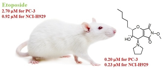
| 6 | 0.47 ± 0.01 | 0.20 ± 0.07 | 3.25 ± 0.64 | 10.11 ± 0.5 | 4.00 ± 0.17 |
| 7 | 1.65 ± 0.63 | 3.65 ± 0.64 | 1.80 ± 0.44 | 12.22 ± 0.2 | 22.30 ± 0.33 |
| 8 | 2.64 ± 0.05 | 4.53 ± 0.40 | 3.20 ± 0.52 | 9.40 ± 0.14 | 6.70 ± 0.23 |
| Etoposide | 22.00 ± 1.10 | 2.70 ± 0.05 | 9.60 ± 0.27 | >100 | >100 |
| Compound | Suspension Cell Cultures, IC50 (μM) | ||||
|---|---|---|---|---|---|
| К562 | NCI-Н929 | Jurkat | THP-1 | RPMI8228 | |
| PPA | 20.47 ± 1.46 | 6.50 ± 0.30 | 9.70 ± 0.42 | 19.10 ± 0.45 | 9.15 ± 0.64 |
| 1 | 3.25 ± 0.64 | 1.35 ± 0.69 | 2.75 ± 0.21 | 2.25 ± 0.21 | 3.97 ± 0.68 |
| 2 | 5.50 ± 0.57 | 2.05 ± 0.35 | 2.60 ± 0.99 | 2.30 ± 0.57 | 3.50 ± 0.82 |
| 3 | 6.70 ± 0.28 | 1.95 ± 0.21 | 3.15 ± 1.77 | 2.60 ± 0.57 | 2.70 ± 0.28 |
| 4 | 10.48 ± 0.41 | 2.35 ± 0.09 | 3.27 ± 0.07 | 3.32 ± 0.10 | 6.00 ± 0.13 |
| 5 | 14.13 ± 0.48 | 7.73 ± 0,25 | 10.10 ± 0.44 | 15.11 ± 0.36 | 16 ± 0.16 |
| 6 | 0.54 ± 0.03 | 0.23 ± 0.02 | 0.55 ± 0.29 | 2.05 ± 0.21 | 0.63 ± 0.23 |
| 7 | 6.03 ± 0.91 | 2.00 ± 0.26 | 2.73 ± 1.53 | 3.45 ± 0.21 | 3.35 ± 1.48 |
| 8 | 4.90 ± 2.40 | 1.87 ± 0.25 | 2.60 ± 1.13 | 2.10 ± 0.28 | 1.40 ± 0.28 |
| Etoposide | 8.47 ± 0.95 | 0.92 ± 0.03 | 0.88 ± 0.74 | 0.83 ± 0.21 | 4.60 ± 0.28 |
© 2018 by the authors. Licensee MDPI, Basel, Switzerland. This article is an open access article distributed under the terms and conditions of the Creative Commons Attribution (CC BY) license (http://creativecommons.org/licenses/by/4.0/).
Share and Cite
Abzianidze, V.; Beltyukov, P.; Zakharenkova, S.; Moiseeva, N.; Mejia, J.; Holder, A.; Trishin, Y.; Berestetskiy, A.; Kuznetsov, V. Synthesis and Biological Evaluation of Phaeosphaeride A Derivatives as Antitumor Agents. Molecules 2018, 23, 3043. https://0-doi-org.brum.beds.ac.uk/10.3390/molecules23113043
Abzianidze V, Beltyukov P, Zakharenkova S, Moiseeva N, Mejia J, Holder A, Trishin Y, Berestetskiy A, Kuznetsov V. Synthesis and Biological Evaluation of Phaeosphaeride A Derivatives as Antitumor Agents. Molecules. 2018; 23(11):3043. https://0-doi-org.brum.beds.ac.uk/10.3390/molecules23113043
Chicago/Turabian StyleAbzianidze, Victoria, Petr Beltyukov, Sofya Zakharenkova, Natalia Moiseeva, Jennifer Mejia, Alvin Holder, Yuri Trishin, Alexander Berestetskiy, and Victor Kuznetsov. 2018. "Synthesis and Biological Evaluation of Phaeosphaeride A Derivatives as Antitumor Agents" Molecules 23, no. 11: 3043. https://0-doi-org.brum.beds.ac.uk/10.3390/molecules23113043