Article

2-(2-Phenylethyl)-4H-chromen-4-one Derivatives from the Resinous Wood of Aquilaria sinensis with Anti-Inflammatory Effects in LPS-Induced Macrophages

1
School of Pharmacy, College of Pharmacy, Kaohsiung Medical University, Kaohsiung 807, Taiwan
2
Institute of Traditional Medicine, National Yang-Ming University, Taipei 112, Taiwan
3
Bioresource Collection and Research Center (BCRC), Food Industry Research and Development Institute (FIRDI), Hsinchu 300, Taiwan
4
Faculty of Pharmacy, School of Pharmaceutical Sciences, National Yang-Ming University, Taipei 112, Taiwan
5
Department of Medical Research, China Medical University Hospital, China Medical University, Taichung 404, Taiwan
*
Authors to whom correspondence should be addressed.
These authors contributed equally to this work.
Submission received: 6 January 2018 / Revised: 22 January 2018 / Accepted: 27 January 2018 / Published: 30 January 2018
(This article belongs to the Section Natural Products Chemistry)

Abstract

:
The resinous wood of Aquilaria sinensis, known as agarwood (Chen Xiang in Chinese), is traditionally used for the treatment of abdominal pain, vomiting, circulatory disorders, and dyspnea. Four new 2-(2-phenylethyl)-4H-chromen-4-one derivatives, namely 7-methoxy-2-[2-(4′-hydroxy-phenyl)ethyl]chromone (1), 7-hydroxy-2-[2-(4′-methoxyphenyl)ethyl]chromone (2), 5,6-dihydroxy- 2-[2-(3′-hydroxy-4′-methoxyphenyl)ethyl]chromone (3), and 6-hydroxy-5-methoxy-2-(2-phenyl-ethyl)chromone (4), have been isolated from the resinous wood of A. sinensis, together with nine known compounds. The structures of these compounds were determined through spectroscopic and MS analyses. Among the isolated compounds, neopetasan, 7-methoxy-2-(2-phenylethyl)-chromone, 6,7-dimethoxy-2-(2-phenylethyl)chromone, and 6,7-dimethoxy-2-[2-(4′-methoxy-phenyl)ethyl]chromone inhibited NF-κB activation in LPS-stimulated RAW 264.7 macrophages with relative luciferase activity values of 0.55 ± 0.09, 0.54 ± 0.03, 0.31 ± 0.05, and 0.38 ± 0.14, respectively, versus that of vehicle control (1.03 ± 0.02). In addition, 5,6-dihydroxy-2-[2-(3′-hydroxy-4′-methoxyphenyl)ethyl]chromone, 7-methoxy-2-(2-phenylethyl)chromone, 7-dimethoxy-2-(2-phenylethyl)chromone, and 6,7-dimethoxy-2-[2-(4′-methoxyphenyl)ethyl]chromone could suppress LPS-induced NO production in RAW 264.7 cells and did not induce cytotoxicity against RAW 264.7 cells after 24-h treatment.

Graphical Abstract

1. Introduction

Aquilaria sinensis (Lour.) Gilg. (Thymelaeaceae) is an evergreen tree endemic to China. The fragrant resin-infused wood derived from the wounded trees of Aquilaria species is called ‘agarwood’ or ‘eaglewood’. Agarwood has been widely used as a traditional sedative, analgesic, and digestive medicine in China [1]. Various benzenoids [2], flavonoids [2,3], 2-(2-phenylethyl)chromones [2,4,5,6,7], sesquiterpenes [8], steroids [2,7], triterpenoids [5], and their derivatives were isolated from this plant in previous studies. Many of these compounds exhibit anti-acetylcholinesterase [8], anti-inflammatory [2,7], antitumor [3], and cytotoxic [6] activities.
Aberrant activation of macrophages is related to many inflammatory disorders such as sepsis, neurodegenerative disorders, osteoporosis, cardiovascular and metabolic diseases [9]. Nuclear factor κB (NF-κB) is an important transcription factor when stimulating an inflammatory reaction and as a molecular target for anti-inflammatory drug discovery. Lipopolysaccharide (LPS)-stimulated macrophages have been prevalently applied as an in vitro model system to study inflammation and to identify anti-inflammatory compounds [10,11]. In our studies on the anti-inflammatory constituents of Formosan plants and Chinese herbal medicines, many species have been screened for in vitro inhibitory activity against NF-κB, a key transcriptional activator of pro-inflammatory molecules in LPS-activated macrophages, and A. sinensis was found to be an active species. Four new 2-(2-phenylethyl)-4H-chromen-4-one derivatives, namely 7-methoxy-2-[2-(4′-hydroxyphenyl)- ethyl]chromone (1), 7-hydroxy-2-[2-(4′-methoxyphenyl)ethyl]chromone (2), 5,6-dihydroxy-2-[2-(3′-hydroxy-4′-methoxyphenyl)ethyl]chromone (3), and 6-hydroxy-5-methoxy-2-(2-phenylethyl)- chromone (4), and nine known compounds 513 have been isolated from the resinous wood of A. sinensis and identified. Their structures are depicted in Figure 1. This paper describes the structural elucidation of the compounds numbered 1 through 4 and the inhibitory activities of all isolates on LPS-induced NF-κB activation of macrophages.

2. Results and Discussion

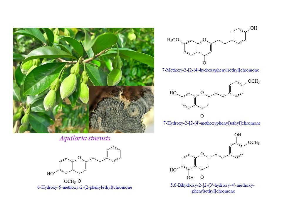
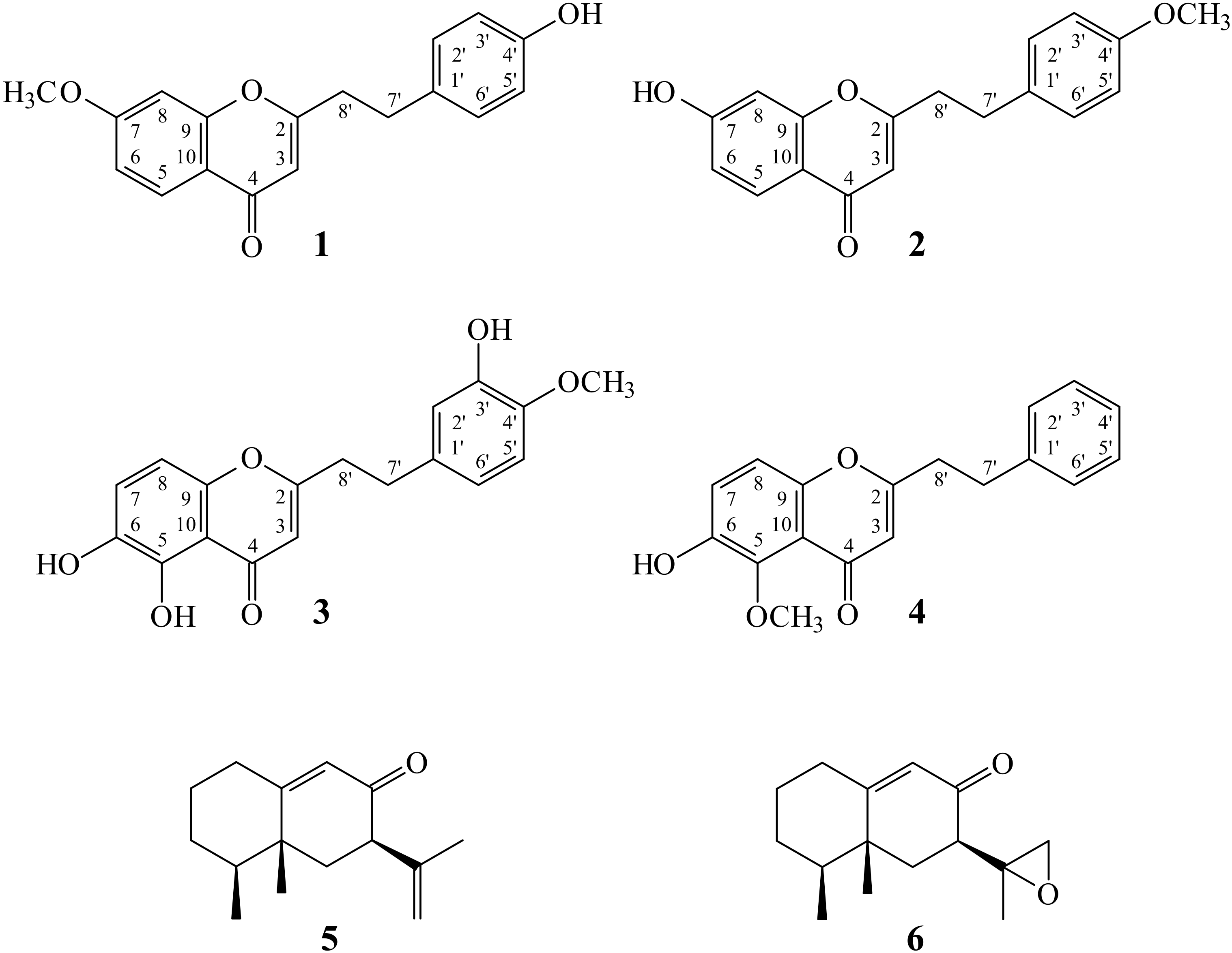
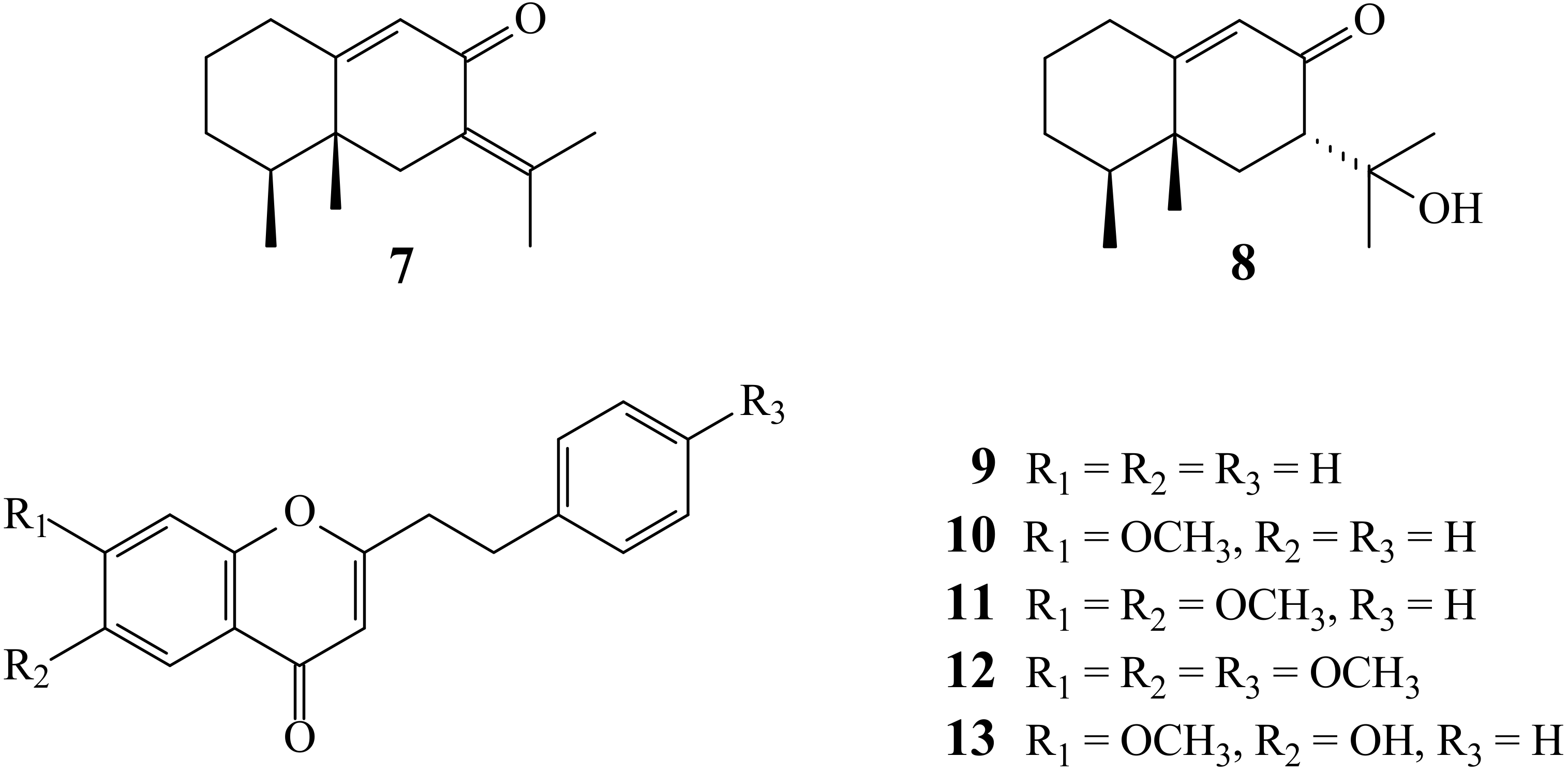
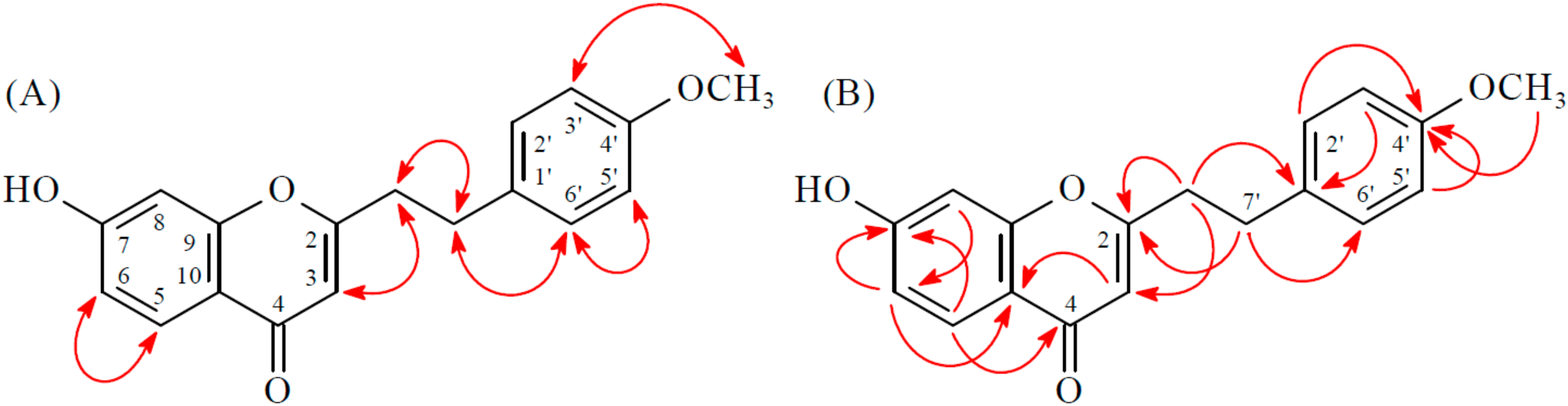
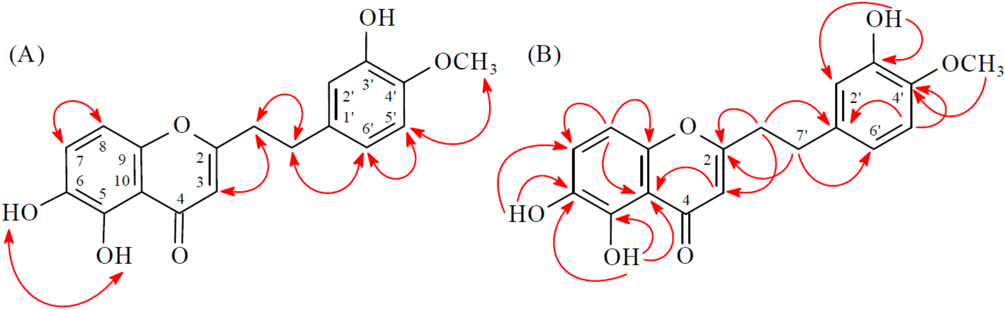
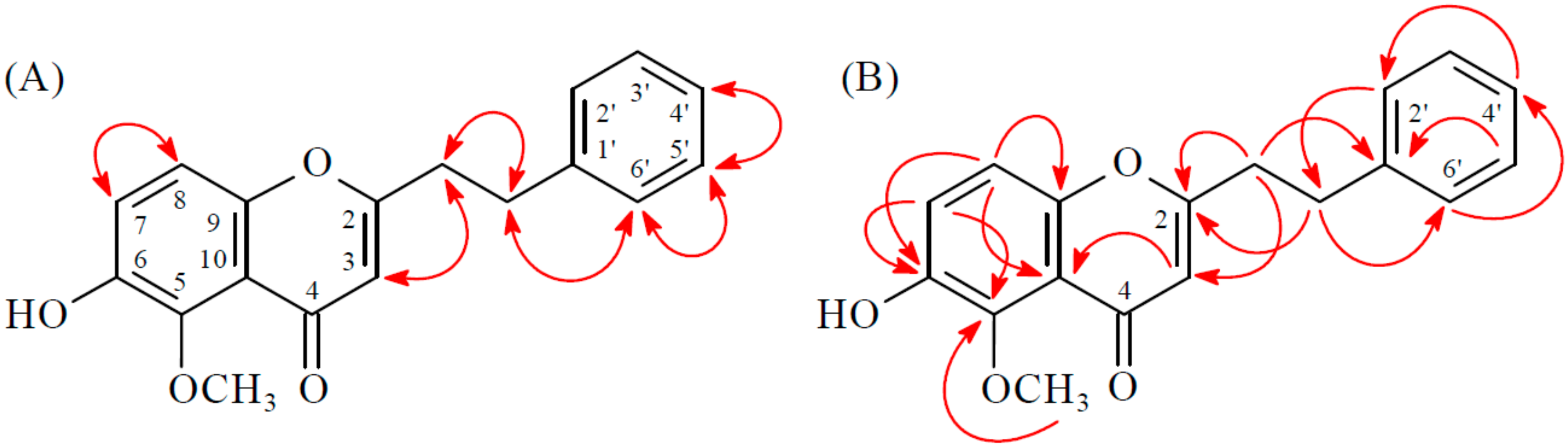
2.1. Isolation and Structural Elucidation

Chromatographic purification of the EtOAc-soluble fraction of a MeOH extract of resinous wood of A. sinensis on a silica gel column and preparative thin-layer chromatography (TLC) afforded four new (14) and nine known compounds (513) (Figure 1).
Compound 1 was obtained as brown plates. The molecular formula, C18H16O4, was deduced from a proton adduct ion at m/z 297.11224 [M + H]+ (calcd. 229.11214) in the HR-ESI-MS spectrum (positive-ion mode) and was supported by the 1H-, 13C-, and DEPT NMR data. IR absorptions for OH (3418 cm−1) and γ-pyrone (1634 and 1590 cm−1) functions were observed. The 1H-NMR spectrum of 1 showed the presence of a methoxy group at δH 3.91 (3H, s, MeO-7), four methylene protons at δH 2.86 (2H, t, J = 8.0 Hz, H-8′) and 2.98 (2H, t, J = 8.0 Hz, H-7′) and a olefinic proton at δH 6.07 (1H, s, H-3). Additionally a set of typical ABX coupling systems at δH 6.83 (1H, d, J = 2.0 Hz, H-8), 6.95 (1H, dd, J = 8.5, 2.0 Hz, H-6) and 8.08 (1H, d, J = 8.5 Hz, H-5) and para-disubstituted benzene protons at δH 6.76 (2H, d, J = 8.5 Hz, H-3′ and H-5′) and δH 7.06 (2H, d, J = 8.5 Hz, H-2′ and H-6′) were observed. The 13C-NMR spectrum of 1 displayed the presence of two methylene groups at δC 32.0 and 36.3, a trisubstituted double bond at δC 109.8 and 168.5, a methoxyl group at δC 56.1, and a carbonyl group at δC 177.9. Based on the above, 1 was deduced to be a 2-(2-phenylethyl)chromone derivative with a methoxyl and a hydroxyl groups. Two signals at δC 164.1 and 154.5 arising from O-bearing aromatic C-atoms were attributable to C-7 and C-4′ respectively, by related HMBC and comparison with a structurally similar compound 10. The HMBC correlations (Figure 2) observed between OMe-7 (δH 3.91) and C-7 (δC 164.1), as well as NOESY correlations (Figure 2) observed between OMe-7 (δH 3.91) and H-8 (δH 6.83) and H-6 (δH 6.95), revealed that the methoxyl group was supposed to be positioned at C-7. Thus, the hydroxyl group should be linked to the last open position at C-4′. Consequently, the structure of 1 was determined to be 7-methoxy-2-[2-(4′-hydroxyphenyl)ethyl]chromone.
Compound 2 was isolated as a yellowish amorphous powder. The ESI-MS afford the quasi-molecular ion [M + H]+ at m/z 297, implying a molecular formula of C18H17O4, which was confirmed by the HR-ESI-MS (m/z 297.11227 [M + H]+, calcd. 297.11214). The IR spectrum showed the presence of OH (3210 cm−1) and γ-pyrone (1635 and 1600 cm−1) groups. The 1H- and 13C-NMR data of 2 were similar to those of 7-methoxy-2-[2-(4′-hydroxyphenyl)ethyl]chromone (1), except that the 7-OH and 4′-OMe [δH 3.79 (3H, s)] groups of 2 replaced the 7-OMe and 4′-OH groups of 1. This was supported by NOESY correlations between OMe-4′ (δH 3.79)/H-3′ (δH 6.83) and OMe-4′ (δH 3.79)/H-5′ (δH 6.83) and by HMBC correlations between OMe-4′ (δH 3.79)/C-4′ (δC 158.3), H-5 (δH 8.02)/C-7 (δC 156.1), and H-6 (δH 7.09)/C-7 (δC 156.1). According to the above data, the structure of 2 was elucidated as 7-hydroxy-2-[2-(4′-methoxyphenyl)ethyl]chromone. This was further confirmed by the 1H–1H-COSY, NOESY (Figure 3), DEPT, HSQC, and HMBC (Figure 3) techniques.
Compound 3 was isolated as a yellowish amorphous powder. Its molecular formula, C18H16O6, was determined on the basis of the positive HRESIMS at m/z 329.10186 [M + H]+ (calcd. 329.10196) and this was supported by the 1H, 13C, and DEPT NMR data. The IR spectrum showed the presence of OH (3418 cm−1) and γ-pyrone (1634 and 1590 cm−1) groups. The 1H- and 13C-NMR data of 3 were similar to those of 3′,6-dihydroxy-4′-methoxy-2-(2-phenylethyl)chromone [7], except that the 5-hydroxy group [δH 12.50 (1H, s, D2O exchangeable)] of 3 replaced H-5 of 3′,6-dihydroxy-4′-methoxy-2-(2-phenylethyl)chromone [7]. This was supported by HMBC correlations between OH-5 (δH 12.50) and C-5 (δC 145.3), C-6 (δC 140.2), and C-10 (δC 111.0). The full assignment of 1H- and 13C-NMR resonances was supported by 1H–1H COSY, DEPT, HSQC, NOESY (Figure 4), and HMBC (Figure 4) spectral analyses. On the basis of the above data, the structure of 3 was elucidated as 5,6-dihydroxy-2-[2-(3′-hydroxy-4′-methoxyphenyl)ethyl]chromone.
Compound 4 was obtained as a yellowish amorphous powder. The molecular formula C18H16O4 was deduced from a sodium adduct ion at m/z 319.09395 [M + Na]+ (calcd. 319.09408) in the HRESI mass spectrum. The presence of a conjugated carbonyl group was revealed by the band at 1633 cm−1 in the IR spectrum, which was confirmed by the resonances at δC 177.5 in the 13C-NMR spectrum. The IR spectrum also revealed a hydroxy absorption at 3340 cm−1. Comparison of the 1H- and 13C-NMR data of 4 with those of 6-hydroxy-2-(2-phenylethyl)chromone (corylifol A) [12] suggested that their structures are closely related, except that the 5-methoxy group [δH 3.98 (3H, s); δC 62.8] of 4 replaced H-5 of 6-hydroxy-2-(2-phenylethyl)chromone [12]. This was supported by HMBC correlation between OMe-5 (δH 3.98) and C-5 (δC 143.5). The structure elucidation of 4 was supported by 1H–1H COSY and NOESY (Figure 5) experiments, and 13C-NMR assignments were confirmed by DEPT, HSQC, and HMBC (Figure 5) techniques.

2.2. Structure Identification of the Known Isolates

The known isolates were readily identified by a comparison of physical and spectroscopic data (UV, IR, 1H-NMR, [α]D, and MS) with corresponding authentic samples or literature values, and this included four sesquiterpenes, neopetasane (5) [13], 7α-H-9(10)-ene-11,12-epoxy-8-oxoeremphilane (6) [13], dehydrokaranone (7) [5], and ligudicin C (8) [14], and five 2-(2-phenylethyl)-4H-chromen-4-one derivatives, 2-(2-phenylethyl)chromone (9) [15], 7-methoxy-2-(2-phenylethyl)chromone (10) [16], 6,7-dimethoxy-2-(2-phenylethyl)chromone (11) [2], 6,7-dimethoxy-2-[2-(4′-methoxyphenyl)ethyl]chro-mone (12) [17], and 6-hydroxy-7-methoxy-2-(2-phenylethyl)chromone (13) [18].

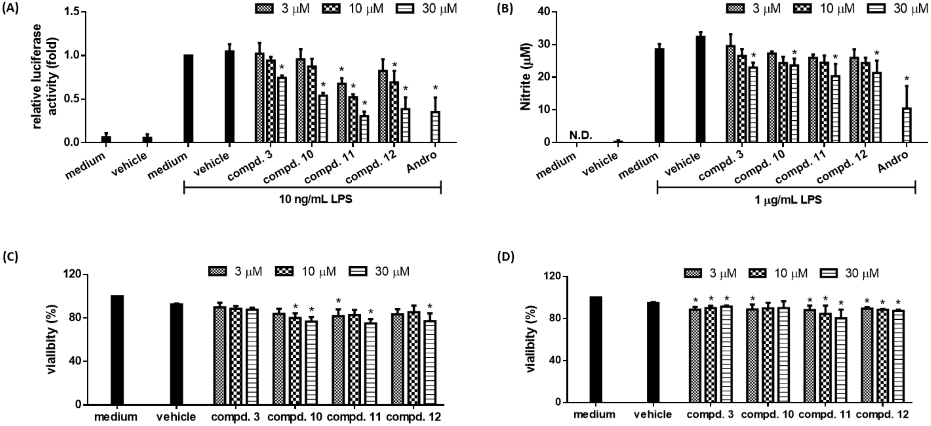
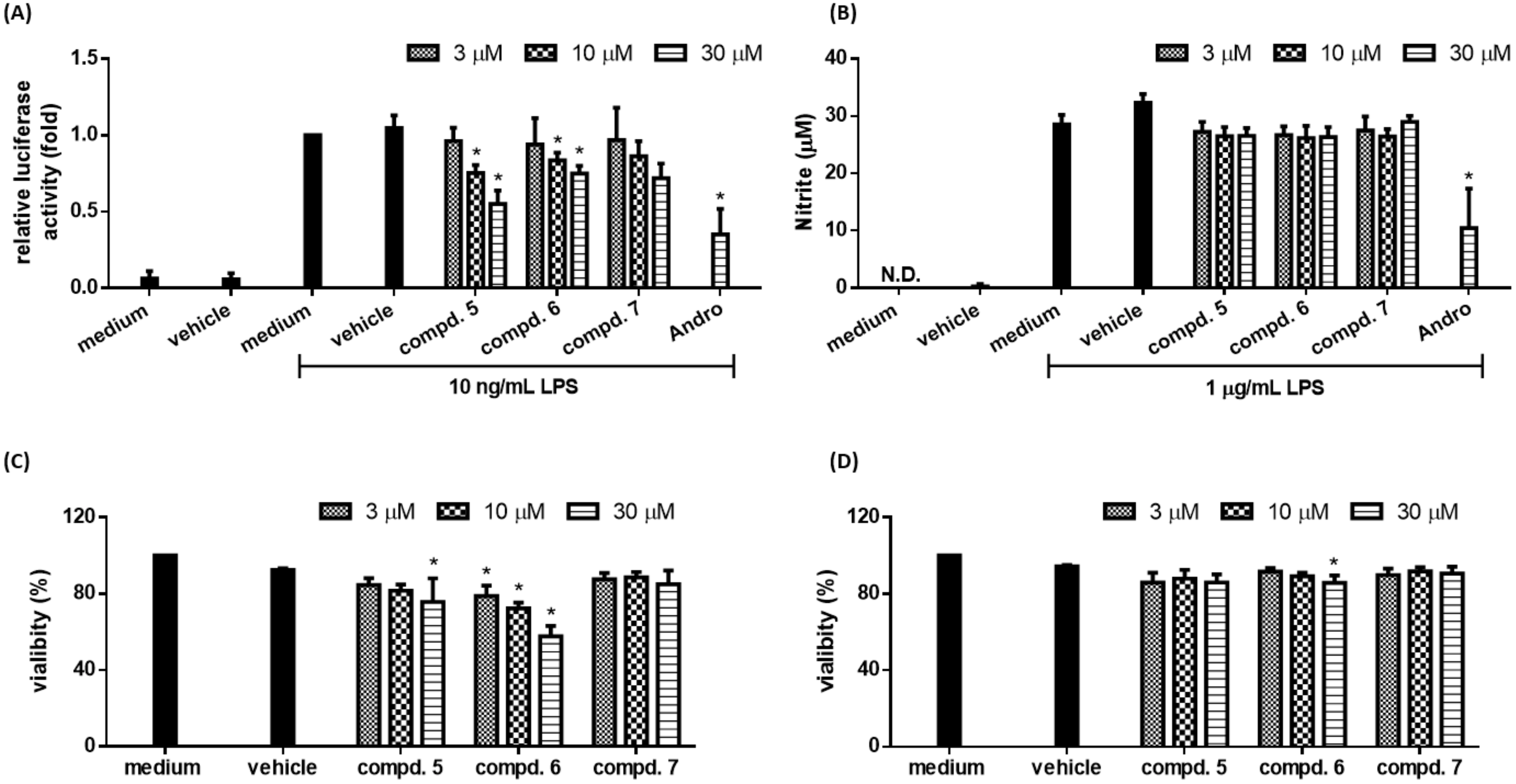
2.3. Biological Studies

LPS-activated macrophage serves as an in vitro model system to study inflammation [19]. A LPS-responsive macrophage cell clone RAW264.7/Luc-P1 was previously established, in which the activity of NF-κB correlates with the expression of reporter gene (luciferase) [10]. This RAW 264.7/Luc-P1 cell line has been successfully applied to identify anti-inflammatory compounds [10,19]. Furthermore, LPS-mediated NF-κB activation leads to upregulation of pro-inflammatory molecules, such as NO, in macrophages [20]. Thus, NO production is a hallmark of inflammatory responses. The anti-inflammatory activities of compounds isolated from the resinous wood of A. sinensis were evaluated by their abilities to suppress NF-κB activation in RAW 264.7/Luc-P1 cell line, their inhibitory activities are summarized in Table 1. Andrographolide was used as positive control. Based on the results of our bioactivity assays, the following conclusions can be drawn: (a) 5,6-Dihydroxy-2-[2-(3′-hydroxy-4′-methoxyphenyl)ethyl]chromone (3), neopetasane (5), 7α-H-9(10)-ene-11,12-epoxy-8-oxoeremphilane (6), dehydrokaranone (7), 7-methoxy-2-(2-phenylethyl)chromone (10), 6,7-dimethoxy-2-(2-phenylethyl)chromone (11), and 6,7-dimethoxy-2-[2-(4′-methoxyphenyl)ethyl]chro-mone (12) can significantly inhibit LPS-induced NF-κB activation and did not show cytotoxicity against RAW 264.7/Luc-P1 cells after 24 h treatment (except compound 6 with dose-dependent cytotoxicity in RAW 264.7/Luc-P1 cells) (Figure 6A,C and Figure 7A,C); (b) among the chromone derivatives, 5,6-dihydroxy-2-[2-(3′-hydroxy-4′-methoxyphenyl)ethyl]chromone (3), 7-methoxy-2-(2-phenylethyl)chromone (10), 7-dimethoxy-2-(2-phenylethyl)chromone (11), and 6,7-dimethoxy-2-[2-(4′-methoxyphenyl)ethyl]chromone (12) could suppress LPS-induced NO production in RAW264.7 macrophages (Figure 6B) and did not induce cytotoxicity against RAW 264.7 cells after 24-h treatment (Figure 6D); (c) among the sesquiterpene analogues, neopetasan (5), 7α-H-9(10)-ene-11,12-epoxy-8-oxoeremphilane (6), dehydrokaranone (7) displayed no inhibitory activity on NO production in RAW264.7 macrophages (Figure 7B) and did not cause significant cytotoxicity (Figure 7D); (d) 6,7-dimethoxy-2-(2-phenylethyl)chromone (11) (with a 6-methoxy moiety) exhibited stronger inhibition than its analogue, 6-hydroxy-7-methoxy-2-(2-phenylethyl)chromone (13) (with a 6-hydroxy group); (e) 7-methoxy-2-(2-phenylethyl)chromone (10) (without the 4′-hydroxy moiety) exhibited more effective inhibition than its analogue, 7-methoxy-2-[2-(4′-hydroxyphenyl)ethyl]chromone (1) (with a 4′-hydroxy group); (f) 2-(2-phenylethyl)chromone (9) (without any substituents) exhibited no inhibitory activity; (g) among the sesquiterpene analogues 5, 6, and 8, neopetasane (5) (with a prop-1-en-2-yl moiety at C-7) exhibited stronger inhibition than the analogues 6 and 8; (h) 6,7-dimethoxy-2-(2-phenylethyl)chromone (11) was the most effective among the isolated compounds, with a relative luciferase activity value of 0.31 ± 0.05, against LPS-induced NF-κB activation in RAW 264.7/Luc-P1 cells.
It is noted that the inhibition on NO production is not always in a tight correlation with NF-κB activation (e.g., compounds 5, 6, and 7 in Figure 7B), which may be due to the discrepancy in experimental conditions and the sensitivity of assays used for measuring these two mediators. Alternatively, natural products are commonly known to be multiple-targeted, these compounds may be involved in NF-κB-independent NO regulation.

3. Experimental Section

3.1. General Procedures

Melting points were determined on a Yanaco micro-melting point apparatus (Yanaco, Tokyo, Japan) and were uncorrected. Optical rotations were measured using a Jasco DIP-370 polarimeter (Japan Spectroscopic Corporation, Tokyo, Japan) in CHCl3. Ultraviolet (UV) spectra were obtained on a Jasco UV-240 spectrophotometer. Infrared (IR) spectra (KBr or neat) were recorded on a Perkin Elmer 2000 FT-IR spectrometer (Perkin Elmer Corporation, Norwalk, CT, USA). Nuclear magnetic resonance (NMR) spectra, including nuclear Overhauser effect spectrometry (NOESY), correlation spectroscopy (COSY), heteronuclear single-quantum coherence (HSQC), and heteronuclear multiple-bond correlation (HMBC) experiments, were acquired using a Varian VNMRS-600 or a Varian Inova 500 spectrometer (Varian Inc., Palo Alto, CA, USA) operating at 600 or 500 MHz (1H) and 150 or 125 MHz (13C), respectively, with chemical shifts given in ppm (δ) using tetramethylsilane (TMS) as an internal standard. Electrospray ionisation (ESI) and high-resolution electrospray ionization (HRESI)-mass spectra were recorded on a VG Platform Electrospray ESI/MS mass spectrometer (Fison, Villeurbanne, France) or a Bruker APEX II (Bruker, Bremen, Germany). Silica gel (70–230, 230–400 mesh, Merck, Darmstadt, Germany) was used for column chromatography (CC). Silica gel 60 F-254 (Merck, Darmstadt, Germany) was used for preparative thin-layer chromatography (PTLC) and thin-layer chromatography (TLC).

3.2. Plant Material

The resinous wood of Aquilaria sinensis (Lour.) Gilg. (Thymelaeaceae) were collected from Chiayi County, Taiwan, in August 2015 and identified by J.-J. Chen. A voucher specimen (AS-201308) was deposited in the Faculty of Pharmacy, National Yang-Ming University, Taipei, Taiwan.

3.3. Extraction and Isolation

The dried resinous wood (600 g) of A. sinensis was pulverized and extracted three times with MeOH (15 L each) for 3 days. The extract was concentrated under reduced pressure at 35 °C, and the residue (110 g) was partitioned between EtOAc and H2O (1:1) to provide the EtOAc-soluble fraction (fraction A; 65 g). Fraction A (65 g) was purified by CC (3.3 kg of SiO2, 70–230 mesh; n-hexane/acetone gradient) to afford 9 fractions: A1–A9. Fraction A1 (3.8 g) was subjected to CC (190 g of SiO2, 230–400 mesh; n-hexane/EtOAc 15:1–0:1, 650 mL-fractions) to give 10 subfractions: A1-1–A1-10. Part (70 mg) of fraction A1-6 was further purified by preparative TLC (SiO2; CHCl3/EtOAc 60:1) to obtain neopetasane (5, 5.2 mg) (Rf = 0.69). Part (85 mg) of fraction A1-10 was further purified by preparative TLC (SiO2; CH2Cl2/EtOAc 15:1) to afford 7α-H-9(10)-ene-1,12-epoxy-8-oxoeremphilane (6, 5.4 mg) (Rf = 0.67). Fraction A2 (4.8 g) was subjected to CC (240 g of SiO2, 230–400 mesh; n-hexane/EtOAc 10:1–0:1, 750 mL-fractions) to give 10 subfractions: A2-1–A2-10. Fraction A2-2 (327 mg) was purified by CC (16.5 g of SiO2, 230–400 mesh, CHCl3/acetone (30:1–0:1), 250 mL-fractions) to give eight sub-fractions: A2-2-1–A2-2-8. Fraction A2-2-2 (35 mg) was further purified by preparative TLC (SiO2; CH2Cl2/EtOAc 30:1) to yield dehydrokaranone (7, 4.1 mg) (Rf = 0.60) and 2-(2-phenylethyl)chromone (9, 4.6 mg) (Rf = 0.36). Fraction A2-8 (235 mg) was purified by CC (11.8 g of SiO2, 230–400 mesh, CHCl3/acetone (25:1–0:1), 200 mL-fractions) to give nine subfractions: A2-8-1–A2-8-9. Fraction A2-8-4 (30 mg) was further purified by preparative TLC (SiO2; CH2Cl2/EtOAc 15:1) to afford 7-methoxy-2-(2-phenylethyl)chromone (10, 3.4 mg) (Rf = 0.43). Fraction A4 (7.3 g) was subjected to CC (365 g of SiO2, 230–400 mesh; CH2Cl2/acetone 10:1–0:1, 800 mL-fractions) to afford 12 subfractions: A4-1–A4-12. Part (125 mg) of fraction A4-7 was further purified by preparative TLC (SiO2; n-hexane/EtOAc 1:1) to yield 6,7-dimethoxy-2-(2-phenylethyl)chromone (11, 5.4 mg) (Rf = 0.48) and ligudicin C (8, 5.7 mg) (Rf = 0.35). Fraction A5 (7.7 g) was subjected to CC (385 g of SiO2, 230–400 mesh; n-hexane/EtOAc 5:1–0:1, 700 mL-fractions) to give 11 subfractions: A5-1–A5-11. Part (110 mg) of fraction A5-6 was further purified by preparative TLC (SiO2; CHCl3/acetone 20:1) to obtain 6,7-dimethoxy-2-[2-(4′-methoxyphenyl)ethyl]chromone (12, 8.2 mg) (Rf = 0.62). Fraction A7 (7.2 g) was subjected to CC (360 g of SiO2, 230–400 mesh; n-hexane/acetone 4:1–0:1, 700 mL-fractions) to give 10 subfractions: A7-1–A7-10. Fraction A7-7 (515 mg) was purified by CC (25.5 g of SiO2, 230–400 mesh, CH2Cl2/EtOAc (10:1–0:1), 500 mL-fractions) to give eight subfractions: A7-7-1–A7-7-8. Fraction A7-7-2 (33 mg) was further purified by preparative TLC (SiO2; n-hexane/acetone 3:2) to obtain 7-methoxy-2-[2-(4′-hydroxy-phenyl)ethyl]chromone (1, 2.5 mg) (Rf = 0.53), 7-hydroxy-2-[2-(4′-methoxyphenyl)ethyl]chromone (2, 2.8 mg) (Rf = 0.47), and 6-hydroxy-7-methoxy-2-(2-phenylethyl)chromone (13, 3.9 mg) (Rf = 0.43). Part (95 mg) of fraction A7-9 was further purified by preparative TLC (SiO2; CH2Cl2/MeOH 20:1) to yield 5,6-dihydroxy-2-[2-(3′-hydroxy-4′-methoxyphenyl)ethyl]chromone (3, 3.7 mg) (Rf = 0.55). Fraction A8 (8.6 g) was subjected to CC (435 g of SiO2, 230–400 mesh; hexane/acetone 2:1–0:1, 900 mL-fractions) to afford 13 subfractions: A8-1–A8-13. Fraction A8-2 (620 mg) was purified by CC (31 g of SiO2, 230–400 mesh, n-hexane/EtOAc (3:1–0:1), 400 mL-fractions) to give seven subfractions: A8-2-1–A8-2-7. Part (82 mg) of fraction A8-2-6 was further purified by preparative TLC (SiO2; n-hexane/EtOAc 1:1) to obtain 6-hydroxy-5-methoxy-2-(2-phenylethyl)chromone (4, 4.4 mg) (Rf = 0.48).
7-Methoxy-2-[2-(4′-hydroxyphenyl)ethyl]chromone (1): brown plates; m.p. 155–157 °C (CH2Cl2-MeOH); UV (MeOH): λmax (log ε) = 219 (4.64), 247 (4.41), 282 (4.31), 302 (4.21) nm; IR (KBr): υmax = 3418 (OH), 1634, 1590 (γ-pyrone) cm−1; 1H-NMR (CDCl3, 500 MHz): see Table 2; 13C-NMR (CDCl3, 125 MHz): see Table 3; ESI-MS: m/z = 297 [M + H]+; HR-ESI-MS: m/z = 297.11224 [M + H]+ (calcd. for C18H17O6, 297.11214).
7-Hydroxy-2-[2-(4′-methoxyphenyl)ethyl]chromone (2): yellowish amorphous powder; UV (MeOH): λmax (log ε) = 222 (4.69), 251 (4.42), 303 (4.16) nm; IR (KBr): υmax = 3210 (OH), 1635, 1600 (γ-pyrone) cm−1; 1H-NMR (CDCl3, 500 MHz): see Table 2; 13C-NMR (CDCl3, 125 MHz): see Table 3; ESI-MS: m/z = 297 [M + Na]+; HR-ESI-MS: m/z = 297.11227 [M + H]+ (calcd. for C18H17O6: 297.11214).
5,6-Dihydroxy-2-[2-(3′-hydroxy-4′-methoxyphenyl)ethyl]chromone (3): yellowish amorphous powder; UV (MeOH): λmax (log ε) = 204 (4.83), 235 (4.52), 358 (3.68) nm; IR (KBr): υmax = 3418 (OH), 1634, 1590 (γ-pyrone) cm−1; 1H-NMR (CDCl3, 500 MHz): see Table 2; 13C-NMR (CDCl3, 125 MHz): see Table 3; ESI-MS: m/z = 329 [M + Na]+; HR-ESI-MS: m/z = 329.10186 [M + H]+ (calcd. for C18H17O6: 329.10196).
6-Hydroxy-5-methoxy-2-(2-phenylethyl)chromone (4): yellowish amorphous powder; UV (MeOH): λmax (log ε) = 203 (4.40), 238 (4.37), 334 (3.71) nm; IR (KBr): υmax = 3340 (OH), 1633, 1609 (γ-pyrone) cm−1; 1H-NMR (CDCl3, 600 MHz): see Table 2; 13C-NMR (CDCl3, 150 MHz): see Table 3; ESI-MS: m/z = 319 [M + Na]+; HR-ESI-MS: m/z = 319.09395 [M + Na]+ (calcd. for C18H16O4Na: 319.09408).

3.4. Biological Assay

The inhibitory effects of the isolated compounds on LPS-induced NF-κB activation in RAW 264.7/Luc-P1 macrophage were evaluated by measuring the luminescence resulted from luciferase activity in a concentration-dependent manner. The purity of the tested compounds was >98% as identified by NMR and MS.

3.4.1. Cells and Culture Medium

RAW 264.7/Luc-P1 cell was an LPS-responsive cell line with an integrated reporter gene (pELAM1-Luc). The cell line was cultured under conditions as described previously [10]. The RAW 264.7/Luc-P1 cells were regularly cultured at 37 °C in 5% CO2 incubator in DMEM supplemented with 10% heat-inactivated BCS, 100 U/mL penicillin, 100 μg/mL streptomycin, 2 μM L-glutamine, and 1 mM sodium pyruvate.

3.4.2. Luciferase Reporter Assay

The RAW 264.7/Luc-P1 cells (1.5 × 10⁵ cells in 24-well plates) were treated with isolated compounds, vehicle (0.1% DMSO) or the positive control (30 μM andrographolide) for 1 h and then LPS (10 ng/mL) for 23 h, collected, and analyzed using luciferase assays (Promega, Madison, WI, USA). Cell lysates (20 μL) were then mixed with 100 μL luciferin right before luminescence detection [11]. The luminescence was measured with an Infinite® 200 PRO (Tecan Group Ltd., Männedorf, Switzerland).

3.4.3. Cytotoxicity Assay

The MTT assay was used according to a previously described method [21]. RAW 264.7 cells or RAW 264.7/Luc-P1 cells (1 × 10⁴ cells) were seeded into 96-well plates and treated with compounds (3, 57, and 1012) and 0.1% DMSO for 24 h.

3.4.4. Determination of Nitric Oxide (NO) Production

RAW 264.7 cells (4 × 10⁴ cells) were seeded into 96-well plates and treated with compounds (3, 57, and 1012) and 0.1% DMSO for 1 h, then incubated with LPS (1 μg/mL) for 23 h. The 100 μL of cell culture medium was incubated with 100 μL of Griess reagent at room temperature for 10 min. The absorbance was measured at 550 nm against a calibration curve with sodium nitrite (NaNO2) standards.

3.4.5. Statistical Analysis

Data are shown as mean ± SD of three independent experiments. Statistical analysis was performed using ANOVA followed by post hoc Dunnett’s test to compare between-group differences. Differences were considered as statistically significant when p < 0.05.

4. Conclusions

Thirteen compounds, including four new 2-(2-phenylethyl)-4H-chromen-4-one derivatives 14 were isolated from the resinous wood of A. sinensis. The structures of these compounds were established on the basis of spectroscopic data. The activity assays revealed that compounds 3, 57, and 1012 inhibited LPS-stimulated NF-κB activation with relative luciferase activity values of 0.74 ± 0.03, 0.55 ± 0.09, 0.75 ± 0.05, 0.72 ± 0.10, 0.54 ± 0.03, 0.31 ± 0.05, and 0.38 ± 0.14, respectively. Moreover, chromone derivatives could suppress LPS-induced NO production without causing significant cytotoxicity. Our study suggests that agarwood and its isolates (especially compound 11) are worthy of further biomedical investigation and could be developed as potential candidates for the treatment or prevention of various inflammatory diseases. The structure-and-activity relationship (SAR) of these isolated compounds in term of anti-inflammatory activity certainly merits further investigation.

Supplementary Materials

Supplementary materials are available online, Figures S1–S8: MS, 1D, and 2D-NMR spectra for 7-methoxy-2-[2-(4′-hydroxyphenyl)ethyl]chromone (1), Figures S9–S16: MS, 1D, and 2D-NMR spectra for 7-hydroxy-2-[2-(4′-methoxyphenyl)ethyl]chromone (2), Figures S17–S24: MS, 1D, and 2D-NMR spectra for 5,6-dihydroxy-2-[2-(3′-hydroxy-4′-methoxyphenyl)ethyl]chromone (3), Figures S25–S32: MS, 1D, and 2D-NMR spectra for 6-hydroxy-5-methoxy-2-(2-phenylethyl)chromone (4).

Acknowledgments

This research was supported by grants from the Ministry of Science and Technology, Taiwan (No. MOST 106-2320-B-010-033-MY3 and MOST 103-2320-B-010-007-MY3), awarded to Jih-Jung Chen and Shu-Ling Fu.

Author Contributions

Sin-Ling Wang performed the isolation and structure elucidation of the constituents, and manuscript writing. Yun-Chen Tsai, Shu-Ling Fu, and Ming-Jen Cheng conducted the bioassay and analyzed the data. Jih-Jung Chen and Mei-Ing Chung planned, designed, and organized all of the research of this study and the preparation of the manuscript. All authors read and approved the final version of the manuscript.

Conflicts of Interest

The authors declare no conflict of interest.

References

  1. National Pharmacopoeia Commission. Pharmacopoeia of the People’s Republic of China; China Medical Science and Technology Press: Beijing, China, 2010; pp. 172–173. [Google Scholar]
  2. Wang, S.L.; Hwang, T.L.; Chung, M.I.; Sung, P.J.; Shu, C.W.; Cheng, M.J.; Chen, J.J. New flavones, a 2-(2-phenylethyl)-4H-chromen-4-one derivative, and anti-inflammatory constituents from the stem barks of Aquilaria sinensis. Molecules 2015, 20, 20912–20925. [Google Scholar] [CrossRef] [PubMed]
  3. Wang, H.G.; Zhou, M.H.; Lu, J.J.; Yu, B.Y. Antitumor constituents from the leaves of Aquilaria sinensis (Lour.) Gilg. Linchan Huaxue Yu Gongye 2008, 28, 1–5. [Google Scholar]
  4. Yang, J.S.; Wang, Y.L.; Su, Y.L. Studies on the chemical constituents of Aquilaria sinensis (Lour.) Gilg. IV. Isolation and characterization of 2-(2-phenylethyl)chromone derivatives. Yaoxue Xuebao 1989, 24, 678–683. [Google Scholar]
  5. Lin, L.D.; Qi, S.Y. Triterpenoid from Chinese eaglewood (Aquilaria sinensis). Zhongcaoyao 2000, 31, 89–90. [Google Scholar]
  6. Liu, J.; Wu, J.; Zhao, Y.X.; Deng, Y.Y.; Mei, W.L.; Dai, H.F. A new cytotoxic 2-(2-phenylethyl)chromone from Chinese eaglewood. Chin. Chem. Lett. 2008, 19, 934–936. [Google Scholar] [CrossRef]
  7. Chen, D.; Xu, Z.; Chai, Z.; Zeng, K.; Jia, Y.; Bi, D.; Ma, Z.; Tu, P. Nine 2-(2-phenylethyl)chromone derivatives from the resinous wood of Aquilaria sinensis and their inhibition of LPS-induced NO production in RAW 264.7 cells. Eur. J. Org. Chem. 2012, 27, 5389–5397. [Google Scholar] [CrossRef]
  8. Yang, D.L.; Wang, H.; Guo, Z.K.; Li, W.; Mei, W.L.; Dai, H.F. Fragrant agarofuran and eremophilane sesquiterpenes in agarwood ‘Qi-Nan’ from Aquilaria sinensis. Phytochem. Lett. 2014, 8, 121–125. [Google Scholar] [CrossRef]
  9. Jou, I.M.; Lin, C.F.; Tsai, K.J.; Wei, S.J. Macrophage-mediated inflammatory disorders. Mediat. Inflamm. 2013, 2013, 316482. [Google Scholar] [CrossRef] [PubMed]
  10. Fu, S.L.; Hsu, Y.H.; Lee, P.Y.; Hou, W.C.; Hung, L.C.; Lin, C.H.; Chen, C.M.; Huang, Y.J. Dioscorin isolated from Dioscorea alata activates TLR4-signaling pathways and induces cytokine expression in macrophages. Biochem. Biophys. Res. Commun. 2006, 339, 137–144. [Google Scholar] [CrossRef] [PubMed]
  11. Tsai, Y.C.; Chen, S.H.; Lin, L.C.; Fu, S.L. Anti-inflammatory principles from Sarcandra glabra. J. Agric. Food Chem. 2017, 65, 6497–6505. [Google Scholar] [CrossRef] [PubMed]
  12. Shimada, Y.; Tominaga, T.; Konishi, T.; Kiyosawa, S. Studies on the agarwood (Jinko). I. Structures of 2-(2-phenylethyl)chromone derivatives. Chem. Pharm. Bull. 1982, 30, 3791–3795. [Google Scholar] [CrossRef]
  13. Xia, F.; Sun, J.; Jiang, Y.; Tu, P.F. Further chemical investigation of leaves of Aquilaria Sinensis. Zhongguo Zhongyao Zazhi 2013, 38, 3299–3303. [Google Scholar] [PubMed]
  14. Riclea, R.; Dickschat, J.S. Identification of intermediates in the biosynthesis of PR toxin by Penicillium roqueforti. Angew. Chem. Int. Ed. 2015, 54, 12167–12170. [Google Scholar] [CrossRef] [PubMed]
  15. Hashimoto, K.; Nakahara, S.; Inoue, T.; Sumida, Y.; Takahashi, M.; Masada, Y. A new chromone from agarwood and pyrolysis products of chromone derivatives. Chem. Pharm. Bull. 1985, 33, 5088–5091. [Google Scholar] [CrossRef]
  16. Ismail, K.A.; Abd El Aziem, T. Synthesis and biological evaluation of some novel 4H-benzopyran-4-one derivatives as nonsteroidal antiestrogens. Eur. J. Med. Chem. 2001, 36, 243–253. [Google Scholar] [CrossRef]
  17. Iwagoe, K.; Konishi, T.; Kiyosawa, S.; Shimada, Y.; Miyahara, K.; Kawasaki, T. Studies on the agarwood (Jinko). VII. Structures of phenylethylchromone derivatives AH7, AH8 and AH9. Chem. Pharm. Bull. 1988, 36, 2417–2422. [Google Scholar] [CrossRef]
  18. Konishi, T.; Konoshima, T.; Shimada, Y.; Kiyosawa, S. Six new 2-(2-phenylethyl)chromones from agarwood. Chem. Pharm. Bull. 2002, 50, 419–422. [Google Scholar] [CrossRef] [PubMed]
  19. Liu, S.H.; Lin, C.H.; Hung, S.K.; Chou, J.H.; Chi, C.W.; Fu, S.L. Fisetin inhibits lipopolysaccharide-induced macrophage activation and dendritic cell maturation. J. Agric. Food Chem. 2010, 58, 10831–10839. [Google Scholar] [CrossRef] [PubMed]
  20. MacMicking, J.; Xie, Q.W.; Nathan, C. Nitric oxide and macrophage function. Annu. Rev. Immunol. 1997, 15, 323–350. [Google Scholar] [CrossRef] [PubMed]
  21. Socolsky, C.; Plietker, B. Total synthesis and absolute configuration assignment of MRSA active garcinol and isogarcinol. Chem.-Eur. J. 2015, 21, 3053–3061. [Google Scholar] [CrossRef] [PubMed]
Sample Availability: Samples of the compounds are available from the authors.
Figure 1. The chemical structures of compounds 113 isolated from A. sinensis.
Molecules 23 00289 g001aMolecules 23 00289 g001b
Figure 2. Key NOESY (A) and HMBC (B) correlations of 1.
Molecules 23 00289 g002
Figure 3. Key NOESY (A) and HMBC (B) correlations of 2.
Molecules 23 00289 g003
Figure 4. Key NOESY (A) and HMBC (B) correlations of 3.
Molecules 23 00289 g004
Figure 5. Key NOESY (A) and HMBC (B) correlations of 4.
Molecules 23 00289 g005
Figure 6. The anti-inflammatory effects of chromone derivatives 3 and 1012 on LPS-stimulated macrophages. (A) RAW264.7/Luc-P1 macrophages (1.5 × 10⁵ cells in 24-well plates) were treated with indicated chromone derivatives or 0.1% DMSO for 1 h, followed by LPS (1 μg/mL) treatment for 23 h. The luciferase activity of treated groups was measured; (B) RAW 264.7 macrophages (4 × 10⁴ cells in 96-well plates) were treated with indicated compounds or 0.1% DMSO for 1 h, followed by LPS (1 μg/mL) treatment 23 h. Culture supernatants were measured for the production of NO using Griess assays. Andro (andrographolide) is the positive control. Data are expressed as the mean ± SD from three independent experiments. Asterisk (*) indicates significant difference versus LPS-treated vehicle control (p < 0.05). The cell viability of RAW 264.7/Luc-P1 cells (1 × 10⁴ cells in 96-well plates) (C) and RAW 264.7 cells (1 × 10⁴ cells in 96-well plates) (D) incubated with chromone derivatives for 24 h was measured using MTT assay. Data are expressed as the mean ± SD from three independent experiments. Asterisk (*) indicates significant difference versus vehicle control (p < 0.05).
Molecules 23 00289 g006
Figure 7. The anti-inflammatory effects of sesquiterpene analogues 57 on LPS-stimulated macrophages. (A) RAW264.7/Luc-P1 macrophages (1.5 × 10⁵ cells in 24-well plates) were treated with indicated sesquiterpenes analogs or 0.1% DMSO for 1 h, followed by LPS (1 μg/mL) treatment for 23 h. The luciferase activity of treated groups was measured; (B) RAW 264.7 macrophages (4 × 10⁴ cells in 96-well plates) were treated with indicated compounds or 0.1% DMSO for 1 h, followed by LPS (1 μg/mL) treatment 23 h. Culture supernatants were measured for the production of NO using Griess assays. Andro (andrographolide) is the positive control. Data are expressed as the mean ± SD from three independent experiments. Asterisk (*) indicates significant difference versus LPS-treated vehicle control (p < 0.05). The cell viability of RAW 264.7/Luc-P1 cells (1 × 10⁴ cells in 96-well plates) (C) and RAW 264.7 cells (1 × 10⁴ cells in 96-well plates) (D) incubated with sesquiterpene analogs for 24 h was measured using MTT assay. Data are expressed as the mean ± SD from three independent experiments. Asterisk (*) indicates significant difference versus vehicle control (p < 0.05).
Molecules 23 00289 g007
Table 1. The effects of compounds 113 from the resinous wood of A. sinensis on NF-κB activation in RAW 264.7/Luc-P1 cells.
Compounds aRelative Luciferase Activity
Mean ± SD c
10.89 ± 0.05
20.93 ± 0.06
30.74 ± 0.03 *
40.92 ± 0.01
50.55 ± 0.09 *
60.75 ± 0.05 *
70.72 ± 0.16 *
80.98 ± 0.07
91.26 ± 0.40
100.54 ± 0.03 *
110.31 ± 0.05 *
120.38 ± 0.14 *
131.09 ± 0.21
LPS-treated vehicle control b1.03 ± 0.02
Andrographolide d0.35 ± 0.17 *
a Compounds 113: 30 μM. b Vehicle control: 0.1% DMSO. c Data are expressed as the mean ± SD from three independent experiments. * indicates significant difference versus LPS (1 μg/mL)-treated vehicle control (p < 0.05). d Andrographolide (30 μM) is the positive control.
Table 2. 1H-NMR data for compounds 14 (δ in ppm, J in Hz).
Position1 a2 a3 a4 b
36.07 s6.16 s6.02 s6.06 s
58.08 d (8.5)8.02 d (9.0)
66.95 dd (8.5, 2.0)7.09 dd (9.0, 2.5)
77.28 d (9.0)7.30 d (9.3)
86.83 d (2.0)6.84 d (2.5)6.86 d (9.0)7.14 d (9.3)
2′7.06 d (8.5)7.13 d (8.5)6.78 d (2.0)7.20 d (7.8)
3′6.76 d (8.5)6.83 d (8.5)7.30 t (7.8)
4′7.22 t (7.8)
5′6.76 d (8.5)6.83 d (8.5)6.76 d (8.0)7.30 t (7.8)
6′7.06 d (8.5)7.13 d (8.5)6.64 dd (8.0, 2.0)7.20 d (7.8)
7′2.98 t (8.0)3.05 t (8.0)2.96 t (7.0)3.04 t (7.8)
8′2.86 t (8.0)2.96 t (8.0)2.88 t (7.0)2.88 t (7.8)
OH-512.50 s
OH-65.43 s
OH-3′5.58 s
OMe-53.98 s
OMe-73.91 s
OMe-4′3.793.87 s
a Measured in CDCl3 at 500 MHz. b Measured in CDCl3 at 600 MHz.
Table 3. 13C-NMR data for compounds 14 (δ in ppm).
Position1 a2 a3 a4 b
2168.5168.1170.4167.0
3109.8110.3108.2110.5
4177.9177.1183.2177.5
5127.2125.2145.3143.5
6116.5113.8140.2146.1
7164.1156.1121.6120.7
8100.1103.9110.7114.1
9158.1156.6144.0151.4
10115.4118.4111.0117.9
1′131.5131.5129.5139.8
2′129.4129.2115.9128.3
3′115.6114.1144.6128.7
4′154.5158.3145.9126.6
5′115.6114.1112.0128.7
6′129.4129.2123.9128.3
7′32.031.930.332.9
8′36.336.134.735.6
OMe-562.8
OMe-756.1
OMe-4′55.356.3
a Measured in CDCl3 at 125 MHz. b Measured in CDCl3 at 150 MHz.

