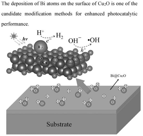
Passivating Surface States on Water Splitting Cuprous Oxide Photocatalyst with Bismuth Decoration


School of Physical Science and Technology, Southwest University, Chongqing 400715, China
*
Author to whom correspondence should be addressed.
Molecules 2019, 24(22), 4156; https://0-doi-org.brum.beds.ac.uk/10.3390/molecules24224156
Submission received: 23 September 2019
/
Revised: 9 November 2019
/
Accepted: 12 November 2019
/
Published: 16 November 2019
(This article belongs to the Special Issue Nanomaterials for Catalysis)
Abstract
:To enhance the visible light photocatalystic activity of Cu2O(100) surface, we performed first-principles calculations on the structural, electronic and optical properties of a bismuth (Bi)-decorated Cu2O(100) surface (Bi@Cu2O(100)). It is shown that the Bi prefer to be loaded to the hollow sites among four surface oxygen atoms and tend to individual dispersion instead of aggregating on the surface due to the lowest formation energy and larger distance between two Bi atoms at the surface than the Bi clusters; the coverage of around 0.25 monolayer Bi atoms can effectively eliminate the surface states and modify the band edges to satisfy the angular momentum selection rules for light excited transition of electrons, and the loaded Bi atoms contribute to the separation of photogenerated electron-holes. The relative positions between the band edges and the redox potentials are suitable for photocatalytic hydrogen production from the redox water, and moreover, the optical absorption spectrum indicates a positive response of the Bi0.25@Cu2O(100) to visible light, implying that the Bi0.25@Cu2O(100) is a promising visible light photocatalyst.
1. Introduction
Cuprous oxide (Cu2O) is a promising candidate as a native p-type oxide semiconductor with a direct band gap of 2.17 eV in the field of gas sensors [1,2,3], solar energy conversion [4,5,6,7] and photocatalysis [8,9,10,11]. Cu2O is characterized by low toxicity, environmental acceptability, and low-cost elemental compositions because they are very abundant in the crust, which makes it promising for achieving large-scale industrial production to solve environmental and energy problems [10,12,13,14]. The photocatalytic technology based on semiconductor photocatalyst is considered as an ideal way to solve the energy and environmental problems fundamentally by using solar energy [15,16]. The ideal photocatalytic materials should be able to make full use of visible light for electrochemical splitting of water and decomposition of organic pollutants [17,18,19]. As a semiconductor photocatalyst, Cu2O has the advantages of absorption of visible light, proper edge position and good optical stability [20,21]. Visible light accounts for the majority of sunlight, which suggest that Cu2O can make full use of solar energy. However, the even-parity symmetry of the valence band maximum and conduction band minimum states in Cu2O prohibits the band edge radiative transition, which has hindered its potential use in optical applications [10,22,23].
Many Cu2O architectures with various morphologies have been successfully synthesized with the development of nanomaterials science and nanotechnology [24,25,26,27,28]. Perspective and progress on polyhedral Cu2O nanostructures also have received extensive attention [25,29,30]. Moreover, it was reported by Huang et al. [31] that the stability of Cu2O crystal planes follows the order of in weak acid solution (pH = 3.5). However, the development of Cu2O semiconductor photocatalyst has been an ongoing challenge because catalytic properties of nanocrystals are highly related to their exposed surfaces, which is the main place for the photocatalytic reaction. For the experimentally facet-dependent photocatalytic activity of Cu2O, we give an explanation by calculating the electronic structure of the Cu2O low-index surface. Especially, the surface states of the Cu2O(100) facet hinders its photocatalytic activity. Passivating surface states has been successful in modifying the surface of hematite [32,33,34], as far as we know, there is no report about the surface state passivation of Cu2O(100).
In this paper, we propose a strategy to modify the electronic structure of Cu2O(100) surface with depositing Bi atoms to investigate whether the surface decoration can improve the photocatalytic performance of Cu2O(100) surface, after analyzing the electronic structures of the Cu2O(100) surface. The electronic and optical properties of the Bi-loaded Cu2O(100) surface is investigated by using hybrid density functional theory. In addition, we also discuss the adsorption structure, adsorption energy, band edge potential and optical properties.
2. Computational Detail
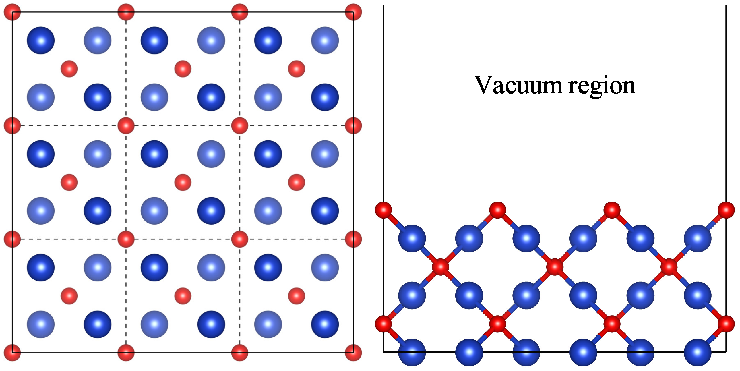
The Cu2O(100) surface is modeled by periodic six-layer slab models, and each slab is separated by a 15 Å vacuum layer to minimize interactions between the slabs. The surface structures are modeled with p (2 × 2) and p (3 × 3) supercells (Figure 1) to simulate different surface coverages. All periodic slab calculations based on density functional theory are performed using the Vinenna ab initio simulation package (VASP) [35,36]. The exchange and correlation interactions are modeled using the Perdew-Burke-Ernzerhof functional [37] within the generalized gradient approximation (GGA) in geometry optimization and total energy calculations [38]. The projector-augmented-wave (PAW) method is used for the description of electron-ion interaction. The wave functions of the valence electrons are expanded using a plane-wave basis set within a specified kinetic cutoff energy of 400 eV. The surface Brillouin Zone integrations are performed using 3 × 3 × 1 Monkhorst-Pack [39] k-points, the energy convergence criterion of 10−5 eV, and the force convergence thresholds of 0.02 eV/Å have been found to be sufficient for structural relaxation. To accurately describe the electronic structures and optical properties, the hybrid density functional as prescribed by Heyd-Scuseria-Ernzerhof (HSE06) [40,41] has been used in the present work. During the geometry optimization, the three topmost layers are allowed to fully relax, whereas the bottom three layers are fixed. To compensate the dipole effects along the z-direction, a dipole vector with the same value in the opposite direction is introduced.
3. Results and Discussion
3.1. Surface Property
In order to investigate the photocatalytic activity, we first calculate the electronic structures of the low-index surface of Cu2O. Figure 2 presents the projected density of states (PDOS) for Cu2O blocks and three low-index surfaces. They have a band gap of about 2 eV, which is good for meeting the photocatalytic requirements of visible light. One noteworthy thing is their band edge component, which is rarely considered a factor that affects the photocatalytic activity. As shown in Figure 2a, the band edges of Cu2O bulk are mainly composed of d-orbitals. Because the limitation of the dipole transition, only a small amount of electrons can transit to the p-orbitals of the conduction bandedge under light irradiation, which become the main factor to suppress the photocatalytic activity. In the bandgap of the (100) and (110) surfaces in Figure 2b,c, there are some empty bands which are not occupied by electrons, due to the reduction of the coordination number of the surface atoms. These empty bands can induce recombination of carriers to further reduce the quantum yield. For the (111) surface in Figure 2d, there are no empty states in the bandgap, but still face similar dipole transition problems.
3.2. Adsorption Energy
The adsorption structures of the Bi on different symmetry sites of the Cu2O(100) surface are investigated. In our calculations, five different adsorption sites on the Cu2O(100) surface are modeled: the top site of the surface oxygen atoms (O-top), the bridge site of the surface oxygen atoms (O-bridge), the hollow site among four surface oxygen atoms (hollow), the top site of the subsurface copper (Cu1-top) atoms and the top site of the fourth layer copper (Cu2-top) atoms (Figure 3). We now examine the energetic and thermodynamical stability of the Bi loaded Cu2O(100) surface (Bi@Cu2O(100)) by calculating the cohesive energy per unit area,
and the adsorption energy pere unit area [42],
Here, is the total number of Cu/O/Bi atoms in the decorated system, is the total energy of the isolated Cu/O/Bi atom, S is the area of the surface, and are the total energies of the Cu2O(100) surface with Bi adatoms and the clean Cu2O(100) surface, respectively.
The calculated and are presented in Table 1 for the Bi loaded Cu2O(100) surface. From the second to sixth row of Table 1, one Bi atom is absorbed at five possible sites of the surface. The of these systems are all negative, which indicates that they are stable structures. The adsorption of Bi atom on the hollow site is the most stable due to the lowest adsorption energy of −0.08 eV. Such system is more stable than the pristine Cu2O(100), given that its is 0.08 eV lower than that of pristine Cu2O(100). Moreover, it can be seen from the last four rows of Table 1 that the absolute adsorption energy increases as the surface coverage increases from 0.11 to 0.33 ML, which reflects that the structural stability of the Bi loaded Cu2O(100) surface can be improved by increasing the surface coverage. In the last two rows of Table 1, the two and three Bi atoms are simultaneously adsorbed at the hollow sites on the surface oxygen atoms, respectively. The of these systems is very negative, which again reflects the structural stability of the Bi@Cu2O(100) on hollow site, in agreement with the tend in which is 0.08 eV favorable than that of the pristine Cu2O(100) surface. Meanwhile, the distances between the two Bi atoms are respectively 5.27 and 4.798 Å in the case of complete relaxation, which are much larger than the atomic bond length in the Bi clusters [43]. Therefore, the Bi@Cu2O(100) on hollow site is more stable than the parent Cu2O(100) surface and Bi atoms tend to individual dispersion instead of aggregation on the Cu2O(100) surface.
3.3. Electronic Structure
In order to study the effect of Bi atom adsorption on the electronic structure of Cu2O(100) surface, we calculate the PDOS of the clear and Bi-decorated Cu2O(100) surface. As a comparison, the PDOS of the clean Cu2O(100) surface is shown in Figure 4a. Close to the right side of the Fermi level, there are significant surface states that are not occupied by electrons and they are mainly composed of Cu d-orbitals and O p-orbitals. This trapping state as a carrier recombination center is not conducive to the photocatalytic reaction [33,34]. Meanwhile, this trapping state grabs electrons from the Cu atoms and oxidizes to , which causes Cu2O to gradually lose its photocatalytic ability. This is also one of the microscopic mechanisms of photocorrosion of Cu2O. Figure 4b shows the PDOS of a single Bi atom adsorbed on the p(3 × 3) surface. For the adsorption of 0.11 monolayer (ML) of Bi atoms, the surface states still exist, but the conduction band edge changes from the d-orbitals character of Cu atoms to the p-orbitals character of the Bi atom, while the valence band edge keeps the d-orbitals character of the Cu atoms. This indicates that the adsorption of Bi atoms successfully change the band edge composition, enabling the band edges to meet the angular momentum selection rules for the light excited transition of electrons. As shown in Figure 4c, when the adsorption concentration is 0.22 monolayer, the surface state is significantly reduced and meanwhile the composition of the band edges also satisfy the transition selection criteria. As is expected, the surface state will be further reduced, when the concentration is increased. As shown in Figure 4d, with a coverage of 0.25 monolayer, the surface state of cuprous oxide is almost completely eliminated, and moreover, the significant d-orbitals of Cu atoms at the valence band and the p-orbitals of Bi toms at the conduction band edges meets the light excited electronic transition. Finally, Figure 4e shows the PDOS of Bi absorbed Cu2O(100) surface with a coverage of 0.33 monolayer. Since the Bi atomic adsorption concentration is too high, the new impurity states, which are mainly contributed by the p-orbital of the Bi atom, appears in the range of 0–0.9 eV. The above analysis of PDOS reveals a trend: with the increase of Bi atom adsorption concentration, the passivation effect of the surface states becomes stronger when the Bi absorption concentration is less than 0.33 ML. To summarize, Bi decoration does not change the bandgap of Cu2O(100) surface; however, it can increase the charge carrier density at the band edge, and more importantly it can eliminate the surface states, reducing the carrier recombination and change band edge composition, allowing the transition of d-electrons at VBM to p-orbitals at CBM under visible light irradiation. It should be pointed out that the modified electronic structure means that the photogenerated electrons will concentrate on the Bi atoms, while the photogenerated holes will concentrate on the Cu atoms. As shown in Figure 5, the Bi atoms become sites of hydrogen evolution reactions to produce hydrogen (H2) and the Cu atoms act as sites of oxygen evolution reactions to generate hydroxyl radicals (·OH). Bi decoration successfully separated oxidation reaction and reduction reaction, which reduces the interference of the two reaction processes. This should reduce the recombination rate of photogenerated electron-hole and improve the photocatalytic efficiency. Therefore, the Bi0.25@Cu2O(100) is the promising photocatalyst.
The difference of charge density can be obtained with the expression: , where , and are the charge densities of the absorbed system, Cu2O substrate and Bi adsorbate, respectively. The charge density difference for Bi0.25@Cu2O displays in Figure 6. It can be seen that the charge transfer from the Bi atom to the Cu atoms and the O atoms on the surface. The Bader charge analysis shows that the charge of the adsorbed Bi is +1.36e. The charge transfer indicates that Bi atoms form robust chemical adsorption rather than physical adsorption. The strong electron transfer also indicates that the p-orbital electrons of Bi atoms hybridize with the d-orbital electrons of copper atoms, which passivate successfully the surface trapping states. Meanwhile, the density of states at the conduct band edge increase. Unfortunately, the intensity of electron density weaken at the valence band edge.
3.4. Band Edge
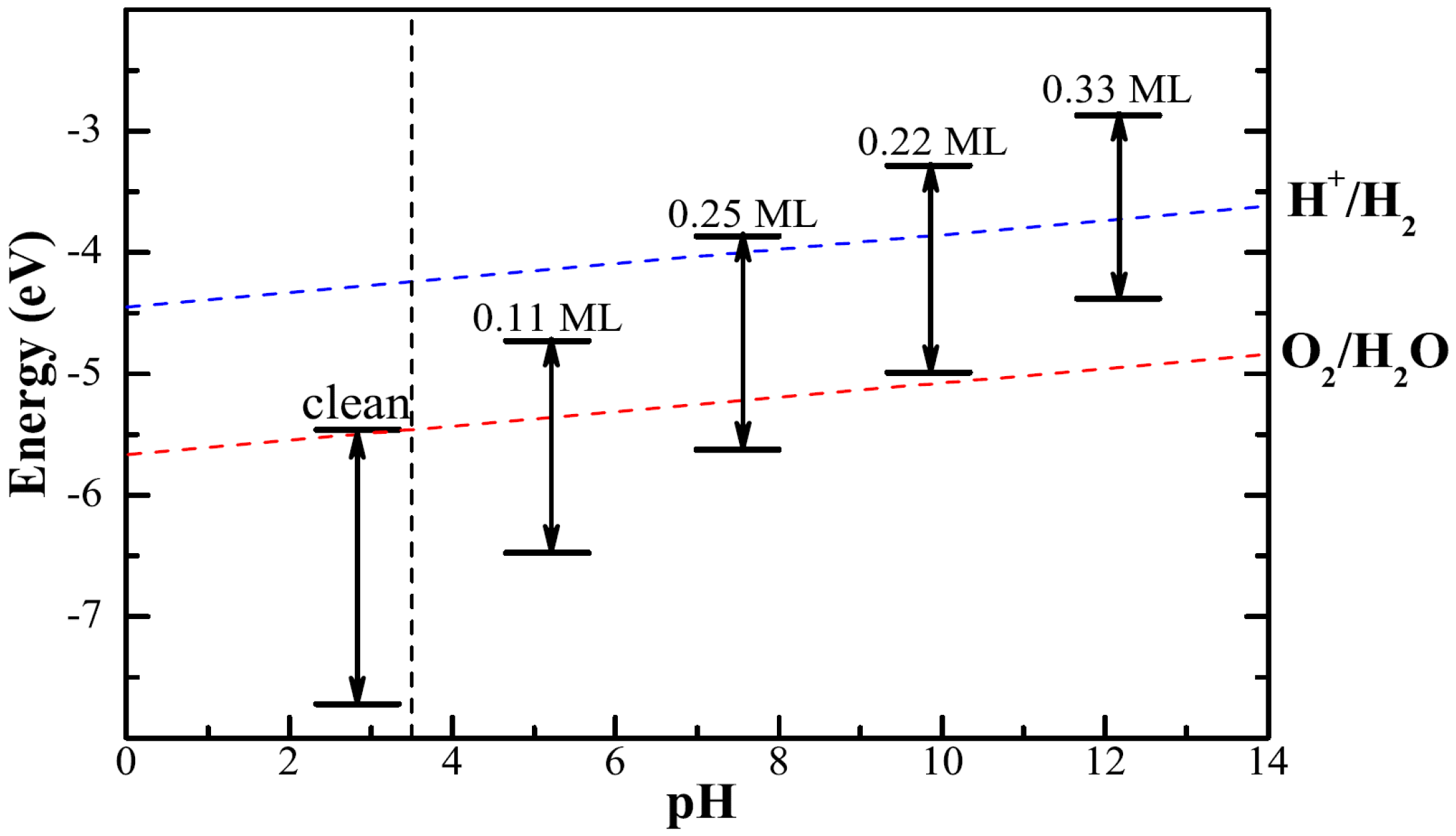
For hydrogen production via photocatalytic water splitting, the band edges of semiconductor need to be placed appropriately relative to the reaction redox potentials. In order to evaluate the photocatalytic performance, we plot the relative position of the calculated band edge energy levels with respect to water redox potential. In this calculation, the potential in the vacuum region is defined as the reference vacuum level. The voltage for the water splitting reaction is 1.23 V, which is the potential difference between the anodic oxygen evolution reaction and the cathodic hydrogen evolution reaction [44]. The reduction potential of is eV with reference to the vacuum level [45]. Then the oxidation potential of is eV. The theoretical band edge values of the clean and adsorbed surface are illustrated in Figure 7. For the synthesis of stable Cu2O(100) surface, the weak acid conditions (pH = 3.5) [31] required are indicated by vertical dashed line. Cu2O(100) surface is not enough to produce hydrogen from photocatalytic water-splitting because of its too low bandedge position. Bi decoration successfully adjusted the position of bandedges. The band edges of Bi0.25Cu2O straddle the redox potentials of water, making it a desirable band edge position as an excellent photocatalyst for hydrogen generation from water splitting.
3.5. Optical Property
The optical absorption spectrum of the material is also a quality factor that reflects the photocatalytic performance. The absorption coefficient is determined by the real and imaginary parts of the frequency dependent complex dielectric function according to the following equation [46]:
The imaginary part of the dielectric function is calculated by [47]
where , , , and are the volume, photon frequencies, the vector defining the polarization of the incident electric field, the occupied and unoccupied wave functions at point k in reciprocal space, respectively. The real part can be evaluated from imaginary part by the Kramer–Kronig relationship [48]:
where p is the principal value of the integral. The calculated optical properties of all the systems are shown in Figure 8. The optical absorption curves indicate that the adsorption of Bi atoms on the Cu2O(100) surface has little effect on the optical absorption. Around 400 nm, which corresponds to visible light, there is an absorption-curve platform suggesting that the system still maintains a positive visible light response. The residual impurity states in the band gap cause some small absorption peaks above 500 nm and cause the absorption limit to move to the infrared region. The introduction of Bi atoms does not damage the good optical properties of the cuprous oxide, and the adsorption of Bi atoms is likely to be a kind of reliable means of modification for cuprous oxide.
4. Conclusions
The first-principles calculations based on hybrid density functional theory are performed to investigate the electronic structure of pure and Bi atoms deposited Cu2O(100) surface. The calculation results reveal the presence of surface trapping states on the clean Cu2O(100) surface. The surface trapping states promote the recombination of the excited electron- hole and inhibit the photocatalytic efficiency in practical application. In order to improve the photocatalytic activity of Cu2O, we propose a strategy to passivate trapping states by depositing Bi atoms on Cu2O(100) surface. The adsorption of Bi atom on the hollow site among four surface oxygen atoms is most stable. For the different surface coverage, the absolute adsorption energy increases as the surface coverage increases. In the case of low surface coverage, the even-parity symmetry of the band edges has changed to meet the angular momentum selection rules for the light excited transition of electrons; But the surface trapping states are still maintained, indicating that the passivation effect is not sufficient. When the adsorption concentration increases to 0.25 ML, not only the surface trapping states are passivated, but also the band edges become satisfactory. Moreover, our scheme helps to reduce the recombination rate of photogenerated electron-hole and improve photocatalytic efficiency. We attempted to continue increasing the Bi atomic adsorption concentration and found a new impurity state from Bi atoms at a surface coverage of 0.33 ML. In addition, the band edges straddle the reaction redox potentials with a surface coverage of 0.25 ML. Finally, the analysis of optical absorption spectrum shows that these systems still retains their response to visible light. Therefore, the surface coverage of 0.25 ML is an ideal adsorption concentration in theoretical calculations for the highly efficiency of visible light photocatalysis. We believe that our findings will provide a new possibility for photocatalytic experiments.
Author Contributions
Conceptualization, H.C. and Y.H.; methodology, H.C.; software, Y.H.; validation, Y.H., H.C. and H.Y.; formal analysis, Y.H.; investigation, Y.H.; resources, H.C.; data curation, Y.H.; writing—original draft preparation, Y.H.; writing—review and editing, H.C.; visualization, Y.H.; supervision, H.Y.; project administration, H.C.; funding acquisition, H.C. and Y.H.
Funding
This research was partially funded by he National Natural Science Foundation of China under grant numbers 11875226 and 11874306.
Conflicts of Interest
The authors declare no conflict of interest.
References
- Zhang, H.G.; Zhu, Q.H.; Zhang, Y.; Wang, Y.; Zhao, L.; Yu, B. One-pot synthesis and hierarchical assembly of hollow Cu2O microspheres with nanocrystals-composed porous multishell and their gas-sensing properties. Adv. Funct. Mater. 2007, 17, 2766–2771. [Google Scholar] [CrossRef]
- Wu, L.; Wu, Y.L.; Jin, S.J.; Zhang, L.; Xun, Z.P. Gas sensitivity and photocatalytic performance of cuprous oxide with novel morphologies. Chem. Phys. Lett. 2016, 662, 47–51. [Google Scholar] [CrossRef]
- Shen, Y.Y.; Tian, F.H.; Chen, S.G.; Ma, Z.Q.; Zhao, L.H.; Jia, X.F. Density functional theory study on the mechanism of CO sensing on Cu2O(111) surface: Influence of the pre-adsorbed oxygen atom. Appl. Surf. Sci. 2014, 288, 452–457. [Google Scholar] [CrossRef]
- Wang, Y.C.; Qin, C.; Lou, Z.R.; Lu, Y.F.; Zhu, L.P. Cu2O photocathodes for unassisted solar water-splitting devices enabled by noble-metal cocatalysts simultaneously as hydrogen evolution catalysts and protection layers. Nanotechnology 2019, 30, 495407. [Google Scholar] [CrossRef] [PubMed]
- Pan, L.F.; Kim, J.H.; Mayer, M.T.; Son, M.K.; Ummadisingu, A.; Lee, J.S.; Hagfeldt, A.; Luo, J.S.; Gratzel, M. Boosting the performance of Cu2O photocathodes for unassisted solar water splitting devices. Nat. Catal. 2018, 1, 412–420. [Google Scholar] [CrossRef]
- Niu, W.Z.; Cai, T.M.W.; Joliat, R.W.; Zhu, L.P.; Tilley, S.D. Extended Light Harvesting with Dual Cu2O-Based Photocathodes for High Efficiency Water Splitting. Adv. Energy Mater. 2017, 8, 1702323. [Google Scholar] [CrossRef]
- Paracchino, A.; Laporte, V.; Sivula, K.; Grätzel, M.; Thimsen, E. Highly active oxide photocathode for photoelectrochemical water reduction. Nat. Mater. 2011, 10, 456–461. [Google Scholar] [CrossRef]
- Yu, X.J.; Zhang, J.; Niu, J.F.; Zhao, J.; Wei, Y.C.; Yao, B.H. Photocatalytic degradation of ciprofloxacin using Zn-doped Cu2O particles: Analysis of degradation pathways and intermediates. Chem. Eng. J. 2019, 374, 316–327. [Google Scholar] [CrossRef]
- Zhan, B.; Liu, Y.; Li, S.Y.; Kaya, C.; Stegmaier, T.; Aliabadi, M.; Han, Z.W.; Ren, L.Q. Fabrication of superwetting Cu@Cu2O cubic film for oil/water emulsion separation and photocatalytic degradation. Appl. Surf. Sci. 2019, 496, 143580. [Google Scholar] [CrossRef]
- Jiang, Y.; Yuan, H.K.; Chen, H. Enhanced visible light photocatalytic activity of Cu2O via cationic-anionic passivated codoping. Phys. Chem. Chem. Phys. 2015, 17, 630–637. [Google Scholar] [CrossRef]
- Tang, L.L.; Du, Y.H.; Kong, C.C.; Sun, S.D.; Yang, Z.M. One-pot synthesis of etched Cu2O cubes with exposed 110 facets with enhanced visible-light-driven photocatalytic activity. Phys. Chem. Chem. Phys. 2015, 17, 29479–29482. [Google Scholar] [CrossRef] [PubMed]
- Sun, S.D. Recent advances in hybrid Cu2O-based heterogeneous nanostructures. Nanoscale 2015, 7, 10850–10882. [Google Scholar] [CrossRef] [PubMed]
- Gao, H.; Zhang, J.Y.; Wang, R.M.; Wang, M. Highly efficient hydrogen production and formaldehyde degradation by Cu2O microcrystals. Appl. Catal. B Environ. 2015, 238, 37–38. [Google Scholar] [CrossRef]
- Hara, M.; Kondo, T.; Komoda, M.; Ikeda, S.; Shinohara, K.; Tanaka, A.; Kondo, J.N.; Domen, K. Cu2O as a photocatalyst for overall water splitting under visible light irradiation. Chem. Commun. 1998, 4, 357–358. [Google Scholar] [CrossRef]
- Fujishima, A.; Honda, K. Electrochemical Photolysis of Water at a Semiconductor Electrode. Nature 1972, 238, 37–38. [Google Scholar] [CrossRef]
- Sun, L.L.; Wang, G.H.; Hao, R.R.; Han, D.Y.; Cao, S. Solvothermal fabrication and enhanced visible light photocatalyticactivity of Cu2O-reduced graphene oxide composite microspheres for photodegradation of Rhodamine B. Appl. Surf. Sci. 2015, 358, 91–99. [Google Scholar] [CrossRef]
- Cai, J.Y.; Liu, W.J.; Li, Z.H. One-pot self-assembly of Cu2O/RGO composite aerogel for aqueousphotocatalysis. Appl. Surf. Sci. 2015, 358, 146–151. [Google Scholar] [CrossRef]
- Liu, Z.F.; Yan, L. High-efficiency p-n junction oxide photoelectrodes for photoelectrochemical water splitting. Phys. Chem. Chem. Phys. 2016, 18, 31230–31237. [Google Scholar] [CrossRef]
- Xu, L.; Xu, H.Y.; Wu, S.B.; Zhang, X.Y. Synergy effect over electrodeposited submicron Cu2O films in photocatalytic degradation of methylene blue. Appl. Surf. Sci. 2012, 258, 4934–4938. [Google Scholar] [CrossRef]
- Bendavid, L.I.; Carter, E.A. First-Principles Predictions of the structure, stability, and photocatalytic potential of Cu2O surfaces. J. Phys. Chem. B 2013, 117, 15750–15760. [Google Scholar] [CrossRef]
- Wang, G.Z.; Chen, H.; Li, Y.; Kuang, A.L.; Yuan, H.K.; Wu, G. A hybrid density functional study on the visible light photocatalytic activity of (Mo,Cr)-N codoped KNbO3. Phys. Chem. Chem. Phys. 2015, 17, 28743–28753. [Google Scholar] [CrossRef]
- Li, Y.F.; Yin, W.J.; Deng, R.; Chen, R.; Chen, J.; Yan, Q.Y.; Yao, B.; Sun, H.D.; Wei, S.; Wu, T. Realizing a SnO2-based ultraviolet light-emitting diode via breaking the dipole-forbidden rule. NPG Asia Mater. 2012, 4, e30. [Google Scholar] [CrossRef]
- Meyer, B.K.; Polity, A.; Reppin, D.; Becker, M.; Hering, P.; Klar, P.J.; Sander, T.; Reindl, C.; Benz, J.; Eickhoff, M.; et al. Binary copper oxide semiconductors: From materials towards devices. Phys. Status Solidi B 2012, 249, 1487–1509. [Google Scholar] [CrossRef]
- Tsai, Y.H.; Chanda, K.; Chu, Y.T.; Chiu, C.Y.; Huang, M.H. Direct formation of small Cu2O nanocubes, octahedra, and octapods for efficient synthesis of triazoles. Nanoscale 2014, 6, 8704–8709. [Google Scholar] [CrossRef] [PubMed]
- Huang, M.H.; Rej, S.; Hsu, S.C. Facet-dependent properties of polyhedral nanocrystals. Chem. Commun. 2014, 50, 1634–1644. [Google Scholar] [CrossRef] [PubMed]
- Nguyen, M.A.; Bedford, N.M.; Ren, Y.; Zahran, E.M.; Goodin, R.C.; Chagani, F.F.; Bachas, L.G.; Knecht, M.R. Direct Synthetic Control over the Size, Composition, and Photocatalytic Activity of Octahedral Copper Oxide Materials: Correlation Between Surface Structure and Catalytic Functionality. ACS Appl. Mater. Interfaces 2015, 7, 13238–13250. [Google Scholar] [CrossRef]
- Li, S.M.; Ge, X.; Jiang, S.N.; Peng, X.N.; Zhang, Z.; Li, W.X.; Yu, S.S. Synthesis of octahedral and cubic Cu2O microcrystals in sub- and super-critical methanol and their photocatalytic performance. J. Mater. Sci. 2015, 50, 4115–4121. [Google Scholar] [CrossRef]
- Liang, Y.H.; Shang, L.; Bian, T.; Zhou, C.; Zhang, D.H.; Yu, H.J.; Xu, H.T.; Shi, Z.; Zhang, T.R.; Wu, L.Z.; et al. Shape-controlled synthesis of polyhedral 50-facet Cu2O microcrystals with high-index facets. CrystEngComm 2012, 14, 4431–4436. [Google Scholar] [CrossRef]
- Sun, S.D.; Yang, Z.M. Recent advances in tuning crystal facets of polyhedral cuprous oxide architectures. RSC Adv. 2014, 4, 3804–3822. [Google Scholar] [CrossRef]
- Kuo, C.H.; Huang, M.H. Morphologically controlled synthesis of Cu2O nanocrystals and their properties. Nano Today 2010, 5, 106–116. [Google Scholar] [CrossRef]
- Hua, Q.; Shang, D.L.; Zhang, W.H.; Chen, K.; Chang, S.J.; Ma, Y.S.; Jiang, Z.Q.; Yang, J.L.; Huang, W.X. Morphological Evolution of Cu2O Nanocrystals in an Acid Solution: Stability of Different Crystal Planes. Langmuir 2011, 27, 665–671. [Google Scholar] [CrossRef] [PubMed]
- Ulman, K.; Nguyen, M.T.; Seriani, N.; Gebauer, R. Passivation of surface states of α-Fe2O3(0001) surface by deposition of Ga2O3 overlayers: A density functional theory study. J. Chem. Phys. 2016, 144, 094701. [Google Scholar] [CrossRef] [PubMed]
- Le Fomal, F.; Tetreault, N.; Cornuz, M.; Moehl, T.; Grätzel, M.; Sivula, K. Passivating surface states on water splitting hematite photoanodes with alumina overlayers. Chem. Sci. 2011, 2, 737–743. [Google Scholar] [CrossRef]
- Liu, R.; Zheng, Z.; Spurgeon, J.; Yang, X.G. Enhanced photoelectrochemical water-splitting performance of semiconductors by surface passivation layers. Energy Environ. Sci. 2014, 7, 2504–2517. [Google Scholar] [CrossRef] [Green Version]
- Kresse, G.; Joubert, D. From ultrasoft pseudopotentials to the projector augmented-wave method. Phys. Rev. B 1999, 59, 1758–1775. [Google Scholar] [CrossRef]
- Kresse, G.; Furthmüller, J. Efficient iterative schemes for ab initio total-energy calculations using a plane-wave basis set. Phys. Rev. B 1996, 54, 11169–11186. [Google Scholar] [CrossRef]
- Ernzerhof, M.; Scuseria, G.E. Assessment of the Perdew-Burke-Ernzerhof exchange-correlation functional. J. Chem. Phys. 1999, 110, 5029–5036. [Google Scholar] [CrossRef] [Green Version]
- White, J.A.; Bird, D.M. Implementation of gradient-corrected exchange-correlation potentials in Car-Parrinello total-energy calculations. Phys. Rev. B 1994, 50, 4954–4957. [Google Scholar] [CrossRef]
- Monkhorst, H.J.; Pack, J.D. Special points for Brillonin-zone integrations. Phys. Rev. B 1976, 13, 5188–5192. [Google Scholar] [CrossRef]
- Heyd, J.; Scuseria, G.E.; Ernzerhof, M. Hybrid functionals based on a screened Coulomb potential. J. Chem. Phys. 2003, 118, 8207–8215. [Google Scholar] [CrossRef] [Green Version]
- Heyd, J.; Scuseria, G.E.; Ernzerhof, M. Erratum: Hybrid functionals based on a screened Coulomb potential. J. Chem. Phys. 2006, 118, 8207–8215, Correction in 2006, 124, 219906. [Google Scholar] [CrossRef] [Green Version]
- Soon, A.; Todorova, M.; Delley, B.; Stampfl, C. Thermodynamic stability and structure of copper oxide surfaces: A first-principles investigation. Phys. Rev. B 2007, 75, 125420. [Google Scholar] [CrossRef] [Green Version]
- Yuan, H.K.; Chen, H.; Kuang, A.L.; Miao, Y.; Xiong, Z.H. Density-functional study of small neutral and cationic bismuth clusters and (n = 2–24). J. Chem. Phys. 2008, 128, 094305. [Google Scholar] [CrossRef] [PubMed]
- Artrith, N.; Sailuam, W.; Limpijumnong, S.; Kolpak, A.M. Reduced overpotentials for electrocatalytic water splitting over Fe- and Ni-modified BaTiO3. Phys. Chem. Chem. Phys. 2016, 18, 29561–29570. [Google Scholar] [CrossRef]
- Chakrapani, V.; Angus, J.C.; Anderson, A.B.; Wolter, S.D.; Stoner, B.R.; Sumanasekera, G.U. Charge Transfer Equilibria Between Diamond and an Aqueous Oxygen Electrochemical Redox Couple. Science 2007, 318, 1424–1430. [Google Scholar] [CrossRef] [Green Version]
- Saha, S.; Sinha, T.P. Electronic structure, chemical bonding, and optical properties of paraelectric BaTiO3. Phys. Rev. B 2000, 62, 8828–8834. [Google Scholar] [CrossRef]
- Tian, F.H.; Liu, C.B. DFT Description on Electronic Structure and Optical Absorption Properties of Anionic S-Doped Anatase TiO2. J. Chem. Phys. B 2006, 110, 17866–17871. [Google Scholar] [CrossRef]
- Fu, Q.; He, T.; Li, J.L.; Yang, G.W. Band-engineered SrTiO3 nanowires for visible light photocatalysis. J. Appl. Phys. 2012, 112, 104322. [Google Scholar] [CrossRef]
Figure 1.
The 3 × 3 × 1 supercell structure for clean Cu2O(100) surface. The red and blue spheres represent oxygen atoms and copper atoms, respectively.
Figure 1.
The 3 × 3 × 1 supercell structure for clean Cu2O(100) surface. The red and blue spheres represent oxygen atoms and copper atoms, respectively.

Figure 2.
The projected density of states (PDOS) for Cu2O low-index surfaces.

Figure 3.
Five different adsorption sites on the Cu2O(100) surface, including O-top, O-bridge, hollow, Cu1-top, Cu2-top. The red and blue spheres represent oxygen atoms and copper atoms, respectively.
Figure 3.
Five different adsorption sites on the Cu2O(100) surface, including O-top, O-bridge, hollow, Cu1-top, Cu2-top. The red and blue spheres represent oxygen atoms and copper atoms, respectively.

Figure 4.
The PDOS of clean Cu2O(100) surface and Bi adsorbed Cu2O(100) surface. The vertical dashed lines at energy zero indicate the Fermi levels.
Figure 4.
The PDOS of clean Cu2O(100) surface and Bi adsorbed Cu2O(100) surface. The vertical dashed lines at energy zero indicate the Fermi levels.

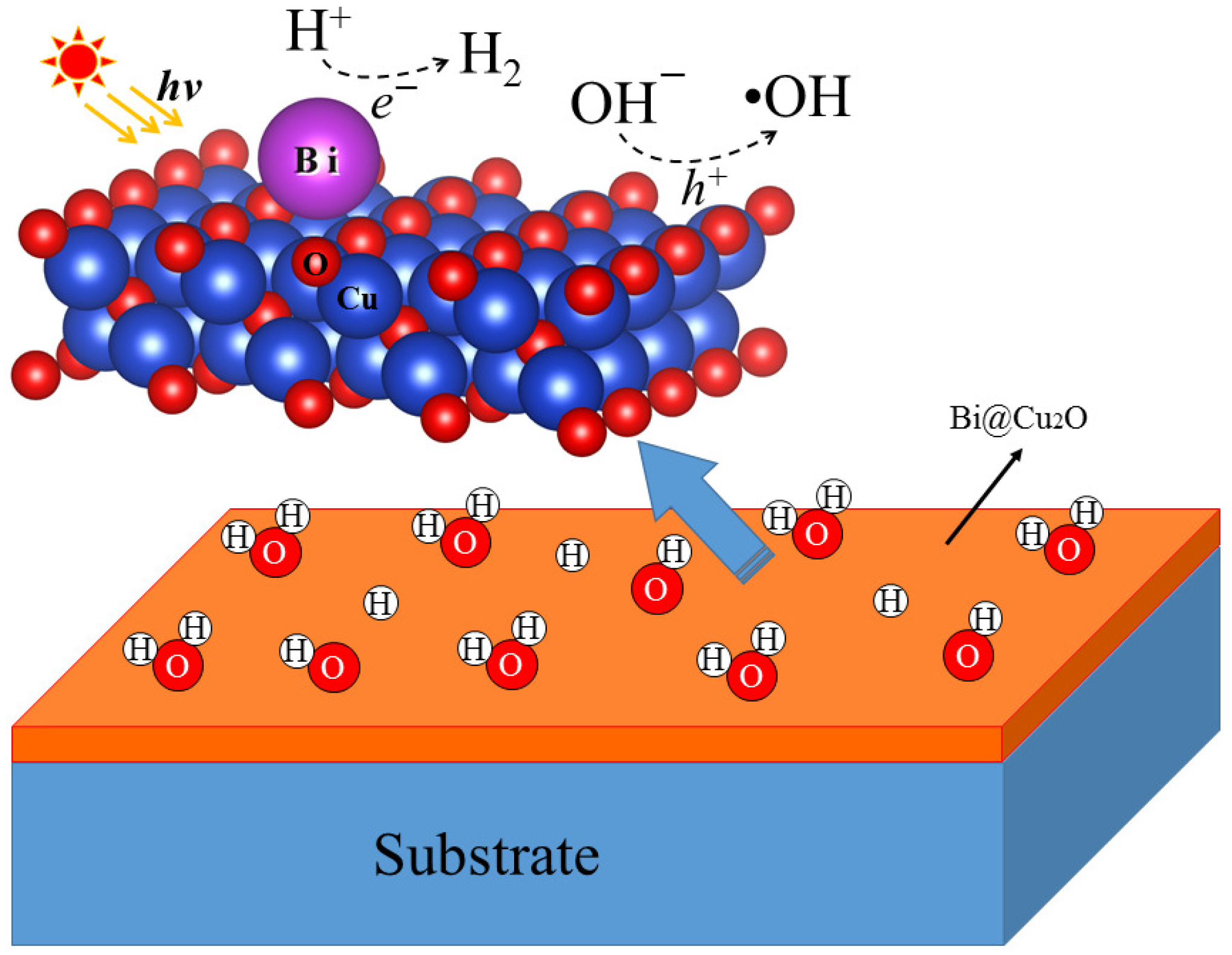
Figure 5.
The photocatalytic oxidation and reduction on the Cu2O(100) surface.

Figure 6.
The charge density difference of Bi adsorbed Cu2O(100) surface with a coverage of 0.25 ML. Electron accumulation and depletion are represented by yellow and cyan-blue areas, respectively. The isosurface value is set as 0.008 e/Bohr3.
Figure 6.
The charge density difference of Bi adsorbed Cu2O(100) surface with a coverage of 0.25 ML. Electron accumulation and depletion are represented by yellow and cyan-blue areas, respectively. The isosurface value is set as 0.008 e/Bohr3.

Figure 7.
The band edges of clean and adsorbed Cu2O(100) surface with respect to the redox potential of water. The blue and red dashed lines represent the reduction potential and the oxidation potential of water as function of pH value, respectively. The black vertical dashed line is at a pH = 3.5.
Figure 7.
The band edges of clean and adsorbed Cu2O(100) surface with respect to the redox potential of water. The blue and red dashed lines represent the reduction potential and the oxidation potential of water as function of pH value, respectively. The black vertical dashed line is at a pH = 3.5.

Figure 8.
The calculated optical absorption curves of Cu2O bulk, clean and Bi adsorbed Cu2O(100) surface.
Figure 8.
The calculated optical absorption curves of Cu2O bulk, clean and Bi adsorbed Cu2O(100) surface.


Table 1.
Adsorption energy and surface cohesion energy for bismuth on the Cu2O(100) surface.
| Systems | Adsorption Site | |||
|---|---|---|---|---|
| (ML) | (eV/Å2) | (ev/Å2) | ||
| Cu2O(100) | 0 | - | - | −2.01 |
| p (2 × 2) | 0.25 | O-top | −0.04 | −2.05 |
| p (2 × 2) | 0.25 | O-Bridge | −0.07 | −2.08 |
| p (2 × 2) | 0.25 | Cu1-top | −0.05 | −2.06 |
| p (2 × 2) | 0.25 | Cu2-top | −0.05 | −2.06 |
| p (2 × 2) | 0.25 | Hollow | −0.08 | −2.09 |
| p (3 × 3) | 0.11 | Hollow | −0.04 | −2.05 |
| p (3 × 3) | 0.22 | Hollow | −0.08 | −2.09 |
| p (3 × 3) | 0.33 | Hollow | −0.09 | −2.10 |
© 2019 by the authors. Licensee MDPI, Basel, Switzerland. This article is an open access article distributed under the terms and conditions of the Creative Commons Attribution (CC BY) license (http://creativecommons.org/licenses/by/4.0/).
Share and Cite
MDPI and ACS Style
Huang, Y.; Yuan, H.; Chen, H. Passivating Surface States on Water Splitting Cuprous Oxide Photocatalyst with Bismuth Decoration. Molecules 2019, 24, 4156. https://0-doi-org.brum.beds.ac.uk/10.3390/molecules24224156
AMA Style
Huang Y, Yuan H, Chen H. Passivating Surface States on Water Splitting Cuprous Oxide Photocatalyst with Bismuth Decoration. Molecules. 2019; 24(22):4156. https://0-doi-org.brum.beds.ac.uk/10.3390/molecules24224156
Chicago/Turabian StyleHuang, Yuhong, Hongkuan Yuan, and Hong Chen. 2019. "Passivating Surface States on Water Splitting Cuprous Oxide Photocatalyst with Bismuth Decoration" Molecules 24, no. 22: 4156. https://0-doi-org.brum.beds.ac.uk/10.3390/molecules24224156