
Recent Advances in Polymer Nanomaterials for Drug Delivery of Adjuvants in Colorectal Cancer Treatment: A Scientific-Technological Analysis and Review


Abstract
:1. Introduction
2. Scientific-Technological Analysis
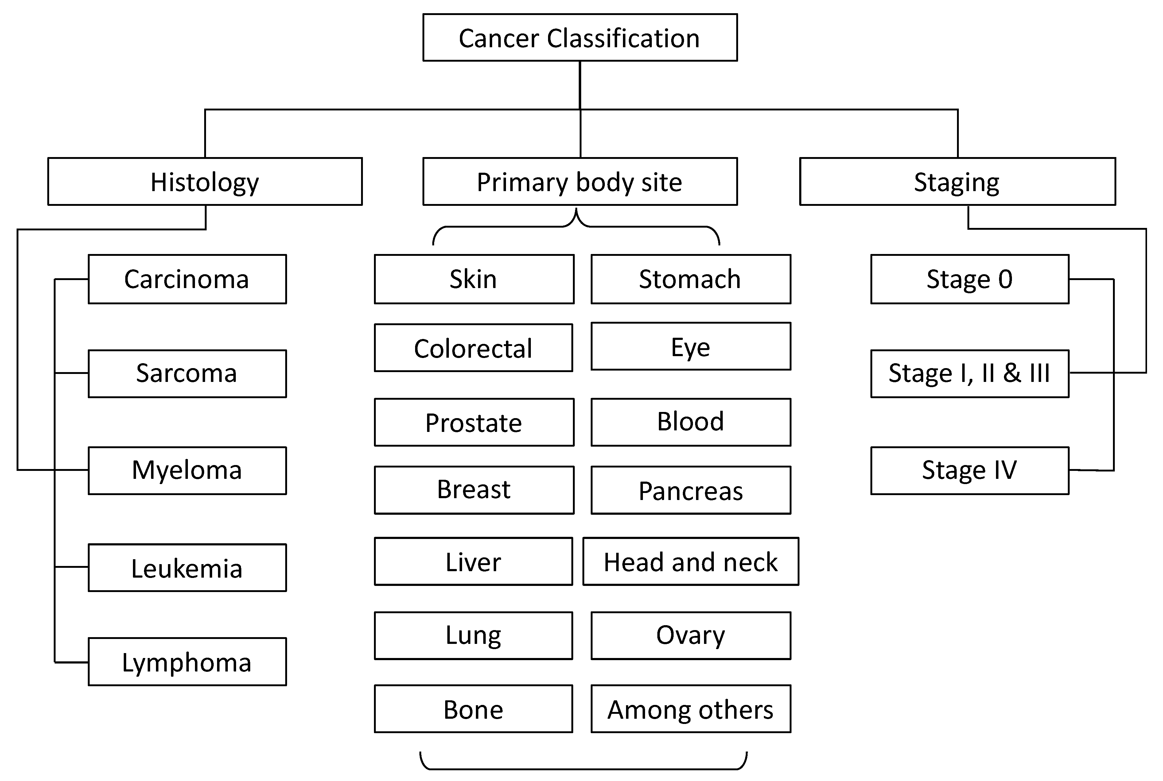
3. Cancer
4. Colorectal Cancer
5. Colorectal Cancer Treatments
5.1. Surgery
5.2. Adjuvant Therapy
5.2.1. Radiation Therapy
5.2.2. Hormone Therapy
5.2.3. Targeted Therapy
5.2.4. Immunotherapy
5.2.5. Chemotherapy
6. Polymer-Based Drug Delivery Systems for Adjuvants for Colorectal Cancer
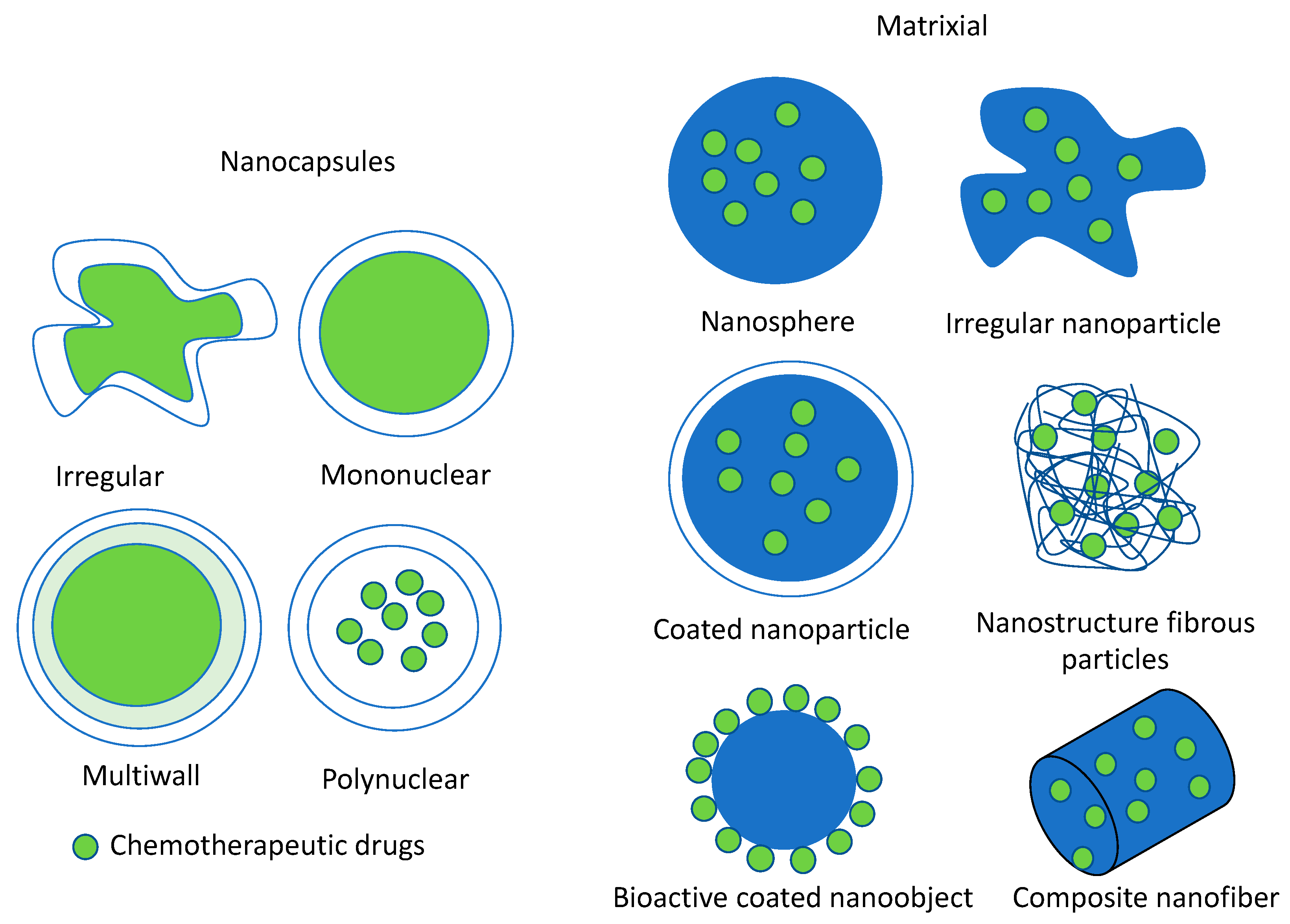
6.1. Nanoencapsulation
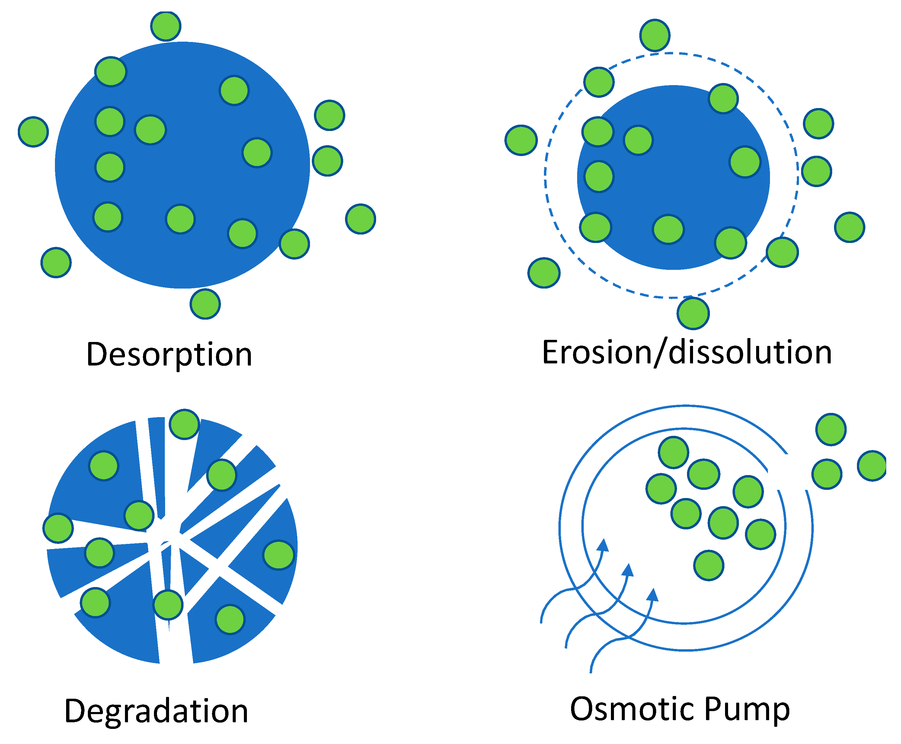
6.2. Release Mechanism
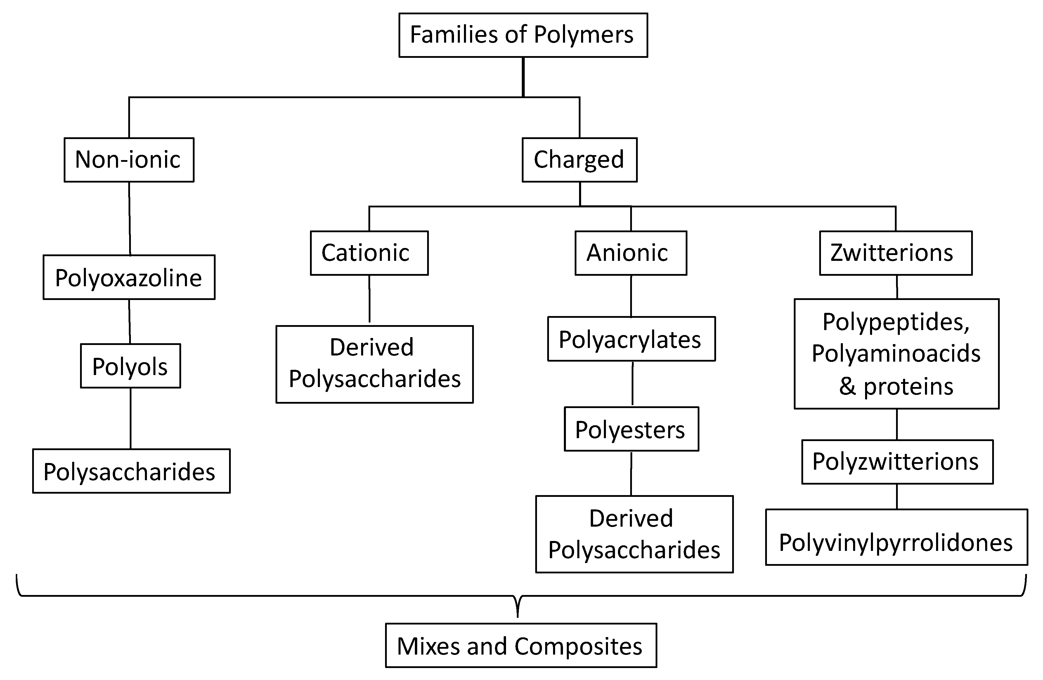
6.3. Polymers for Oral Drug Delivery Systems
6.4. Polymers for Encapsulating Antioxidants for Colorectal Cancer
6.4.1. Polysaccharides and Derived Polysaccharides
6.4.2. Polyacrylates
6.4.3. Polyols
6.4.4. Polyzwitterions
6.4.5. Polyoxazoline
6.4.6. Polypeptides and Polyaminoacids
6.4.7. Polyesters
6.4.8. Poly(vinylpyrrolidones)
6.4.9. Composites, Copolymers, and Mixtures
6.5. Polymers for Encapsulating Synthetics and Hybrid Adjuvants for CRC
7. Conclusions
Author Contributions
Funding
Acknowledgments
Conflicts of Interest
References
- Roser, M.; Ritchie, H. Cancer. Available online: https://ourworldindata.org/cancer (accessed on 25 February 2019).
- Becker, D.; Hershman, D.L. Adjuvant Therapy for Elderly Patients with Breast, Colon, and Lung Cancer. In Management of Cancer in the Older Patient; Elsevier: Philadelphia, PA, USA, 2012; pp. 79–88. [Google Scholar]
- Institute for Health Metrics and Evaluation (IHME) Global Burden of Disease Collaborative Network. Available online: http://ghdx.healthdata.org/gbd-results-tool.%0D%0A (accessed on 25 February 2019).
- Chiavarini, M.; Bertarelli, G.; Minelli, L.; Fabiani, R. Dietary intake of meat cooking-related mutagens (HCAs) and risk of colorectal adenoma and cancer: A systematic review and meta-analysis. Nutrients 2017, 9, 514. [Google Scholar] [CrossRef] [PubMed] [Green Version]
- Romieu, I.; Bagnardi, V.; Scotti, L.; Negri, E.; La Vecchia, C.; Tramacere, I.; Rota, M.; Fedirko, V.; Straif, K.; Jenab, M.; et al. Alcohol drinking and colorectal cancer risk: An overall and dose-response meta-analysis of published studies. Ann. Oncol. 2011, 22, 1958–1972. [Google Scholar] [CrossRef]
- Lewis, R.; Burden, S. Colorectal cancer and nutrition. Adv. Nutr. Diet. Gastroenterol. 2014, 255–262. [Google Scholar] [CrossRef]
- Ornitz, D.M.; Itoh, N. Protein family review Fibroblast growth factors Gene organization and evolutionary history. Genome Biol. 2001, 2, 1–12. [Google Scholar] [CrossRef] [PubMed] [Green Version]
- Reyes-Aldasoro, C.C. The proportion of cancer-related entries in PubMed has increased considerably; Is cancer truly “the Emperor of All Maladies”? PLoS ONE 2017, 12, 1–15. [Google Scholar] [CrossRef] [Green Version]
- Luke, M. Intestinal obstruction from cancer of the rectum, operation for artificial anus in the left groin; death in six hours. Lancet 1860, 6, 243. [Google Scholar] [CrossRef]
- Dunphy, J.E. Recurrent Cancer of the Colon and Rectum. N. Engl. J. Med. 2010, 237, 1–3. [Google Scholar] [CrossRef]
- Adams, J.T.; Schwartz, S.I.; Rubin, P.; Rob, C.G. Intralymphatic 5-Fluorouracil and radioactive gold as an adjuvant to surgical operation for colorectal carcinoma. Dis. Colon Rectum 1970, 13, 201–206. [Google Scholar] [CrossRef]
- Grand View Research Vaccine Adjuvants Market sie and Analysis. Available online: https://www.grandviewresearch.com/industry-analysis/vaccine-adjuvants-market (accessed on 17 March 2019).
- Gaudry, B.K.; Rothwell, R. The Unpredictable Prospects of Patenting Cancer Innovation. Available online: https://www.ipwatchdog.com/2018/03/01/unpredictable-patenting-cancer-innovation/id=94280/ (accessed on 17 March 2019).
- Vainio, H.; Morgan, G. Aspirin for the second hundred years: New uses for an old drug. Pharmacol. Toxicol. 1997, 81, 151–152. [Google Scholar] [CrossRef]
- X. Garcia-Albeniz, A.T.C. Aspirin for the prevention of colorectal cancer. Best Pr. Res Clin Gastroenterol. 2012, 1–18. [Google Scholar] [CrossRef]
- Chan, A.T. Aspirin Use and Survival After Diagnosis of Colorectal Cancer. Jama 2009, 302, 649–659. [Google Scholar] [CrossRef] [PubMed]
- Solomon, V.R.; Lee, H. Anti-breast cancer activity of heteroaryl chalcone derivatives. Biomed. Pharmacother. 2012, 66, 213–220. [Google Scholar] [CrossRef] [PubMed]
- R. Solomon, V.; Lee, H. Quinoline as a Privileged Scaffold in Cancer Drug Discovery. Curr. Med. Chem. 2011, 18, 1488–1508. [Google Scholar] [CrossRef] [PubMed]
- World Health Organization Cancer. Available online: https://www.who.int/cancer/en/ (accessed on 5 March 2019).
- Hecker, E. Definitions and terminology in cancer (tumor) etiology. Bull. World Health Organ. 1976, 54, 463. [Google Scholar] [CrossRef]
- National Institutes of Health (US); Biological Sciences Curriculum Study. Understanding Cancer. NIH Curric. Suppl. Ser. 2007. [Google Scholar]
- Pelengaris, S.; Khan, M. The Molecular Biology of Cancer: A Bridge from Bench to Bedside, 2nd ed.; Pelengaris, S., Khan, M., Eds.; Wiley: West Sussex, UK, 2013; Volume 28. [Google Scholar]
- NIH National Cancer Institutes Cancer Classification. Available online: https://training.seer.cancer.gov/disease/categories/classification.html (accessed on 11 March 2019).
- Gajdos, C.; Tartter, P.I.; Bodian, C.; Brower, S.T.; Bleiweiss, I.J. Stage 0 to stage III breast cancer in young women. J. Am. Coll. Surg. 2003, 190, 523–529. [Google Scholar] [CrossRef]
- Edge, S.B.; Compton, C.C. The american joint committee on cancer: The 7th edition of the AJCC cancer staging manual and the future of TNM. Ann. Surg. Oncol. 2010, 17, 1471–1474. [Google Scholar] [CrossRef] [PubMed]
- Idikio, H.A. Human cancer classification: A systems biology-based model integrating morphology, cancer stem cells, proteomics, and genomics. J. Cancer 2011, 2, 107–115. [Google Scholar] [CrossRef] [PubMed]
- Ali, H.R.; Rueda, O.M.; Aparicio, S.A.; Caldas, C.; Dunning, M.J.; Curtis, C. Genome-driven integrated classification of breast cancer validated in over 7,500 samples. Genome Biol. 2014, 15, 1–14. [Google Scholar] [CrossRef]
- Hoadley, K.A.; Yau, C.; Hinoue, T.; Wolf, D.M.; Lazar, A.J.; Drill, E.; Shen, R.; Taylor, A.M.; Cherniack, A.D.; Thorsson, V.S.; et al. Cell-of-Origin Patterns Dominate the Molecular Classification of 10,000 Tumors from 33 Types of Cancer. Cell 2018, 173, 291–304. [Google Scholar] [CrossRef] [Green Version]
- Lee, Z.J. An integrated algorithm for gene selection and classification applied to microarray data of ovarian cancer. Artif. Intell. Med. 2008, 42, 81–93. [Google Scholar] [CrossRef] [PubMed]
- Shen, L.; Toyota, M.; Kondo, Y.; Lin, E.; Zhang, L.; Guo, Y.; Hernandez, N.S.; Chen, X.; Ahmed, S.; Konishi, K.; et al. Integrated genetic and epigenetic analysis identifies three different subclasses of colon cancer. Proc. Natl. Acad. Sci. 2007, 104, 18654–18659. [Google Scholar] [CrossRef] [PubMed] [Green Version]
- Bookshelf, N. Colorectal Cancer: Overview. Available online: https://0-www-ncbi-nlm-nih-gov.brum.beds.ac.uk/books/NBK279198/?report=printable (accessed on 13 March 2018).
- Cooper, G.M.; Hausman, R. The Cell: A Molecular Approach, 6th ed.; Cooper, G.M., Hausman, R., Eds.; ASM Press: Boston, MA, USA, 2013; ISBN 978-0878939640. [Google Scholar]
- Jolly, T.A.; Williams, G.R.; Bushan, S.; Pergolotti, M.; Nyrop, K.A.; Jones, E.L.; Muss, H.B. Adjuvant treatment for older women with invasive breast cancer. Women’s Heal. 2016, 12, 129–146. [Google Scholar] [CrossRef]
- Ilson, D.H. Adjuvant therapy in colon cancer: Less is more. Lancet Oncol. 2018, 19, 442–443. [Google Scholar] [CrossRef] [Green Version]
- Kazemi, T.; Younesi, V.; Jadidi-Niaragh, F.; Yousefi, M. Immunotherapeutic approaches for cancer therapy: An updated review. Artif. Cells, Nanomedicine Biotechnol. 2016, 44, 769–779. [Google Scholar] [CrossRef] [PubMed]
- Kumari, S.; Badana, A.K.; Murali Mohan, G.; Shailender, G.; Malla, R.R. Reactive Oxygen Species: A Key Constituent in Cancer Survival. Biomark. Insights 2018, 13. [Google Scholar] [CrossRef] [Green Version]
- Shi, J.; Fang, G.; Sheng, Y. Neo-adjuvant chemotherapy for breast cancer. Zhonghua Zhong Liu Za Zhi 2001, 23, 423–425. [Google Scholar] [CrossRef]
- Jaffray, D.; Gospodarowicz, M. Radiation Therapy for Cancer. In Cancer: Disease Control Priorities; Gelband, H., Jha, P., Sankaranarayanan, R., Eds.; The International Bank for Reconstruction and Development: Washington, DC, USA, 2015; p. 10. [Google Scholar]
- Häfner, M.F.; Debus, J. Radiotherapy for colorectal cancer: Current standards and future perspectives. Visc. Med. 2016, 32, 172–177. [Google Scholar] [CrossRef]
- Skowronek, J. Current status of brachytherapy in cancer treatment–short overview. J. Contemp. Brachytherapy 2017, 9, 581–589. [Google Scholar] [CrossRef]
- Johnson, J.R.; Lacey, J.V., Jr.; Lazovich, D.; Geller, M.A.; Schairer, C.; Schatzkin, A.; Flood, A. Menopausal Hormone Therapy and Risk of Colorectal Cancer. Cancer Epidemiol Biomarkers Prev. 2009, 18, 196–203. [Google Scholar] [CrossRef] [Green Version]
- Lin, J.H.; Zhang, S.M.; Rexrode, K.M.; Manson, J.E.; Chan, A.T.; Wu, K.; Tworoger, S.S.; Hankinson, S.E.; Fuchs, C.; Gaziano, J.M.; et al. Association between sex hormones and colorectal cancer risk in men and women. Clin. Gastroenterol. Hepatol. 2014, 11, 419–424.e1. [Google Scholar] [CrossRef] [PubMed] [Green Version]
- Rennert, G.; Rennert, H.S.; Pinchev, M.; Lavie, O.; Gruber, S.B. Use of hormone replacement therapy and the risk of colorectal cancer. J. Clin. Oncol. 2009, 27, 4542–4547. [Google Scholar] [CrossRef] [PubMed] [Green Version]
- Venning, G.; Lange, S. Hormone Replacement Therapy. Available online: https://0-www-ncbi-nlm-nih-gov.brum.beds.ac.uk/books/NBK493191/?report=printable (accessed on 2 July 2019).
- Machluf, M.; Orsola, A.; Atala, A. Controlled release of therapeutic agents: Slow delivery and cell encapsulation. World J. Urol. 2000, 18, 80–83. [Google Scholar] [CrossRef]
- Ohhara, Y.; Fukuda, N.; Takeuchi, S.; Honma, R.; Shimizu, Y.; Kinoshita, I.; Dosaka-Akita, H. Role of targeted therapy in metastatic colorectal cancer. World J. Gastrointest. Oncol. 2016, 8, 642. [Google Scholar] [CrossRef]
- Hainsworth, J.; Heim, W.; Berlin, J.; Baron, A.; Griffing, S.; Holmgren, E.; Ph, D.; Ferrara, N.; Fyfe, G.; Rogers, B.; et al. Bevacizumab plus Irinotecan, Fluorouracil, and Leucovorin for Metastatic Colorectal Cancer. N. Engl. J. Med. 2004, 350, 2335–2342. [Google Scholar]
- Longley, D.B.; Harkin, D.P.; Johnston, P.G. 5-Fluorouracil: Mechanisms of Action and Clinical Strategies. Nat. Rev. Cancer 2003, 3, 330–338. [Google Scholar] [CrossRef]
- Zouhairi, M.E.; Charabaty, A.; Pishvaian, M.J. Molecularly targeted therapy for metastatic colon cancer: Proven treatments and promising new agents. Gastrointest. Cancer Res. 2011, 4, 15–21. [Google Scholar]
- Pitt, J.M.; Kroemer, G.; Zitvogel, L.; Pitt, J.M.; André, F.; Amigorena, S.; Soria, J.; Eggermont, A. Dendritic cell–derived exosomes for cancer therapy Find the latest version: Dendritic cell–derived exosomes for cancer therapy. J. Clin. Investig. 2016, 126, 1224–1232. [Google Scholar] [CrossRef]
- Adams, G.P.; Weiner, L.M. Monoclonal antibody therapy of cancer. Nat. Biotechnol. 2005, 23, 1147–1157. [Google Scholar] [CrossRef]
- Anderson, R.J.; Schneider, J. Plasmid DNA and viral vector-based vaccines for the treatment of cancer. Vaccine 2007, 25, 24–34. [Google Scholar] [CrossRef]
- Diakos, C.I.; Charles, K.A.; McMillan, D.C.; Clarke, S.J. Cancer-related inflammation and treatment effectiveness. Lancet Oncol. 2014, 15, e493–e503. [Google Scholar] [CrossRef]
- Lianos, G.D.; Alexiou, G.A.; Mangano, A.; Mangano, A.; Rausei, S.; Boni, L.; Dionigi, G.; Roukos, D.H. The role of heat shock proteins in cancer. Cancer Lett. 2015, 360, 114–118. [Google Scholar] [CrossRef] [PubMed]
- Institue for Quality and Efficiency in Health Care How Does Chemotherapy Work? Available online: http://0-www-ncbi-nlm-nih-gov.brum.beds.ac.uk/pubmedhealth/PMH0041062/ (accessed on 2 July 2019).
- Gustavsson, B.; Carlsson, G.; MacHover, D.; Petrelli, N.; Roth, A.; Schmoll, H.J.; Tveit, K.M.; Gibson, F. A review of the evolution of systemic chemotherapy in the management of colorectal cancer. Clin. Colorectal Cancer 2015, 14, 1–10. [Google Scholar] [CrossRef] [PubMed] [Green Version]
- Petrelli, N.; Herrera, L.; Rustum, Y.; Burke, P.; Creaven, P.; Stulc, J.; Emrich, L.J.; Mittelman, A. A prospective randomized trial of 5-fluorouracil versus 5-fluorouracil and high-dose leucovorin versus 5-fluorouracil and methotrexate in previously untreated patients with advanced colorectal carcinoma. J. Clin. Oncol. 1987, 5, 1559–1565. [Google Scholar] [CrossRef]
- Goldberg, R.M.; Sargent, D.J.; Morton, R.F.; Fuchs, C.S.; Ramanathan, R.K.; Williamson, S.K.; Findlay, B.P.; Pitot, H.C.; Alberts, S.R. A Randomized Controlled Trial of Fluorouracil Plus Leucovorin, Irinotecan, and Oxaliplatin Combinations in Patients With Previously Untreated Metastatic Colorectal Cancer. J. Clin. Oncol. 2004, 22, 23–30. [Google Scholar] [CrossRef]
- Tveit, K.M.; Guren, T.; Glimelius, B.; Pfeiffer, P.; Sorbye, H.; Pyrhonen, S.; Sigurdsson, F.; Kure, E.; Ikdahl, T.; Skovlund, E.; et al. Phase III Trial of Cetuximab With Continuous or Intermittent Fluorouracil, Leucovorin, and Oxaliplatin (Nordic FLOX) Versus FLOX Alone in First-Line Treatment of Metastatic Colorectal Cancer: The NORDIC-VII Study. J. Clin. Oncol. 2012, 30, 1755–1762. [Google Scholar] [CrossRef]
- Yokoyama, C.; Sueyoshi, Y.; Ema, M.; Mori, Y.; Takaishi, K.; Hisatomi, H. Induction of oxidative stress by anticancer drugs in the presence and absence of cells. Oncol. Lett. 2017, 14, 6066–6070. [Google Scholar] [CrossRef] [Green Version]
- Abbas, M.; Saeed, F.; Anjum, F.M.; Afzaal, M.; Tufail, T.; Bashir, M.S.; Ishtiaq, A.; Hussain, S.; Suleria, H.A.R. Natural polyphenols: An overview. Int. J. Food Prop. 2017, 20, 1689–1699. [Google Scholar] [CrossRef] [Green Version]
- Jin, X.; Che, D.B.; Zhang, Z.H.; Yan, H.M.; Jia, Z.Y.; Jia, X. Bin Ginseng consumption and risk of cancer: A meta-analysis. J. Ginseng Res. 2016, 40, 269–277. [Google Scholar] [CrossRef] [Green Version]
- Wong, A.S.T.; Cheb, C.-M.; Leunga, K.-W. Ginseng as cancer therapeutics: Recent advances in functional and mechanistic overview. Nat. Prod. Rep. 2015. [Google Scholar] [CrossRef]
- Chong-zhi, W.; Anderson, S.; Wei, D.U.; Tong-chuan, H.E.; Chun-su, Y. Red ginseng and cancer treatment. Chin. J. Nat. Med. 2016, 14, 7–16. [Google Scholar] [CrossRef]
- Tang, Y.C.; Zhang, Y.; Zhou, J.; Zhi, Q.; Wu, M.Y.; Gong, F.R.; Shen, M.; Liu, L.; Tao, M.; Shen, B.; et al. Ginsenoside Rg3 targets cancer stem cells and tumor angiogenesis to inhibit colorectal cancer progression in vivo. Int. J. Oncol. 2018, 52, 127–138. [Google Scholar] [CrossRef]
- Kim, E.J.; Kwon, K.A.; Lee, Y.E.; Kim, J.H.; Kim, S.H.; Kim, J.H. Korean Red Ginseng extract reduces hypoxia-induced epithelial-mesenchymal transition by repressing NF-κB and ERK1/2 pathways in colon cancer. J. Ginseng Res. 2018, 42, 288–297. [Google Scholar] [CrossRef] [PubMed]
- Girisa, S.; Shabnam, B.; Monisha, J.; Fan, L.; Halim, C.E.; Arfuso, F.; Ahn, K.S.; Sethi, G.; Kunnumakkara, A.B. Potential of zerumbone as an anti-cancer agent. Molecules 2019, 24, 734. [Google Scholar] [CrossRef] [PubMed] [Green Version]
- Sithara, T.; Dhanya, B.P.; Arun, K.B.; Sini, S.; Dan, M.; Kokkuvayil Vasu, R.; Nisha, P. Zerumbone, a Cyclic Sesquiterpene from Zingiber zerumbet Induces Apoptosis, Cell Cycle Arrest, and Antimigratory Effects in SW480 Colorectal Cancer Cells. J. Agric. Food Chem. 2018, 66, 602–612. [Google Scholar] [CrossRef] [PubMed]
- Kim, H.J.; Kang, S.; Kim, D.Y.; You, S.; Park, D.; Oh, S.C.; Lee, D.H. Diallyl disulfide (DADS) boosts TRAIL-Mediated apoptosis in colorectal cancer cells by inhibiting Bcl-2. Food Chem. Toxicol. 2019, 125, 354–360. [Google Scholar] [CrossRef]
- Roy, N.; Nazeem, P.A.; Babu, T.D.; Abida, P.S.; Narayanankutty, A.; Valsalan, R.; Valsala, P.A.; Raghavamenon, A.C. EGFR gene regulation in colorectal cancer cells by garlic phytocompounds with special emphasis on S-Allyl-l-Cysteine Sulfoxide. Interdiscip. Sci. Comput. Life Sci. 2018, 10, 686–693. [Google Scholar] [CrossRef]
- Li, H.Y.; Li, M.; Luo, C.C.; Wang, J.Q.; Zheng, N. Lactoferrin Exerts Antitumor Effects by Inhibiting Angiogenesis in a HT29 Human Colon Tumor Model. J. Agric. Food Chem. 2017, 65, 10464–10472. [Google Scholar] [CrossRef]
- Sugihara, Y.; Zuo, X.; Takata, T.; Jin, S.; Miyauti, M.; Isikado, A.; Imanaka, H.; Tatsuka, M.; Qi, G.; Shimamoto, F. Inhibition of DMH-DSS-induced colorectal cancer by liposomal bovine lactoferrin in rats. Oncol. Lett. 2017, 14, 5688–5694. [Google Scholar] [CrossRef]
- Yang, C.S.; Wang, H. Cancer preventive activities of tea catechins. Molecules 2016, 21, 1679. [Google Scholar] [CrossRef]
- Gómez-Juaristi, M.; Martínez-López, S.; Sarria, B.; Bravo, L.; Mateos, R. Absorption and metabolism of yerba mate phenolic compounds in humans. Food Chem. 2018, 240, 1028–1038. [Google Scholar] [CrossRef] [PubMed] [Green Version]
- Amigo-Benavent, M.; Wang, S.; Mateos, R.; Sarriá, B.; Bravo, L. Antiproliferative and cytotoxic effects of green coffee and yerba mate extracts, their main hydroxycinnamic acids, methylxanthine and metabolites in different human cell lines. Food Chem. Toxicol. 2017, 106, 125–138. [Google Scholar] [CrossRef] [PubMed] [Green Version]
- Schmit, S.L.; Rennert, H.S.; Rennert, G.; Gruber, S.B. Coffee consumption and the risk of colorectal cancer. Cancer Epidemiol. Biomarkers Prev. 2016, 25, 634–639. [Google Scholar] [CrossRef] [Green Version]
- Scafuri, B.; Marabotti, A.; Carbone, V.; Minasi, P.; Dotolo, S.; Facchiano, A. A theoretical study on predicted protein targets of apple polyphenols and possible mechanisms of chemoprevention in colorectal cancer. Sci. Rep. 2016, 6, 1–13. [Google Scholar] [CrossRef] [PubMed]
- Darband, S.G.; Kaviani, M.; Yousefi, B.; Sadighparvar, S.; Pakdel, F.G.; Attari, J.A.; Mohebbi, I.; Naderi, S.; Majidinia, M. Quercetin: A functional dietary flavonoid with potential chemo-preventive properties in colorectal cancer. J. Cell. Physiol. 2018, 233, 6544–6560. [Google Scholar] [CrossRef] [PubMed]
- Huang, Y.-F.; Zhu, D.-J.; Chen, X.-W.; Chen, Q.-K.; Luo, Z.-T.; Liu, C.-C.; Wang, G.-X.; Zhang, W.-J.; Liao, N.-Z. Curcumin enhances the effects of irinotecan on colorectal cancer cells through the generation of reactive oxygen species and activation of the endoplasmic reticulum stress pathway. Oncotarget 2017, 8, 40264–40275. [Google Scholar] [CrossRef] [PubMed] [Green Version]
- Ravindranathan, P.; Pasham, D.; Balaji, U.; Cardenas, J.; Gu, J.; Toden, S.; Goel, A. A combination of curcumin and oligomeric proanthocyanidins offer superior anti-tumorigenic properties in colorectal cancer. Sci. Rep. 2018, 8, 1–12. [Google Scholar] [CrossRef] [Green Version]
- Marjaneh, R.M.; Rahmani, F.; Hassanian, S.M.; Rezaei, N.; Hashemzehi, M.; Bahrami, A.; Ariakia, F.; Fiuji, H.; Sahebkar, A.; Avan, A.; et al. Phytosomal curcumin inhibits tumor growth in colitis-associated colorectal cancer. J. Cell. Physiol. 2018, 233, 6785–6798. [Google Scholar] [CrossRef]
- Carlos, D.A.; Sandra, A.; Fabián, C.-M.; Benjamín, R.; Maria, E.M. Antiproliferative and pro-apoptotic effects of Andean berry juice (Vaccinium meridionale Swartz) on human colon adenocarcinoma SW480 cells. J. Med. Plants Res. 2017, 11, 393–402. [Google Scholar] [CrossRef] [Green Version]
- Buhrmann, C.; Yazdi, M.; Popper, B.; Shayan, P.; Goel, A.; Aggarwal, B.B.; Shakibaei, M. Resveratrol chemosensitizes TNF-β-induced survival of 5-FU-treated colorectal cancer cells. Nutrients 2018, 10, 888. [Google Scholar] [CrossRef] [Green Version]
- Buhrmann, C.; Yazdi, M.; Popper, B.; Shayan, P.; Goel, A.; Aggarwal, B.B.; Shakibaei, M. Evidence that TNF-β induces proliferation in colorectal cancer cells and resveratrol can down-modulate it. Exp. Biol. Med. 2019, 244, 1–12. [Google Scholar] [CrossRef] [PubMed]
- Chang, C.Y.; Ho, B.Y.; Pan, T.M. Lactobacillus paracasei subsp. paracasei NTU 101-fermented skim milk as an adjuvant to uracil-tegafur reduces tumor growth and improves chemotherapy side effects in an orthotopic mouse model of colorectal cancer. J. Funct. Foods 2019, 55, 36–47. [Google Scholar] [CrossRef]
- Turan, I.; Demir, S.; Kilinc, K.; Yaman, S.O.; Misir, S.; Kara, H.; Genc, B.; Mentese, A.; Aliyazicioglu, Y.; Deger, O. Cytotoxic effect of Rosa canina extract on human colon cancer cells through repression of telomerase expression. J. Pharm. Anal. 2018, 8, 394–399. [Google Scholar] [CrossRef] [PubMed]
- Aguilera, O.; Muñoz-Sagastibelza, M.; Torrejón, B.; Borrero-Palacios, A.; del Puerto-Nevado, L.; Martínez-Useros, J.; Rodriguez-Remirez, M.; Zazo, S.; García, E.; Fraga, M.; et al. Vitamin C uncouples the Warburg metabolic switch in KRAS mutant colon cancer. Oncotarget 2016, 7. [Google Scholar] [CrossRef]
- Semeraro, P.; Chimienti, G.; Altamura, E.; Fini, P.; Rizzi, V.; Cosma, P. Chlorophyll a in cyclodextrin supramolecular complexes as a natural photosensitizer for photodynamic therapy (PDT) applications. Mater. Sci. Eng. C 2018, 85, 47–56. [Google Scholar] [CrossRef]
- Bantal, V.; Ghanta, P.; Tejasvi, P. Piperine Presents Chemo-preventive Property Against 1, 2-Dimethyl Hydrazine Induced Colon Cancer in Mice: Biochemical and Physiological Evidences. Pharmacologia 2018. [Google Scholar] [CrossRef]
- Tan, B.L.; Norhaizan, M.E. Manilkara zapota (L.) P. Royen leaf water extract triggered apoptosis and activated caspase-dependent pathway in HT-29 human colorectal cancer cell line. Biomed. Pharmacother. 2019, 110, 748–757. [Google Scholar] [CrossRef]
- Brglez Mojzer, E.; Knez Hrnčič, M.; Škerget, M.; Knez, Ž.; Bren, U. Polyphenols: Extraction Methods, Antioxidative Action, Bioavailability and Anticarcinogenic Effects. Molecules 2016, 21, 901. [Google Scholar] [CrossRef]
- Bamia, C.; Lagiou, P.; Buckland, G.; Grioni, S.; Agnoli, C.; Taylor, A.J.; Dahm, C.C.; Overvad, K.; Olsen, A.; Tjønneland, A.; et al. Mediterranean diet and colorectal cancer risk: Results from a European cohort. Eur. J. Epidemiol. 2013, 28, 317–328. [Google Scholar] [CrossRef]
- Tibbitt, M.W.; Dahlman, J.E.; Langer, R. Emerging Frontiers in Drug Delivery. J. Am. Chem. Soc. 2016, 138, 704–717. [Google Scholar] [CrossRef]
- Ferreira, I.; Rocha, S.; Coelho, M. Encapsulation of Antioxidants by Spray-Drying. Mater. Eng. 2005, 11, 713–717. [Google Scholar]
- Kothamasu, P.; Kanumur, H.; Ravur, N.; Maddu, C.; Parasuramrajam, R.; Thangavel, S. Nanocapsules: The weapons for novel drug delivery systems. BioImpacts 2012, 2, 71–81. [Google Scholar] [CrossRef] [PubMed]
- Contreras-Cáceres, R.; Cabeza, L.; Perazzoli, G.; Díaz, A.; López-Romero, J.M.; Melguizo, C.; Prados, J. Electrospun Nanofibers: Recent Applications in Drug Delivery and Cancer Therapy. Nanomaterials 2019, 9, 656. [Google Scholar] [CrossRef] [PubMed] [Green Version]
- Suganya, V.; Anuradha, V. Microencapsulation and Nanoencapsulation: A Review. Int. J. Pharm. Clin. Res. 2017, 9, 233–239. [Google Scholar] [CrossRef] [Green Version]
- Trucillo, P.; Campardelli, R.; Reverchon, E. Production of liposomes loaded with antioxidants using a supercritical CO2 assisted process. Powder Technol. 2018, 323, 155–162. [Google Scholar] [CrossRef]
- Paini, M.; Daly, S.R.; Aliakbarian, B.; Fathi, A.; Tehrany, E.A.; Perego, P.; Dehghani, F.; Valtchev, P. An efficient liposome based method for antioxidants encapsulation. Colloids Surfaces B Biointerfaces 2015, 136, 1067–1072. [Google Scholar] [CrossRef]
- Li, Q.; Cai, T.; Huang, Y.; Xia, X.; Cole, S.P.C.; Cai, Y. A review of the structure, preparation, and application of NLCs, PNPs, and PLNs. Nanomaterials 2017, 7, 122. [Google Scholar] [CrossRef]
- Thakur, R.P.; Rai, K.N. Advances and Implications in Nanotechnology for Lung Cancer Management. Curr. drug Metab. 2017, 18. [Google Scholar] [CrossRef]
- Bazylińska, U.; Wawrzyńczyk, D.; Kulbacka, J.; Fraçkowiak, R.; Cichy, B.; Bednarkiewicz, A.; Samoć, M.; Wilk, K.A. Polymeric nanocapsules with up-converting nanocrystals cargo make ideal fluorescent bioprobes. Sci. Rep. 2016, 6. [Google Scholar] [CrossRef]
- Ibrahim, M.M.; El-Zawawy, W.K.; Nassar, M.A. Synthesis and characterization of polyvinyl alcohol/nanospherical cellulose particle films. Carbohydr. Polym. 2010, 79, 694–699. [Google Scholar] [CrossRef]
- Hirai, A.; Inui, O.; Horii, F.; Tsuji, M. Phase separation behavior in aqueous suspensions of bacterial cellulose nanocrystals prepared by sulfuric acid treatment. Langmuir 2009, 25, 497–502. [Google Scholar] [CrossRef] [PubMed]
- Franco, R.A.; Nguyen, T.H.; Lee, B.T. Electro-spinning of PLGA/PCL blends for tissue engineering and their biocompatibility. J. Mater. Sci. Mater. Med. 2011, 22, 2207–2218. [Google Scholar] [CrossRef] [PubMed]
- Asabuwa Ngwabebhoh, F.; Ilkar Erdagi, S.; Yildiz, U. Pickering emulsions stabilized nanocellulosic-based nanoparticles for coumarin and curcumin nanoencapsulations: In vitro release, anticancer and antimicrobial activities. Carbohydr. Polym. 2018, 201, 317–328. [Google Scholar] [CrossRef] [PubMed]
- Choi, J.Y.; Gupta, B.; Ramasamy, T.; Jeong, J.H.; Jin, S.G.; Choi, H.G.; Yong, C.S.; Kim, J.O. PEGylated polyaminoacid-capped mesoporous silica nanoparticles for mitochondria-targeted delivery of celastrol in solid tumors. Colloids Surfaces B Biointerfaces 2018, 165, 56–66. [Google Scholar] [CrossRef] [PubMed]
- Anandharamakrishnan, C. Techniques for Nanoencapsulation of Food Ingredients, 1st ed.; Anandharamakrishnan, C., Ed.; Springer: Madison, WI, USA, 2014. [Google Scholar]
- Siepmann, J.; Peppas, N.A. Modeling of drug release from delivery systems based on hydroxypropyl methylcellulose (HPMC). Adv. Drug Deliv. Rev. 2001, 48, 139–157. [Google Scholar] [CrossRef]
- Fenton, O.S.; Olafson, K.N.; Pillai, P.S.; Mitchell, M.J.; Langer, R. Advances in Biomaterials for Drug Delivery. Adv. Mater. 2018, 30, 1–29. [Google Scholar] [CrossRef]
- Montagne, L.; Pluske, J.; Hampson, D. A review of interactions between dietary fibre and the intestinal mucosa, and their consequences on digestive health in young non-ruminant animals. Anim. Feed Sci. Technol. 2003, 108, 95–117. [Google Scholar] [CrossRef]
- Tian, Z.; Yu, Q.; Xie, Y.; Li, F.; Lu, Y.; Dong, X.; Zhao, W.; Qi, J.; Wu, W. Controlling Release of Integral Lipid Nanoparticles Based on Osmotic Pump Technology. Pharm. Res. 2016, 33, 1988–1997. [Google Scholar] [CrossRef]
- Quideau, S.; Deffieux, D.; Douat-Casassus, C.; Pouységu, L. Plant polyphenols: Chemical properties, biological activities, and synthesis. Angew. Chemie - Int. Ed. 2011, 50, 586–621. [Google Scholar] [CrossRef]
- Kiokias, S.; Proestos, C.; Varzakas, T. A Review of the Structure, Biosynthesis, Absorption of Carotenoids-Analysis and Properties of their Common Natural Extracts. Curr. Res. Nutr. Food Sci. J. 2016, 4, 25–37. [Google Scholar] [CrossRef]
- Liyana-Pathirana, C.M.; Shahidi, F. Antioxidant activity of commercial soft and hard wheat (Triticum aestivum L.) as affected by gastric pH conditions. J. Agric. Food Chem. 2005, 53, 2433–2440. [Google Scholar] [CrossRef]
- Pettinato, M.; Aliakbarian, B.; Casazza, A.A.; Perego, P. Encapsulation of Antioxidants from Spent Coffee Ground Extracts by Spray Drying. Chem. Eng. Trans. 2017, 57, 1219–1224. [Google Scholar] [CrossRef]
- Yoon, S.H.; Jin, H.-J.; Kook, M.-C.; Pyun, Y.R. Electrically conductive bacterial cellulose by incorporation of carbon nanotubes. Biomacromolecules 2006, 7, 1280–1284. [Google Scholar] [CrossRef] [PubMed]
- McMurry, J. Quimica Organica, 6th ed.; Thomson: Mexico City, Mexico, 2004; ISBN 970 686 3545. [Google Scholar]
- Kyatanwar, A.U. Self micro-emulsifying drug delivery system (SMEDDS): Review. J. Pharm. Res. 2010, 7, 9. [Google Scholar] [CrossRef]
- Shrestha, H.; Bala, R.; Arora, S. Lipid-Based Drug Delivery Systems. J. Pharm. 2014, 2014, 1–10. [Google Scholar] [CrossRef] [Green Version]
- García-Pinel, B.; Porras-Alcalá, C.; Ortega-Rodríguez, A.; Sarabia, F.; Prados, J.; Melguizo, C.; López-Romero, J.M. Lipid-Based Nanoparticles: Application and Recent Advances in Cancer Treatment. Nanomaterials 2019, 9, 638. [Google Scholar] [CrossRef] [PubMed] [Green Version]
- Aisner, J. Overview of the changing paradigm in cancer treatment: Oral chemotherapy. Am. J. Heal. Pharm. 2007, 64, 4–7. [Google Scholar] [CrossRef]
- Kerz, T.; Paret, G.; Herff, H. Routes of drug administration. Int. J. Pharm. Stud. Res. 2007, I, 614–638. [Google Scholar] [CrossRef]
- Osorio-Delgado, M.A.; Henao-Tamayo, L.J.; Velásquez-Cock, J.A.; Cañas-Gutierrez, A.I.; Restrepo-Múnera, L.M.; Gañán-Rojo, P.F.; Zuluaga-Gallego, R.O.; Ortiz-Trujillo, I.C.; Castro-Herazo, C.I. Biomedical applications of polymeric biomaterials. DYNA 2017, 84. [Google Scholar] [CrossRef]
- Han, U.; Seo, Y.; Hong, J. Effect of pH on the structure and drug release profiles of layer-by-layer assembled films containing polyelectrolyte, micelles, and graphene oxide. Sci. Rep. 2016, 6, 1–10. [Google Scholar] [CrossRef]
- Guarino, V.; Causa, F.; Taddei, P.; di Foggia, M.; Ciapetti, G.; Martini, D.; Fagnano, C.; Baldini, N.; Ambrosio, L. Polylactic acid fibre-reinforced polycaprolactone scaffolds for bone tissue engineering. Biomaterials 2008, 29, 3662–3670. [Google Scholar] [CrossRef]
- Mathew, A.P.; Oksman, K.; Pierron, D.; Harmad, M.F. Crosslinked fibrous composites based on cellulose nanofibers and collagen with in situ pH induced fibrillation. Cellulose 2012, 19, 139–150. [Google Scholar] [CrossRef]
- Mérillon, K.G.; RamawatJ, J.-M. Polysaccharides: Bioactivity and Biotechnology, 1st ed.; Mérillon, K.G., RamawatJ, J.-M., Eds.; Springer: Zug, Switzerland, 2015; ISBN 978-3-319-16297-3. [Google Scholar]
- Kosaraju, S.L. Colon targeted delivery systems: Review of polysaccharides for encapsulation and delivery. Crit. Rev. Food Sci. Nutr. 2005, 45, 251–258. [Google Scholar] [CrossRef]
- Osorio, M.A.; Restrepo, D.; Velásquez-Cock, J.A.; Zuluaga, R.O.; Montoya, U.; Rojas, O.; Gañán, P.F.; Marin, D.; Castro, C.I. Synthesis of thermoplastic starch-bacterial cellulose nanocomposites via in situ fermentation. J. Braz. Chem. Soc. 2014, 25. [Google Scholar] [CrossRef]
- Velásquez-Cock, J.; Castro, C.; Gañán, P.; Osorio, M.; Putaux, J.L.; Serpa, A.; Zuluaga, R. Influence of the maturation time on the physico-chemical properties of nanocellulose and associated constituents isolated from pseudostems of banana plant c.v. Valery. Ind. Crops Prod. 2016, 83, 551–560. [Google Scholar] [CrossRef]
- Sunasee, R.; Despres, H.W.; Nunez, K.D.; Ckless, K.; Pacherille, A.; Carson, M. Analysis of the Immune and Antioxidant Response of Cellulose Nanocrystals Grafted with β -Cyclodextrin in Myeloid Cell Lines. J. Nanomater. 2019, 2019, 1–9. [Google Scholar] [CrossRef] [Green Version]
- Li, X.; Liu, Y.; Yu, Y.; Chen, W.; Liu, Y.; Yu, H. Nanoformulations of quercetin and cellulose nanofibers as healthcare supplements with sustained antioxidant activity. Carbohydr. Polym. 2019, 207, 160–168. [Google Scholar] [CrossRef]
- Ching, Y.C.; Gunathilake, T.M.S.U.; Chuah, C.H.; Ching, K.Y.; Singh, R.; Liou, N.S. Curcumin/Tween 20-incorporated cellulose nanoparticles with enhanced curcumin solubility for nano-drug delivery: Characterization and in vitro evaluation. Cellulose 2019, 26, 5467–5481. [Google Scholar] [CrossRef]
- Elieh-Ali-Komi, D.; Hamblin, M.R. Chitin and Chitosan: Production and Application of Versatile Biomedical Nanomaterials. Int. J. Adv. Res. 2016, 4, 411–427. [Google Scholar] [CrossRef]
- Kumar, S.P.; Birundha, K.; Kaveri, K.; Devi, K.T.R. Antioxidant studies of chitosan nanoparticles containing naringenin and their cytotoxicity effects in lung cancer cells. Int. J. Biol. Macromol. 2015, 78, 87–95. [Google Scholar] [CrossRef]
- Jeong, H.; Samdani, K.J.; Yoo, D.H.; Lee, D.W.; Kim, N.H.; Yoo, I.S.; Lee, J.H. Resveratrol cross-linked chitosan loaded with phospholipid for controlled release and antioxidant activity. Int. J. Biol. Macromol. 2016, 93, 757–766. [Google Scholar] [CrossRef] [PubMed]
- Shi, M.; Bai, J.; Zhao, L.; Yu, X.; Liang, J.; Liu, Y.; Nord, W.; Li, Y. Co-loading and intestine-specific delivery of multiple antioxidants in pH-responsive microspheres based on TEMPO-oxidized polysaccharides. Carbohydr. Polym. 2017, 157, 858–865. [Google Scholar] [CrossRef]
- Sookkasem, A.; Chatpun, S.; Yuenyongsawad, S.; Wiwattanapatapee, R. Alginate beads for colon specific delivery of self-emulsifying curcumin. J. Drug Deliv. Sci. Technol. 2015, 29, 159–166. [Google Scholar] [CrossRef]
- Wang, H.; Gong, X.; Guo, X.; Liu, C.; Fan, Y.Y.; Zhang, J.; Niu, B.; Li, W. Characterization, release, and antioxidant activity of curcumin-loaded sodium alginate/ZnO hydrogel beads. Int. J. Biol. Macromol. 2019, 121, 1118–1125. [Google Scholar] [CrossRef]
- Elgegren, M.; Kim, S.; Cordova, D.; Silva, C.; Noro, J.; Cavaco-paulo, A.; Nakamatsu, J. Ultrasound-Assisted Encapsulation of Sacha Inchi (Plukenetia volubilis Linneo.) Oil in Alginate-Chitosan Nanoparticles Mariela. Polymers (Basel). 2019, 11, 1245. [Google Scholar] [CrossRef] [Green Version]
- Al-Ogaidi, I. Evaluation of the Antioxidant and Anticancer Effects of Biodegradable / Biocompatible Chitosan – Alginate Nanoparticles Loaded with. Int. J. Pharm. Res. Allied Sci. 2018, 7, 189–197. [Google Scholar]
- Min, J.-Y.; Ahn, S.-I.; Lee, Y.-K.; Kwak, H.-S.; Chang, Y.H. Optimized conditions to produce water-in-oil-in-water nanoemulsion and spray-dried nanocapsule of red ginseng extract. Food Sci. Technol. 2018, 38, 485–492. [Google Scholar] [CrossRef] [Green Version]
- Pan, K.; Chen, H.; Seung Joon, B.; Zhong, Q. Self-assembled curcumin-soluble soybean polysaccharide nanoparticles: Physicochemical properties and in vitro anti- proliferation activity against cancer cells Kang. Food Chem. 2019, 344, 1173–1178. [Google Scholar] [CrossRef]
- Li, X.; Li, M.; Liu, J.; Ji, N.; Liang, C.; Sun, Q.; Xiong, L. Preparation of hollow biopolymer nanospheres employing starch nanoparticle templates for enhancement of phenolic acid antioxidant activities. J. Agric. Food Chem 2017, 48. [Google Scholar] [CrossRef] [Green Version]
- Assis, R.Q.; Lopes, S.M.; Costa, T.M.H.; Flôres, S.H.; Rios, A.d.O. Active biodegradable cassava starch films incorporated lycopene nanocapsules. Ind. Crops Prod. 2017, 109, 818–827. [Google Scholar] [CrossRef]
- Ahmad, M.; Mudgil, P.; Gani, A.; Hamed, F.; Masoodi, F.A.; Maqsood, S. Nano-encapsulation of catechin in starch nanoparticles: Characterization, release behavior and bioactivity retention during simulated in-vitro digestion. Food Chem. 2019, 270, 95–104. [Google Scholar] [CrossRef]
- Jana, S.; Maiti, S.; Jana, S. Stimuli-responsive guar gum composites for colon-specific drug delivery. In Biopolymer-Based Composites: Drug Delivery and Biomedical Applications; Sougata, J., Sabyasachi, M., Subrata, J., Eds.; Elsevier Ltd: Cambridge, MA, USA, 2017; pp. 61–79. ISBN 9780081019153. [Google Scholar]
- Saffarzadeh-Matin, S.; Shahbazi, M. Maltodextrine nanoparticles loaded with polyphenolic extract from apple industrial waste:Preparation, optimization and characterization. RSM Optim. S. Saffarzadeh-Matin al. / J. Part. Sci. Technol. 2017, 3, 197–209. [Google Scholar] [CrossRef]
- Garay-Jimenez, J.C.; Turos, E. A convenient method to prepare emulsified polyacrylate nanoparticles from for drug delivery applications. Bioorganic Med. Chem. Lett. 2011, 21, 4589–4591. [Google Scholar] [CrossRef]
- Ramalingam, N.; Rajiv, S. Curcumin loaded electrospun poly ( 2-hydroxyethyl methacrylate ) p ( HEMA ) nanofibers as Antioxidant and Anticancer agents. J. Adv. Res. Nanosci. Nanotechnol. 2018, 1, 1–9. [Google Scholar]
- Feuser, P.E.; Arévalo, J.M.C.; Junior, E.L.; Rossi, G.R.; da Silva Trindade, E.; Rocha, M.E.M.; Jacques, A.V.; Ricci-Júnior, E.; Santos-Silva, M.C.; Sayer, C.; et al. Increased cellular uptake of lauryl gallate loaded in superparamagnetic poly(methyl methacrylate) nanoparticles due to surface modification with folic acid. J. Mater. Sci. Mater. Med. 2016, 27. [Google Scholar] [CrossRef]
- Ballestri, M.; Caruso, E.; Guerrini, A.; Ferroni, C.; Banfi, S.; Gariboldi, M.; Monti, E.; Sotgiu, G.; Varchi, G. Core–shell poly-methyl methacrylate nanoparticles covalently functionalized with a non-symmetric porphyrin for anticancer photodynamic therapy. J. Photochem. Photobiol. B Biol. 2018, 186, 169–177. [Google Scholar] [CrossRef]
- Sobh, R.A.; Nasr, H.E.; Moustafa, A.B.; Mohamed, W.S. Tailoring of anticancer drugs loaded in MWCNT/Poly(MMA-co-HEMA) nanosphere composite by using in situ microemulsion polymerization. J. Pharm. Investig. 2019, 49, 45–55. [Google Scholar] [CrossRef]
- Sunoqrot, S.; Abujamous, L. pH-sensitive polymeric nanoparticles of quercetin as a potential colon cancer-targeted nanomedicine. J. Drug Deliv. Sci. Technol. 2019, 52, 670–676. [Google Scholar] [CrossRef]
- Osorio-delgado, M.A.; Henao-tamayo, L.J.; Velásquez-cock, J.A.; Isabel, A.; Restrepo-múnera, L.M.; Gañán-rojo, P.F.; Zuluaga-, R.O.; Ortiz-trujillo, I.C.; Castro-herazo, C.I. Biomedical applications of polymeric biomaterials Aplicaciones biomédicas de biomateriales poliméricos. DYNA 2017, 84, 241–252. [Google Scholar] [CrossRef]
- Kweon, D.K.; Kang, D.O.O.W. Drug-Release Behavior of Chitosan-g-Poly ( vinyl alcohol ). J. Appl. Polym. Sci. 1999, 74, 458–464. [Google Scholar] [CrossRef]
- Osorio, M.; Velásquez-Cock, J.; Restrepo, L.M.; Zuluaga, R.; Gañán, P.; Rojas, O.J.; Ortiz-Trujillo, I.; Castro, C. Bioactive 3D-Shaped Wound Dressings Synthesized from Bacterial Cellulose: Effect on Cell Adhesion of Polyvinyl Alcohol Integrated In Situ. Int. J. Polym. Sci. 2017, 2017. [Google Scholar] [CrossRef]
- Millon, L.E.; Guhados, G.; Wan, W.K. Anisotropic polyvinyl alcohol-bacterial cellulose nanocomposite for biomedical applications. J. Biomed. Mater. Res. Part B Appl. Biomater. 2008, 86, 444–452. [Google Scholar] [CrossRef] [PubMed]
- Li, H.; Yan, L.; Tang, E.K.Y.; Zhang, Z.; Chen, W.; Liu, G.; Mo, J. Synthesis of TPGS/Curcumin Nanoparticles by Thin-Film Hydration and Evaluation of Their Anti-Colon Cancer Efficacy In Vitro and In Vivo. Front. Pharmacol. 2019, 10, 1–12. [Google Scholar] [CrossRef] [Green Version]
- Wen, P.; Hu, T.G.; Wen, Y.; Linhardt, R.J.; Zong, M.H.; Zou, Y.X.; Wu, H. Targeted delivery of phycocyanin for the prevention of colon cancer using electrospun fibers. Food Funct. 2019, 10, 1816–1825. [Google Scholar] [CrossRef]
- Golkar, P.; Kalani, S.; Allafchian, A.R.; Mohammadi, H.; Jalali, S.A.H. Fabrication and characterization of electrospun Plantago major seed mucilage/PVA nanofibers. J. Appl. Polym. Sci. 2019, 136, 1–10. [Google Scholar] [CrossRef]
- Singh, M.; Tarannum, N. Polyzwitterions. In Engineering of Biomaterials for Drug Delivery Systems Beyond Polyethylene Glycol; Parambath, A.B.T.-E., Ed.; Woodhead Publishing: Cambridge, MA, USA, 2018; pp. 69–101. ISBN 978-0-08-101750-0. [Google Scholar]
- Gromadzki, D.; Tzankova, V.; Kondeva, M.; Gorinova, C.; Rychter, P.; Libera, M.; Momekov, G.; Marić, M.; Momekova, D. Amphiphilic core-shell nanoparticles with dimer fatty acid-based aliphatic polyester core and zwitterionic poly(sulfobetaine) shell for controlled delivery of curcumin. Int. J. Polym. Mater. Polym. Biomater. 2017, 66, 915–925. [Google Scholar] [CrossRef]
- Viegas, T.X.; Bentley, M.D.; Harris, J.M.; Fang, Z.; Yoon, K.; Dizman, B.; Weimer, R.; Mero, A.; Pasut, G.; Veronese, F.M. Polyoxazoline: Chemistry, properties, and applications in drug delivery. Bioconjug. Chem. 2011, 22, 976–986. [Google Scholar] [CrossRef]
- Simon, L.; Vincent, M.; Le Saux, S.; Lapinte, V.; Marcotte, N.; Morille, M.; Dorandeu, C.; Devoisselle, J.M.; Bégu, S. Polyoxazolines based mixed micelles as PEG free formulations for an effective quercetin antioxidant topical delivery. Int. J. Pharm. 2019, 118516. [Google Scholar] [CrossRef]
- González-Aramundiz, J.V.; Lozano, M.V.; Sousa-Herves, A.; Fernandez-Megia, E.; Csaba, N. Polypeptides and polyaminoacids in drug delivery. Expert Opin. Drug Deliv. 2012, 9, 183–201. [Google Scholar] [CrossRef]
- Patsula, V.; Moskvin, M.; Siow, W.X.; Konefal, R.; Ma, Y.H.; Horák, D. Antioxidant polymer-modified maghemite nanoparticles. J. Magn. Magn. Mater. 2019, 473, 517–526. [Google Scholar] [CrossRef]
- Hu, X.; Zheng, W.; Luo, Y.; Ou, X.; Song, L.; Zhang, S.; He, T.; Guo, Z.; Zhu, J.; Shi, H.; et al. Arca subcrenata Polypeptides Inhibit Human Colorectal Cancer HT-29 Cells Growth via Suppression of IGF-1R/Akt/mTOR Signaling and ATP Production. Nutr. Cancer 2019, 0, 1–13. [Google Scholar] [CrossRef] [PubMed]
- Lima, A.I.G.; Mota, J.; Monteiro, S.A.V.S.; Ferreira, R.M.S.B. Legume seeds and colorectal cancer revisited: Protease inhibitors reduce MMP-9 activity and colon cancer cell migration. Food Chem. 2016, 197, 30–38. [Google Scholar] [CrossRef] [PubMed]
- Chen, Z.; Li, W.; Santhanam, R.K.; Wang, C.; Gao, X.; Chen, Y.; Wang, C.; Xu, L.; Chen, H. Bioactive peptide with antioxidant and anticancer activities from black soybean [Glycine max (L.) Merr.] byproduct: Isolation, identification and molecular docking study. Eur. Food Res. Technol. 2019, 245, 677–689. [Google Scholar] [CrossRef]
- González-Montoya, M.; Hernández-Ledesma, B.; Silván, J.M.; Mora-Escobedo, R.; Martínez-Villaluenga, C. Peptides derived from in vitro gastrointestinal digestion of germinated soybean proteins inhibit human colon cancer cells proliferation and inflammation. Food Chem. 2018, 242, 75–82. [Google Scholar] [CrossRef] [PubMed] [Green Version]
- Zhang, M.; Mu, T.H. Contribution of different molecular weight fractions to anticancer effect of sweet potato protein hydrolysates by six proteases on HT-29 colon cancer cells. Int. J. Food Sci. Technol. 2018, 53, 525–532. [Google Scholar] [CrossRef]
- Luo, T.; Yang, L.; Wu, J.; Zheng, Z.; Li, G.; Wang, X.; Kaplan, D.L. Stabilization of Natural Antioxidants by Silk Biomaterials. ACS Appl. Mater. Interfaces 2016, 8, 13573–13582. [Google Scholar] [CrossRef]
- Lerdchai, K.; Kitsongsermthon, J.; Ratanavaraporn, J.; Kanokpanont, S.; Damrongsakkul, S. Thai Silk Fibroin/Gelatin Sponges for the Dual Controlled Release of Curcumin and Docosahexaenoic Acid for Anticancer Treatment. J. Pharm. Sci. 2016, 105, 221–230. [Google Scholar] [CrossRef]
- Lozano-Pérez, A.A.; Rivero, H.C.; Pérez Hernández, M.d.C.; Pagán, A.; Montalbán, M.G.; Víllora, G.; Cénis, J.L. Silk fibroin nanoparticles: Efficient vehicles for the natural antioxidant quercetin. Int. J. Pharm. 2017, 518, 11–19. [Google Scholar] [CrossRef]
- Alippilakkotte, S.; Sreejith, L. Pectin mediated synthesis of curcumin loaded poly(lactic acid) nanocapsules for cancer treatment. J. Drug Deliv. Sci. Technol. 2018, 48, 66–74. [Google Scholar] [CrossRef]
- Grobelski, B.; Wach, R.A.; Adamus, A.; Olejnik, A.K.; Kowalska-Ludwicka, K.; Kolodziejczyk, M.; Bielecki, S.; Rosiak, J.M.; Pasieka, Z. Biocompatibility of Modified Bionanocellulose and Porous Poly(ϵ-caprolactone) Biomaterials. Int. J. Polym. Mater. Polym. Biomater. 2014, 63, 518–526. [Google Scholar] [CrossRef]
- Seyednejad, H.; Gawlitta, D.; Kuiper, R.V.; De Bruin, A.; Van Nostrum, C.F.; Vermonden, T.; Dhert, W.J.A.; Hennink, W.E. In vivo biocompatibility and biodegradation of 3D-printed porous scaffolds based on a hydroxyl-functionalized poly(ε-caprolactone). Biomaterials 2012, 33, 4309–4318. [Google Scholar] [CrossRef] [PubMed]
- Stoyko, F. Biodegradable Polyesters, 1st ed.; Stoyko, F., Ed.; Wiley: Singapore, 2015; ISBN 9781118837252. [Google Scholar]
- Pereira, M.C.; Oliveira, D.A.; Hill, L.E.; Zambiazi, R.C.; Borges, C.D.; Vizzotto, M.; Mertens-Talcott, S.; Talcott, S.; Gomes, C.L. Effect of nanoencapsulation using PLGA on antioxidant and antimicrobial activities of guabiroba fruit phenolic extract. Food Chem. 2018, 240, 396–404. [Google Scholar] [CrossRef] [PubMed]
- Kariduraganavar, M.Y.; Kittur, A.A.; Kamble, R.R. Polymer Synthesis and Processing. In Handbook of Polymer Synthesis; Kumbar, S.G., Laurencin, C.T., Deng, M.B.T.-N.S.B.P., Eds.; Elsevier: Oxford, UK, 2014; pp. 1–31. ISBN 978-0-12-396983-5. [Google Scholar]
- Sriyanti, I.; Edikresnha, D.; Rahma, A.; Munir, M.M.; Rachmawati, H.; Khairurrijal, K. Correlation between Structures and Antioxidant Activities of Polyvinylpyrrolidone/ Garcinia mangostana L. Extract Composite Nanofiber Mats Prepared Using Electrospinnin. J. Nanomater. 2017, 2017. [Google Scholar] [CrossRef] [Green Version]
- Andjani, D.; Sriyanti, I.; Fauzi, A.; Edikresnha, D.; Munir, M.M.; Rachmawati, H. Khairurrijal Rotary Forcespun Polyvinylpyrrolidone (PVP) Fibers as a Mangosteen Pericarp Extracts Carrier. Procedia Eng. 2017, 170, 14–18. [Google Scholar] [CrossRef]
- Zahra, F.; Fauzi, A.; Miftahul Munir, M.; Khairurrijal, K. Synthesis and Characterization of Rotary Forcespun Polyvinylpyrrolidone Fibers Loaded by Garlic (Allium sativum) Extract. IOP Conf. Ser. Mater. Sci. Eng. 2019, 515. [Google Scholar] [CrossRef]
- Kamaruddin; Sriyanti, I.; Edikresnha, D.; Munir, M.M.; Khairurrijal, K. Electrosprayed polyvinylpyrrolidone (PVP) submicron particles loaded by green tea extracts. IOP Conf. Ser. Mater. Sci. Eng. 2018, 367. [Google Scholar] [CrossRef]
- Guamán-Balcázar, M.C.; Montes, A.; Pereyra, C.; Martínez de la Ossa, E. Production of submicron particles of the antioxidants of mango leaves/PVP by supercritical antisolvent extraction process. J. Supercrit. Fluids 2019, 143, 294–304. [Google Scholar] [CrossRef]
- Contardi, M.; Heredia-Guerrero, J.A.; Guzman-Puyol, S.; Summa, M.; Benítez, J.J.; Goldoni, L.; Caputo, G.; Cusimano, G.; Picone, P.; Di Carlo, M.; et al. Combining dietary phenolic antioxidants with polyvinylpyrrolidone: Transparent biopolymer films based on: P-coumaric acid for controlled release. J. Mater. Chem. B 2019, 7, 1384–1396. [Google Scholar] [CrossRef]
- Thanyacharoen, T.; Chuysinuan, P.; Techasakul, S.; Nooeaid, P.; Ummartyotin, S. Development of a gallic acid-loaded chitosan and polyvinyl alcohol hydrogel composite: Release characteristics and antioxidant activity. Int. J. Biol. Macromol. 2018, 107, 363–370. [Google Scholar] [CrossRef]
- Aluani, D.; Tzankova, V.; Kondeva-Burdina, M.; Yordanov, Y.; Nikolova, E.; Odzhakov, F.; Apostolov, A.; Markova, T.; Yoncheva, K. Evaluation of biocompatibility and antioxidant efficiency of chitosan-alginate nanoparticles loaded with quercetin. Int. J. Biol. Macromol. 2017, 103, 771–782. [Google Scholar] [CrossRef]
- Rahaiee, S.; Hashemi, M.; Shojaosadati, S.A.; Moini, S.; Razavi, S.H. Nanoparticles based on crocin loaded chitosan-alginate biopolymers: Antioxidant activities, bioavailability and anticancer properties. Int. J. Biol. Macromol. 2017, 99, 401–408. [Google Scholar] [CrossRef] [PubMed]
- Huang, X.; Huang, X.; Gong, Y.; Xiao, H.; McClements, D.J.; Hu, K. Enhancement of curcumin water dispersibility and antioxidant activity using core-shell protein-polysaccharide nanoparticles. Food Res. Int. 2016, 87, 1–9. [Google Scholar] [CrossRef] [PubMed] [Green Version]
- Wei, Y.; Yu, Z.; Lin, K.; Sun, C.; Dai, L.; Yang, S.; Mao, L.; Yuan, F.; Gao, Y. Fabrication and characterization of resveratrol loaded zein-propylene glycol alginate-rhamnolipid composite nanoparticles: Physicochemical stability, formation mechanism and in vitro digestion. Food Hydrocoll. 2019, 95, 336–348. [Google Scholar] [CrossRef]
- Arunkumar, R.; Prashanth, K.V.H.; Manabe, Y.; Hirata, T.; Sugawara, T.; Dharmesh, S.M.; Baskaran, V. Biodegradable poly (lactic-co-glycolic acid)-polyethylene glycol nanocapsules: An efficient carrier for improved solubility, bioavailability, and anticancer property of lutein. J. Pharm. Sci. 2015, 104, 2085–2093. [Google Scholar] [CrossRef] [PubMed]
- Jaiswal, S.; Dutta, P.K.; Kumar, S.; Koh, J.; Pandey, S. Methyl methacrylate modified chitosan: Synthesis, characterization and application in drug and gene delivery. Carbohydr. Polym. 2019, 211, 109–117. [Google Scholar] [CrossRef]
- Eatemadi, A.; Daraee, H.; Aiyelabegan, H.T.; Negahdari, B.; Rajeian, B.; Zarghami, N. Synthesis and Characterization of Chrysin-loaded PCL-PEG-PCL nanoparticle and its effect on breast cancer cell line. Biomed. Pharmacother. 2016, 84, 1915–1922. [Google Scholar] [CrossRef]
- Wu, C.S.; Liao, H.T. Interface design and reinforced features of arrowroot (Maranta arundinacea) starch/polyester-based membranes: Preparation, antioxidant activity, and cytocompatibility. Mater. Sci. Eng. C 2016, 70, 54–61. [Google Scholar] [CrossRef]
- Teiten, M.H.; Dicato, M.; Diederich, M. Hybrid curcumin compounds: A new strategy for cancer treatment. Molecules 2014, 19, 20839–20863. [Google Scholar] [CrossRef]
- Abdel Latif, Y.; El-Bana, M.; Hussein, J.; El-Khayat, Z.; Farrag, A.R. Effects of resveratrol in combination with 5-fluorouracil on N-methylnitrosourea-induced colon cancer in rats. Comp. Clin. Path. 2019. [Google Scholar] [CrossRef]
- Carrillo, E.; Navarro, S.A.; Ramírez, A.; García, M.Á.; Griñán-Lisón, C.; Perán, M.; Marchal, J.A. 5-fluorouracil derivatives: A patent review (2012–2014). Expert Opin. Ther. Pat. 2015, 25, 1131–1144. [Google Scholar] [CrossRef]
- Han, R.; Sun, Y.; Kang, C.; Sun, H.; Wei, W. Amphiphilic dendritic nanomicelle-mediated co-delivery of 5-fluorouracil and doxorubicin for enhanced therapeutic efficacy. J. Drug Target. 2017, 25, 140–148. [Google Scholar] [CrossRef] [PubMed]
- Moodley, T.; Singh, M. Polymeric mesoporous silica nanoparticles for enhanced delivery of 5-fluorouracil in vitro. Pharmaceutics 2019, 11, 288. [Google Scholar] [CrossRef] [PubMed] [Green Version]
- Wang, Y.; Li, P.; Chen, L.; Gao, W.; Zeng, F.; Kong, L.X. Targeted delivery of 5-fluorouracil to HT-29 cells using high efficient folic acid-conjugated nanoparticles. Drug Deliv. 2015, 22, 191–198. [Google Scholar] [CrossRef]
- Anirudhan, T.S.; Christa, J. Binusreejayan pH and magnetic field sensitive folic acid conjugated protein–polyelectrolyte complex for the controlled and targeted delivery of 5-fluorouracil. J. Ind. Eng. Chem. 2018, 57, 199–207. [Google Scholar] [CrossRef]
- Hariharan, M.S.; Sivaraj, R.; Ponsubha, S.; Jagadeesh, R.; Enoch, I.V.M.V. 5-Fluorouracil-loaded β-cyclodextrin-carrying polymeric poly(methylmethacrylate)-coated samarium ferrite nanoparticles and their anticancer activity. J. Mater. Sci. 2019, 54, 4942–4951. [Google Scholar] [CrossRef]
- Roth Stefaniak, K.; Epley, C.C.; Novak, J.J.; McAndrew, M.L.; Cornell, H.D.; Zhu, J.; McDaniel, D.K.; Davis, J.L.; Allen, I.C.; Morris, A.J.; et al. Photo-triggered release of 5-fluorouracil from a MOF drug delivery vehicle. Chem. Commun. 2018, 54, 7617–7620. [Google Scholar] [CrossRef] [PubMed]
- Hadjianfar, M.; Semnani, D.; Varshosaz, J. Polycaprolactone/chitosan blend nanofibers loaded by 5-fluorouracil: An approach to anticancer drug delivery system. Polym. Adv. Technol. 2018, 29, 2972–2981. [Google Scholar] [CrossRef]
- Gioumouxouzis, C.I.; Chatzitaki, A.-T.; Karavasili, C.; Katsamenis, O.L.; Tzetzis, D.; Mystiridou, E.; Bouropoulos, N.; Fatouros, D.G. Controlled Release of 5-Fluorouracil from Alginate Beads Encapsulated in 3D Printed pH-Responsive Solid Dosage Forms. AAPS PharmSciTech. 2018, 19, 3362–3375. [Google Scholar] [CrossRef] [Green Version]
- Pal, P.; Pandey, J.P.; Sen, G. Sesbania gum based hydrogel as platform for sustained drug delivery: An ‘in vitro’ study of 5-Fu release. Int. J. Biol. Macromol. 2018, 113, 1116–1124. [Google Scholar] [CrossRef]
- Ma, Z.; Ma, R.; Wang, X.; Gao, J.; Zheng, Y.; Sun, Z. Enzyme and PH responsive 5-flurouracil (5-FU)loaded hydrogels based on olsalazine derivatives for colon-specific drug delivery. Eur. Polym. J. 2019, 118, 64–70. [Google Scholar] [CrossRef]
- Sun, X.; Liu, C.; Omer, A.M.; Lu, W.; Zhang, S.; Jiang, X.; Wu, H.; Yu, D.; Ouyang, X. kun pH-sensitive ZnO/carboxymethyl cellulose/chitosan bio-nanocomposite beads for colon-specific release of 5-fluorouracil. Int. J. Biol. Macromol. 2019, 128, 468–479. [Google Scholar] [CrossRef] [PubMed]
- Cuadra, I.A.; Zahran, F.; Martín, D.; Cabañas, A.; Pando, C. Preparation of 5-fluorouracil microparticles and 5-fluorouracil/poly(l-lactide) composites by a supercritical CO2 antisolvent process. J. Supercrit. Fluids 2019, 143, 64–71. [Google Scholar] [CrossRef]
- Khan, S.; Anwar, N. Highly Porous pH-Responsive Carboxymethyl Chitosan- Grafted -Poly (Acrylic Acid) Based Smart Hydrogels for 5-Fluorouracil Controlled Delivery and Colon Targeting. Int. J. Polym. Sci. 2019, 2019. [Google Scholar] [CrossRef] [Green Version]
- Yan, J.K.; Qiu, W.Y.; Wang, Y.Y.; Wu, L.X.; Cheung, P.C.K. Formation and characterization of polyelectrolyte complex synthesized by chitosan and carboxylic curdlan for 5-fluorouracil delivery. Int. J. Biol. Macromol. 2018, 107, 397–405. [Google Scholar] [CrossRef]
- Wu, X.; He, C.; Wu, Y.; Chen, X. Synergistic therapeutic effects of Schiff’s base cross-linked injectable hydrogels for local co-delivery of metformin and 5-fluorouracil in a mouse colon carcinoma model. Biomaterials 2016, 75, 148–162. [Google Scholar] [CrossRef]
- Wang, Y.; Bai, F.; Luo, Q.; Wu, M.; Song, G.; Zhang, H.; Cao, J.; Wang, Y. Lycium barbarum polysaccharides grafted with doxorubicin: An efficient pH-responsive anticancer drug delivery system. Int. J. Biol. Macromol. 2019, 121, 964–970. [Google Scholar] [CrossRef]
- Dhanavel, S.; Revathy, T.A.; Sivaranjani, T.; Sivakumar, K.; Palani, P.; Narayanan, V.; Stephen, A. 5-Fluorouracil and curcumin co-encapsulated chitosan/reduced graphene oxide nanocomposites against human colon cancer cell lines. Polym. Bull. 2019. [Google Scholar] [CrossRef]


| Keyword Groups | Percentage | |
|---|---|---|
| Cancer Adjuvants | Colorectal Cancer Adjuvants | |
| Chemotherapy | 41.2 | 45.3 |
| Others adjuvant therapies | 23.8 | 13.9 |
| Drugs | 10.4 | 28.0 |
| Radiotherapy | 19.9 | 9.2 |
| Others | 4.8 | 3.6 |
| Natural Antioxidant | Authors | Main Findings | Reference |
|---|---|---|---|
| Ginseng extracts | Jin et al. (2016) | Meta-analysis indicated a significant 16% lower risk of developing cancer in patients who consumed ginseng. | [62] |
| Wong et al. (2015) | Carcinogenic modulation. Cancer cells do not generate resistance to ginseng extracts. Kill cancer cells while exerting low toxicity toward healthy cells. | [63] | |
| Chong-Zin et al. (2016) | Ginseng extract enhanced the antiproliferative effect of 5-FU on human colorectal cancer cells. | [64] | |
| Tang et al. (2018) Kim et al. (2018) | Inhibits metastasis and reduces the invasion of CRC in vitro and in vivo. | [65,66] | |
| Ginger (Zerumbone) | Sithara et al. (2018) Girisa et al. (2019) | Inhibits the proliferation of CRC cells (SW480) and thereby induced apoptosis, which might be due to mitochondria transmembrane dysfunction, translocation of phosphatidylserine, and chromatin condensation. Oral administration of zerumbone at more than 100 ppm for 17 weeks to mice significantly inhibited the multiplicity of and inflammation in colonic adenocarcinomas. | [67,68] |
| Garlic | Kim et al. (2019) | Diallyl disulfide from garlic increased tumor necrosis factor-related apoptosis in CRC cell lines and in vivo. | [69] |
| Roy et al. (2018) | S-allyl-l-cysteine-sulfoxide (SACS)/alliin compounds from garlic showed higher affinity towards EGFR in silico, and in vitro, showed anticancer activity modulating the EGFR in CRC cells. | [70] | |
| Lactoferrin | Li et al. (2017) | Lactoferrin inhibited cell viability, with the 50% concentration of inhibition at 81.3 ± 16.7 mg/mL and 101 ± 23.8 mg/mL for HT29 and HCT8 cells, respectively. Moreover, lactoferrin reduces the relative tumor volume in mouse models compared with negative control. | [71] |
| Sugihara et al. (2017) | Rats given 500 and 1000 mg/kg/day of lactoferrin harbored significantly fewer colon aberrant crypt foci, adenomas, and adenocarcinomas than the rats from the control group, due to lactoferrin inhibiting cell growth and TNF-α mRNA expression. | [72] | |
| Polyphenols | Yang et al. (2016) | Polyphenol (−)-epigallocatechin-3-gallate from green tea inhibited growth and the activation of the VEGF/VEGFR axis in human colorectal cancer cells. | [73] |
| Gómez-Juaristi et al. (2017) | Yerba mate tea flavonoids are highly adsorbed and metabolized by the human body, especially in the colon microbiota. | [74] | |
| Amigo-Benavent et al. (2017) | Yerma mate, its phenolic components, and metabolites decrease cancer cell viability and proliferation; evaluated in vitro in Caco-2 colon cells. | [75] | |
| Schmit et al. (2016) | Coffee consumption was associated with 26% lower odds of developing colorectal cancer. | [76] | |
| Amigo-Benavent et al. (2017) | Green coffee bean, its phenolic components, and metabolites decrease cancer cell viability and proliferation; evaluated in vitro in Caco-2 colon cells. | [75] | |
| Scafuri et al. (2016) | Apple phenolic compounds interfere with the activity of nucleotide metabolism and methylation enzymes, similarly to some classes of anticancer drug. | [77] | |
| Darband et al. (2018) | The polyphenol quercetin poses anticancer effects in colon cancer; it inhibits cell proliferation, angiogenesis, and tumor metastasis, along with promoting apoptosis and autophagy and reducing the drug resistance. | [78] | |
| Huang et al. (2017) | Curcumin enhances the effects of irinotecan on CRC cells through the generation of reactive oxygen species and activation of the endoplasmic reticulum stress pathway. | [79] | |
| Ravindranathan et al. (2018) | Curcumin and oligomeric proanthocyanidins offer superior anti-tumorigenic properties in CRC, affecting DNA replication, the cell cycle, and mismatch repair in CRC cells. | [80] | |
| Marjaneh et al. (2018) | Combination of curcumin with 5-FU dramatically reduced the tumor number and tumor size in both the distal and middle parts of colon in colitis-associated CRC. Additionally, curcumin suppressed colonic inflammation and notably recovered the levels of antioxidant activity. | [81] | |
| Agudelo et al. (2017) | Polyphenols in Vaccinium meridionale Swartz juices showed an apoptotic effect on SW480. The caspase 3 activity was increased in a time-dependent manner in SW480-treated cells; the proapoptotic proteins were increased by 1.6- to 2.0-fold. In addition, SW480 cells significantly increased the production of intracellular ROS, parallel with a reduction in the intracellular content of glutathione (GSH) and consequently a decrease in the GSH/oxidized glutathione (GSSG) ratio. | [82] | |
| Buhrmann et al. (2018) Buhrmann et al. (2019) | Resveratrol suppressed the formation of cancer-like stem cells in two different CRC lines, and this was accompanied with a significant increase in apoptosis. Moreover, resveratrol suppresses the tumor necrosis factor B, which is a pro-carcinogenic compound. | [83,84] | |
| Fermented skim milk | Chang et al. (2019) | Lactobacillus paracasei subsp. paracasei NTU 101-fermented skim milk in combination with chemotherapy for CRC in vivo significantly suppressed tumor growth and metastasis compared to chemotherapy alone via regulating vascular endothelial growth factor, matrix metalloprotein-9, and tissue inhibitor of matrix metalloproteinase-1 levels. | [85] |
| Rosa canina extracts | Turan et al. (2018) | Rosa canina extract exhibited a selective cytotoxic effect on CRC cells compared with normal colon cells. The extract induced cell cycle arrest at the S phase and apoptosis via reducing matrix metalloproteinases in CRC cells. | [86] |
| Vitamin C | Aguilera et al. (2018) | Vitamin C uncouples the Warburg metabolic switch in KRAS mutant colorectal cancer, inducing apoptosis. | [87] |
| Chlorophyll | Semeraro et al. (2018) | Chlorophyll a is a good candidate for photodynamic therapy due to its intense absorption of red and near-infrared light. In combination with β-cyclodextrins, it was demonstrated that it selectively kills CRC via a necrotic mechanism. | [88] |
| Piperin | Bantal et al. (2018) | Piperin at 50 mg/kg reduced CRC’s effects in vivo (mouse model), i.e., inflammation and focal congestion in sub-mucosa and muscularis layers. | [89] |
| Manilkara Zapota extract | Tan et al. (2019) | Manilkara Zapota extract leaf water extract can inhibit the viability of CRC cells in 72 h, at a concentration ranging from 21 to 84 µg/mL. | [90] |
| Polypeptide | Authors | Main Findings | Reference |
|---|---|---|---|
| Arca subcrenata Polypeptides | Hu et al. (2019) | Arca subcrenata polypeptides (PAS) inhibited the growth of HT-29 cells with an LC50 value of 117 µg/mL after 48h treatment, and significantly suppressed the tumor growth in nude mice bearing xenografted HT-29 cells at a dosage of 63mg/kg, with little influence on normal colon cells and normal colonic mucosa. PAS were then inspiringly found to induce apoptosis and G2/M phase arrest in HT-29 cells. The effect’s mechanism involved in the inhibition of IGF-1/IGF-1R signaling activation, which was responsible for inactivating the downstream Akt/mTOR pathway. PAS significantly inhibited the ATP production of HT-29 cells both in vitro and in vivo. | [169] |
| Legume Seed Polypeptides | Lima et al. (2016) | Albumin and globulin fractions from legume seeds were screened for MMP-9 inhibitors (enzymes related to cancer growth and metastasis). Lupin seeds contain the most efficient MMP-9 inhibitors of all legume seeds analyzed, inhibiting both gelatinases and HT29 migration and growth, while pea seeds showed no effect. Results reveal legume protein MMPIs as novel metalloproteinase inhibitors of possible pharmacological interest. | [170] |
| Black Soybean Peptides | Chen et al. (2019) | The peptide fractions that were collected in each step were tested for their antioxidant capacity and anticancer activities against cancer cell lines. The most active fraction with a molecular weight of 455.0 Da showed the highest free radical scavenging and hydroxyl radical scavenging activity with LC50 values of 0.12 and 0.037 µM, respectively. Moreover, it showed high cytotoxic potential against cancer cells. The amino acid sequence was identified as Leu/Ile-Val-Pro-Lys (L/I-VPK). | [171] |
| Polypeptides from Intestinal Digestion of Germinated Soybean | González-montoya et al. (2018) | The protein concentrate from germinated soybean was hydrolyzed with pepsin/pancreatin and fractionated by ultrafiltration. Whole digest and fractions > 10, 5–10, and < 5 kDa caused cytotoxicity to Caco-2, HT-29, and HCT-116 human colon cancer cells, and reduced inflammatory responses caused by lipopolysaccharide in macrophages RAW 264.7. Antiproliferative and anti-inflammatory effects were generally higher in 5–10 kDa fractions. The most potent fraction was mainly composed of β-conglycinin and glycinin fragments rich in glutamine. | [172] |
| Sweet Potato Protein Hydrolysates | Zhang et al. (2018) | Six sweet potato protein hydrolysates (SPPH) showed certain antiproliferative effects on HT-29 cells. Specifically, Alcalase exhibited the highest antiproliferative effect with the lowest LC50 value of 119.72 µg/mL. SPPH by Alcalase were further separated into four fractions (> 10, 5–10, 3–5 and < 3 kDa). Fractions < 3KDa showed the strongest antiproliferative effects, which were 43.87% at 100 µg/mL (p < 0.05). The <3 kDa fractions could cause G2/M cell cycle arrest with increased p21 expression and induce apoptosis via decreasing Bcl-2 expression, increasing Bax expression, and inducing caspase-3 activation in HT-29 cells. In addition, <3 kDa fractions could significantly inhibit the cell migration of HT-29 cells. | [173] |
| Compounds | Polymers | Highlights | References |
|---|---|---|---|
| 5-FU Mixed with Doxorubicin (Dox) | Dendritic nanomicelle of poly lactic acid (core) and polyamidoamine dendron (shell) | The nanocapsule has a diameter of 68.6 ± 3.3 nm and shows a pH-sensitive drug release behavior. The parallel activity of 5-FU and Dox shows synergistic anticancer efficacy. | [201] |
| 5-FU | Core-shell nanocapsules; core: mesoporous silica; shell: chitosan/PEG | Drug loading (0.15–0.18 mg of 5FU/mg capsule). Controlled release profiles (15–65%) over 72 h. Cell specific cytotoxicity in cancer cells. | [202] |
| Nanoparticles of PLGA conjugated with folic acid | Lower LC50 for encapsulated 5-FU against HT-29 cancer cells compared with free 5-FU. Folic acid on the surface of the nanoparticles induces a rapid intake of the nanoparticle into the cell. | [203] | |
| Complex of casein-coated iron oxide nanoparticles and folic acid-conjugated chitosan-graft-poly (2-dimethylaminoethyl methacrylate) | Lower toxicity to normal cells. pH-sensitive nanoparticles. Magnetic-sensitive nanoparticles. | [204,205] | |
| Loaded β-cyclodextrin-carrying polymeric poly(methylmethacrylate)-coated samarium ferrite nanoparticles | |||
| Zirconium metal organic nanoparticles (5-FU encapsulated in the crystal structure of Zirconium) coated with PEG | The encapsulation system is photosensitive, releasing the drug in response to the light | [206,207] | |
| 5FU conjugated with chitosan | |||
| Non-coated and chitosan-coated alginate beads in a 3D printed tablet of polyacrilates | Controlled release of 5-FU from the alginate beads encapsulated within the hollow pH-sensitive tablet matrix at pH values corresponding to the colonic environment. | [208] | |
| Electrospum nanofibers of PCL/chitosan | High chitosan ratios led to increasing the drug release period. The release mechanism for all nanofibers was Fickian diffusion according to Korsmeyer–Peppas model. | [208] | |
| Crosslinked Sesbaniam gum (polyssacharide) | pH-responsive encapsulation system for colon-specific release. | [209,210,211,212,213] | |
| Carboxymethyl chitosan-grafted-poly (Acrylic Acid)-based | |||
| 5-FU poly (l-lactide) composite by supercritical CO2 antisolvent | |||
| ZnO/carboxymethyl cellulose/chitosan nanocomposite beads | |||
| Azo hydrogels consisting in acryloyl chloride copolymerized with polyacrylates | |||
| Carboxylic curdlan and chitosan | Spherical morphology with an average size of about 180 nm and a zeta potential of around 41 mV. Encapsulation efficiency (86.47%) and loading content (10.81%). | [214] | |
| 5-FU and Metformin (ME) | Injectable hydrogels of PEG-b-poly(l-lysine) | In vitro degradation and drug release studies demonstrated that both ME and 5FU were released through hydrogels in a controlled and pH-dependent manner. The hydrogels had synergistic inhibitory effects on the cell cycle progression and cell proliferation in colon cancer cells, resulted from a combination of p53-mediated G1 arrest and apoptosis in C26 cells. | [215] |
| 5-fluorouracil and Oxaliplatin | Poly (3-hydroxybutyrate-co-3-hydroxyvalerate acid)/poly (lactic-co-glycolic acid) | Higher anticancer activity using encapsulated drugs over free drugs. The nanoparticles are hemocompatible. Platform for co-delivery of anticancer compounds. | [210] |
| Doxorubicin | Lycium barbarum polysaccharides | The doxorubicin release from the nanoparticles was pH-dependent and was accelerated by decreasing pH. Cytotoxicity study showed that the loaded nanoparticles have significantly enhanced cytotoxicity in vitro, especially for human cancer cell lines. | [216] |
| 5-FU and Curcumin | Chitosan/reduced graphene oxide nanocomposites | Higher encapsulationn efficiency (>90%). The synergistic cytotoxicity was observed upon addition of 5-FU and curcumin loaded in the nanocomposite, which shows the effectiveness of the system toward the inhibition of growth of HT-29 colon cancer cells. Better cytotoxicity with an LC50 of 23.8 μg/mL was observed for the dual-drug-loaded nanocomposite. | [217] |
© 2020 by the authors. Licensee MDPI, Basel, Switzerland. This article is an open access article distributed under the terms and conditions of the Creative Commons Attribution (CC BY) license (http://creativecommons.org/licenses/by/4.0/).
Share and Cite
Osorio, M.; Martinez, E.; Naranjo, T.; Castro, C. Recent Advances in Polymer Nanomaterials for Drug Delivery of Adjuvants in Colorectal Cancer Treatment: A Scientific-Technological Analysis and Review. Molecules 2020, 25, 2270. https://0-doi-org.brum.beds.ac.uk/10.3390/molecules25102270
Osorio M, Martinez E, Naranjo T, Castro C. Recent Advances in Polymer Nanomaterials for Drug Delivery of Adjuvants in Colorectal Cancer Treatment: A Scientific-Technological Analysis and Review. Molecules. 2020; 25(10):2270. https://0-doi-org.brum.beds.ac.uk/10.3390/molecules25102270
Chicago/Turabian StyleOsorio, Marlon, Estefanía Martinez, Tonny Naranjo, and Cristina Castro. 2020. "Recent Advances in Polymer Nanomaterials for Drug Delivery of Adjuvants in Colorectal Cancer Treatment: A Scientific-Technological Analysis and Review" Molecules 25, no. 10: 2270. https://0-doi-org.brum.beds.ac.uk/10.3390/molecules25102270