Article

Synergistic Effect of Propidium Iodide and Small Molecule Antibiotics with the Antimicrobial Peptide Dendrimer G3KL against Gram-Negative Bacteria

1
Department of Chemistry and Biochemistry, University of Bern, Freiestrasse 3, 3012 Bern, Switzerland
2
Department of Microbiology and Molecular Medicine, University of Geneva, 1211 Geneva, Switzerland
3
Service of Infectious Diseases, Geneva University Hospitals, 1211 Geneva, Switzerland
*
Author to whom correspondence should be addressed.
Submission received: 15 October 2020 / Revised: 24 November 2020 / Accepted: 26 November 2020 / Published: 30 November 2020

Abstract

:
There is an urgent need to develop new antibiotics against multidrug-resistant bacteria. Many antimicrobial peptides (AMPs) are active against such bacteria and often act by destabilizing membranes, a mechanism that can also be used to permeabilize bacteria to other antibiotics, resulting in synergistic effects. We recently showed that G3KL, an AMP with a multibranched dendritic topology of the peptide chain, permeabilizes the inner and outer membranes of Gram-negative bacteria including multidrug-resistant strains, leading to efficient bacterial killing. Here, we show that permeabilization of the outer and inner membranes of Pseudomonas aeruginosa by G3KL, initially detected using the DNA-binding fluorogenic dye propidium iodide (PI), also leads to a synergistic effect between G3KL and PI in this bacterium. We also identify a synergistic effect between G3KL and six different antibiotics against the Gram-negative Klebsiella pneumoniae, against which G3KL is inactive.

Graphical Abstract

1. Introduction

There is an urgent need to develop new antibiotics due to the increasing resistance of bacteria to the current antibiotics. Antibiotic resistance is particularly problematic with Gram-negative bacteria due to the presence of an additional outer membrane, which, in combination with multidrug efflux pumps, represents an efficient barrier against most antimicrobial compounds [1]. Many antimicrobial peptides (AMPs) show strong activities against multidrug-resistant Gram-negative bacteria [2,3,4,5,6,7,8,9]. Most AMPs act by directly disrupting the bacterial outer membrane and sometimes the inner membrane, and have therefore been investigated as permeabilizing agents for other antibiotics. These are typically antibiotics that act on intracellular targets on Gram-positive bacteria but are inactive on Gram-negative bacteria due to lack of cell penetration, leading to synergistic effects [10,11,12,13,14,15,16,17,18,19,20,21,22,23,24,25,26,27,28,29,30].
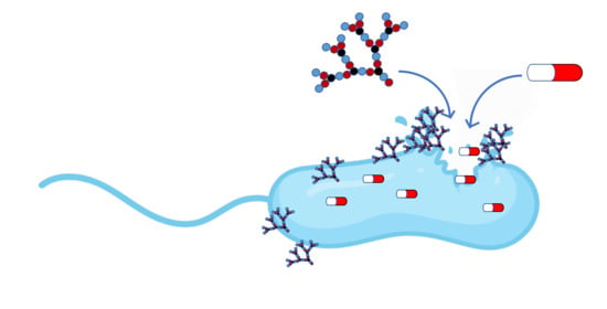
Herein, we report our investigation of possible synergistic effects between antibiotics and peptide dendrimer G3KL as a membrane permeabilizing agent (Figure 1). G3KL is an antimicrobial peptide dendrimer discovered by optimizing an initial combinatorial library hit [31] by sequence design, and exhibiting remarkable activity against Gram-negative bacteria such as Pseudomonas aeruginosa, Acinetobacter baumannii, and Escherichia coli including multidrug-resistant clinical isolates, but no activity against Klebsiella pneumoniae or methicillin-resistant Staphylococcus aureus (MRSA) [32,33]. G3KL selectively disrupts bacterial versus mammalian membrane models as evidenced by vesicle leakage assays [32], displaying pro-angiogenic properties in biological burn-wound bandages [34], anti-biofilm activity [35,36], low toxicity to mammalian cells (IC50 ~1000 μg/mL) [37], and low propensity to resistance development [38]. Our study was motivated by our recent observation that G3KL acts as a rapid membrane permeabilizer and strong membrane disruptor of bacterial cells. In this study, we show that G3KL destabilizes the LPS (lipopolysaccharide) layer, disrupts the outer and the inner membranes, interacts with DNA, and accumulates in Gram-negative bacteria up to an amount of dendrimer corresponding to 10% of the bacterial weight [37].

2. Results and Discussion

2.1. Membrane Permeabilization and Synergy with Propidium Iodide

We previously showed that G3KL permeabilizes the outer and inner membranes of P. aeruginosa cells using fluorescence microscopy and propidium iodide (PI), a fluorogenic DNA-binding dye, which is impermeable to intact cell membranes [37]. Permeabilization of bacterial membranes by G3KL might enable entry of potent antibacterial compounds at sub-inhibitory concentrations of G3KL and the cytotoxic compound. To test the feasibility of this approach and possible synergy between antimicrobials and G3KL, we used the classical checkerboard assay and stained live bacteria with 3-(4,5-dimethylthiazol-2-yl)-2,5-diphenyltetrazolium (MTT) (Figure 2) [32,39,40]. Synergy is a positive interaction in which the effect of the combined drugs is greater than when they are used alone. An additive effect indicates that the effects of the drugs used together are the same as when used independently. An indifferent effect is observed when the combination of the drugs is as efficient as the most potent drug alone. Antagonism is a negative effect observed when the combined effect of the drugs is significantly less than expected [41,42]. Interpretation of the fractional inhibitory concentration index (FICi) was followed as described by Park et al. [43]. We considered a synergistic effect for FICi < 0.5; partial synergy for 0.5 ≤ FICi < 1; additive for FICi = 1; indifferent for 1 < FICi < 4; antagonism for FICi ≥ 4.
We investigated P. aeruginosa, A. baumannii, E. coli, and K. pneumoniae as Gram-negative bacteria and MRSA as Gram-positive bacterium. We first tested whether the permeabilization effect of G3KL observed with PI might result in a synergistic effect with this dye, which binds to DNA and therefore should interfere with bacterial growth [44]. When used alone, PI was essentially inactive against Gram-negative bacteria but showed moderate activity against MRSA (methicillin-resistant Staphylococcus aureus) (Table 1). In the checkerboard assay, we observed a partial synergistic effect with G3KL in P. aeruginosa (FICi = 0.5, Figure 2A), in line with our previous microscopy studies [37]. Partial synergy also occurred with E. coli (FICi = 0.6, Figure S1) and A. baumannii (FICi < 0.53, Figure S2). These data suggest that G3KL permeabilizes the bacterial membranes below its MIC value to facilitate the entry of PI.
In the case of K. pneumoniae, neither PI nor G3KL nor their combination showed any activity (Figure S3). As G3KL permeabilizes K. pneumoniae toward other antibiotics (see below), we interpret these data as an indication that PI is inactive against this bacterium at the highest concentration used, even in the presence of membrane permeabilization. The effect between PI and G3KL was indifferent in the case of MRSA, indicating that G3KL, which is inactive against this bacterium, also does not significantly increase the activity of PI against this bacterium (Figure S4).
By comparison, polymyxin B (PMB), a well-known membrane-disrupting natural antimicrobial cyclic peptide [45,46], showed partial synergistic effects with PI in P. aeruginosa (FICi = 0.6, Figure S5), E. coli (FICi = 0.6, Figure S1), A. baumannii (FICi < 0.8, Figure S2), and MRSA (FICi = 0.8, Figure S4), but showed a surprising antagonistic effect in the case of K. pneumoniae (FICi > 4, Figure S3), which is difficult to rationalize as PMB is known to permeabilize this bacterium [45].

2.2. G3KL Synergizes with Small Molecule Antibiotics against K. pneumoniae

We next conducted a broader survey to test if membrane permeabilization by G3KL might enable synergistic effects with classical antibiotics using the checkerboard assay with P. aeruginosa and K. pneumoniae as Gram-negative bacteria and MRSA as Gram-positive bacterium. We tested vancomycin [47], erythromycin [48], and trimethoprim [49], which are active against Gram-positive bacteria but inactive against Gram-negative bacteria due to inefficient uptake. We also tested novobiocin, which is most active against Gram-positive bacteria but shows activity against some Gram-negative pathogenic strains [50,51], as well as the broad-spectrum antibiotics ciprofloxacin [52], chloramphenicol [53], gentamicin [54], azithromycin [55], sulfamethoxazole, and ampicillin [56].
In P. aeruginosa, the combination of G3KL with ampicillin did not lead to synergy (Figure S6), as expected, due to the presence of the AmpC β-lactamase and the MexAB-OprM efflux pump in this bacterium [57]. We observed partial synergy of G3KL in combination with vancomycin (FICi = 0.6), chloramphenicol (FICi = 0.6), and azithromycin (FICi = 0.5), in line with the partial synergy observed with PI (Figures S6 and S7). Combination of G3KL with erythromycin or sulfamethoxazole led to an additive effect (FICi = 1, Figure S7), indicating that, in both cases, G3KL and the antibiotic act independently on their target without interfering with each other. An indifferent effect was observed between G3KL and novobiocin, ciprofloxacin, gentamicin, and trimethoprim (Figures S8 and S9). In general, the absence of synergy reflects that the concentration of G3KL necessary for membrane permeabilization (1–2 μg/mL) is very close to its MIC value when used alone (4–8 μg/mL). Nevertheless, G3KL was shown to kill P. aeruginosa within two hours by disrupting the outer membrane, the inner membrane, and ultimately by complexing with some negatively charged intracellular components, which probably includes DNA and proteins [37]. Thus, it is possible that G3KL impairs the binding of small molecule drugs to such targets, which would also contribute to the absence of synergistic effects.
In the case of K. pneumoniae, we observed a strong synergy between G3KL and vancomycin (FICi < 0.3, Figure 2B), erythromycin (FICi < 0.3), novobiocin (FICi < 0.2), chloramphenicol (FICi < 0.5), azithromycin (FICi < 0.4), and trimethoprim (FICi < 0.5), and partial synergy with the broad-spectrum antibiotic gentamicin (FICi < 0.6) (Figures S10 and S11, Table 2). The synergistic effects between G3KL and vancomycin, erythromycin, or trimethoprim are particularly striking because these compounds were inactive against K. pneumoniae when used alone. These synergistic effects suggest that G3KL was able to permeabilize K. pneumoniae cells even if it was not active against the bacterium, in line with that close derivatives of G3KL showing significant activity against K. pneumoniae [58,59,60]. Synergies have been reported previously between polymyxin B/colistin and small molecule antibiotics in K. pneumoniae [10,11,12,13,14,30,61]. Synergistic effects involving a permeabilizing but inactive compound have been previously observed with polymyxin B nonapeptide (PMBN), an inactive polymyxin B derivative lacking the fatty acyl chain [11,62].
G3KL did not increase the activity of ampicillin and sulfamethoxazole, which were inactive against K. pneumoniae when used alone (Figure S12). In the case of ampicillin, the antibiotic is probably degraded by the naturally-occurring β-lactamase in K. pneumoniae NCTC 418, rendering permeabilization inefficient [63]. G3KL also did not significantly increase the activity of ciprofloxacin, which is very active and whose activity is not limited by uptake (Figure S12).
Finally, we tested the permeabilization effect of G3KL in a Gram-positive MRSA strain. The results showed a very weak permeabilization effect, in line with G3KL being inactive against this bacterium. We observed a weak synergy between G3KL with erythromycin (FICi < 1), ampicillin (FICi < 1), and azithromycin (FICi < 0.8) (Figure S13) and an indifferent effect with all the other antibiotics (Figures S14–S16), suggesting that G3KL is unable to pass the peptidoglycan layer and reach the membrane, and therefore cannot permeabilize MRSA for the uptake of small molecule drugs.
To confirm the synergistic effect observed in the checkerboard assay, we performed time-kill experiments for the combinations of G3KL with vancomycin (FICi < 0.3), erythromycin (FICi < 0.3), novobiocin (FICi < 0.2), and trimethoprim (FICi < 0.5). Killing kinetics on K. pneumoniae at an initial inoculum of ~106 CFU/mL showed that the pairs G3KL/vancomycin (32 μg/mL/32 μg/mL) and G3KL/trimethoprim (32 μg/mL/16 μg/mL) effectively killed the bacteria after 4 h, and G3KL/erythromycin (32 μg/mL/16 μg/mL) after 8 h, all below the MIC level (Figure 3). The pair G3KL/novobiocin did not show a reduction in bacterial burden but exhibited a growth-inhibiting activity. The low level of surviving bacteria might not have been detected in the checkerboard assay. Similar growth inhibition was observed for novobiocin when used alone at 2 × MIC (16 μg/mL) against K. pneumoniae (Figure 3 and Figure S17). This effect was also observed in previous studies against E. coli, which suggested that novobiocin generally inhibits cell division and induces slower cell growth [50,64]. Note that G3KL, vancomycin, trimethoprim, erythromycin, and novobiocin when used alone at the same concentration as in combination showed no effect on bacterial killing.

3. Materials and Methods

3.1. Compounds

Peptide dendrimer G3KL was synthesized by solid-phase peptide synthesis and purified as described earlier [32]. Vancomycin, ampicillin, novobiocin, azithromycin, sulfamethoxazole, and trimethoprim were purchased from Sigma Aldrich (Buchs, Switzerland), erythromycin and ciprofloxacin were purchased from Acros Organics (Geel, Belgium), and chloramphenicol and gentamicin were purchased from AppliChem (Darmstadt, Germany). All compounds were conditioned as 8 or 10 mg/mL stock solutions in water (G3KL, ampicillin, novobiocin, chloramphenicol, and gentamicin), 1% acetic acid (ciprofloxacin), and DMSO (erythromycin, azithromycin, sulfamethoxazole, and trimethoprim).

3.2. Broth Microdilution Method

Antimicrobial activity was assayed against P. aeruginosa (PAO1), A. baumannii (ATCC19606), E. coli W3110 (TE823), K. pneumoniae (NCTC418), and methicillin-resistant S. aureus (COL). The minimal inhibitory concentration (MIC) was determined by using the broth microdilution method. A colony of bacteria was picked and grown in a Luria-Bertani (LB, Sigma Aldrich, Buchs, Switzerland) medium overnight at 37 °C. Stock solutions of 1 mg/mL of the samples were prepared in sterilized Milli-Q water and diluted to the starting concentration of 128 μg/mL in 300 μL Mueller Hinton (MH) medium. The diluted samples were added to the first well of the 96-well microtiter plate (TPP, untreated, Faust Laborbedarf, AG, Schaffhausen, Switzerland) and diluted serially by ½. Bacteria were quantified by measuring the optical density at 600 nm and diluted to OD600 of 0.022 in MH medium. We used 4 μL of the diluted bacterial solution to inoculate the sample solutions (150 μL) with a final inoculation of about 5 × 10⁵ CFU/mL. The plates were then incubated at 37 °C for 18 h. For each assay, sterility (broth only) and growth control (broth with bacterial inoculum, without antibiotics) were checked with two columns in the plate. The next day, 15 μL of MTT (1 mg/mL stock solution) (Sigma Aldrich, Buchs, Switzerland) was added to each well of the plate. The MIC was defined as the lowest concentration of the peptide dendrimer with a colorless well indicating no bacterial growth.

3.3. Checkerboard Assay

To verify the activity of two drugs in combination, the checkerboard method was used to determine the MICs for each antibiotic alone and in combination. Both G3KL [37] and a paired small molecule (propidium iodide, sulfamethoxazole, chloramphenicol, novobiocin, azithromycin, erythromycin, ciprofloxacin, gentamicin, ampicillin, and trimethoprim) were diluted by 1/2 in a 96 well plate. Stock solutions of 1 mg/mL of the antibiotics were prepared in sterilized Milli-Q water and diluted to the starting concentration with 2–4 × MIC of the corresponding compounds in 300 μL Mueller Hinton (MH) medium. For propidium iodide, the starting concentration was 1000 μg/mL. For checkerboard assay testing the combinations of PI with G3KL, PMB, and ciprofloxacin, PI was diluted across the rows and G3KL, PMB, and ciprofloxacin across the columns. For checkerboard assay testing the combinations of G3KL with small-molecule antibiotics, G3KL was diluted across the rows and the small-molecule antibiotics across the columns. We then added 75 μL containing 2× the final concentration of G3KL, PMB, and ciprofloxacin to each well containing the respective drug (PI and the small-molecule drugs) to be tested in combination, across the columns and down the rows, resulting in a checkerboard of 150 μL final volume with the final wells containing only G3KL or the antibiotics.
Similar to broth microdilution, bacteria were quantified by measuring the optical density at 600 nm and diluted to OD600 of 0.022 in an MH medium. We used 4 μL of the diluted bacterial solution to inoculate the sample solutions (150 μL) with a final inoculation of about 5 × 10⁵ CFU/mL. The plates were then incubated at 37 °C for 18 h. The next day, 15 μL of MTT (1 mg/mL stock solution) was added to each well of the plate so that MIC and MICcomb were defined as the lowest concentration of the peptide dendrimer and/or antibiotics with a colorless well indicating no bacterial growth. The FIC of each antibiotic was calculated as follows:
FICA (MICAcomb/MICA) + FICB (MICBcomb/MICB) = ΣFIC = FICindex (FICi).
A synergistic interaction is defined by an FIC index of <0.5, partial synergy is defined by an FIC index of ≥0.5 and <1, an additive interaction by an FIC index of 1.0, indifferent by an FIC of >1 and <4, and antagonism is defined by an FIC of ≥4 [43].

3.4. Time-Kill Assay

Time-kill kinetics against K. pneumoniae (NCTC418) were performed on G3KL (32 µg/mL), vancomycin (32 μg/mL), erythromycin (16 μg/mL), novobiocin (2 μg/mL), G3KL/vancomycin (32/32 μg/mL), G3KL/trimethoprim (32/16 μg/mL), and G3KL/erythromycin (32/16 μg/mL) and using 2× the concentrations indicated by the checkerboard assay. Untreated bacteria at 1 × 10⁶ CFU/mL were used as a growth control.
A single colony of K. pneumoniae (NCTC418) was picked and grown overnight with shaking (180 rpm) in a 5 mL LB medium (Sigma Aldrich, Buchs, Switzerland) at 37 °C. The overnight bacterial culture was diluted to OD600 0.002 (2 × 10⁶ CFU/mL) in fresh MH (Sigma Aldrich, Buchs, Switzerland) medium. Stock solutions of G3KL and antibiotics (8 mg/mL) were prepared in sterilized Milli-Q water and diluted to two-times more concentrated than the required concentration in the fresh MH (Sigma Aldrich, Buchs, Switzerland) medium. G3KL and antibiotic were pre-mixed when needed. We mixed 100 µL of the adjusted bacteria and 100 µL samples in a 96-well microtiter plate (TPP, untreated, Corning Incorporated, Kennebunk, ME, USA) and the time-kill kinetics started at the moment of mixing. Ninety-six-well microtiter plates were incubated at 37 °C under shaking (180 rpm). Bacterial growth was quantified at 0, 0.5, 1, 2, 3, 4, 5, and 6 h, G3KL/erythromycin (32/16 μg/mL) were also quantified at 7 and 8 h. The quantification was performed by plating 10-fold dilutions of a sample in sterilized 0.9% NaCl on LB agar plates. LB agar plates were incubated at 37 °C for 10 h and the number of individual colonies was counted at each time-point. The assay was performed in triplicate.

4. Conclusions

The experiments above showed that the membrane permeabilizing effects of antimicrobial peptide dendrimer G3KL can be exploited to obtain synergistic effects with other substances. G3KL showed only weak or no synergy with small-molecule antibiotics in the case of P. aeruginosa, a bacterium against which the dendrimer is very active, probably because the concentration of G3KL necessary to permeabilize the membrane is very close to the MIC value. G3KL showed very significant synergistic effects when tested against K. pneumoniae, against which the dendrimer is inactive when used alone, revealing that G3KL is capable of permeabilizing the membrane even if it does not show activity. The effect is particularly striking in combination with vancomycin, erythromycin, or trimethoprim because these antibiotics are inactive against K. pneumoniae when used alone. However, our synergistic study showed no effects against MRSA, indicating that G3KL is not only inactive against this Gram-positive bacterium, but also does not significantly permeabilize its membrane.

Supplementary Materials

The following are available online. Figures S1–S5: Checkerboard microtiter plate assay testing the combination of G3KL and PMB with PI in P. aeruginosa, E. coli, A. baumannii, and K. pneumoniae. Figures S6–S9: Checkerboard microtiter plate assay testing the combination of G3KL with small molecule drugs in P. aeruginosa. Figures S10–S12: Checkerboard microtiter plate assay testing the combination of G3KL with small molecule drugs in K. pneumoniae. Figures S13–S16: Checkerboard microtiter plate assay testing the combination of G3KL with small molecule drugs in MRSA. Figure S17: Static time-kill assay of novobiocin at 2 × MIC (16 μg/mL).

Author Contributions

B.-H.G. designed, performed the entire study, interpreted the data, and wrote the paper. X.C. performed the time-kill experiment, interpreted data, and wrote the paper. S.J. and T.K. designed, supervised, interpreted the data, and wrote the paper. J.-L.R. designed and supervised the entire study, interpreted the data, and wrote the paper. All authors have read and agreed to the published version of the manuscript.

Funding

This research was funded by the Swiss National Science Foundation, Grant no. 407240_167048 and 200020_178998.

Conflicts of Interest

The authors declare no conflict of interest.

References

  1. Li, X.Z.; Nikaido, H. Efflux-mediated drug resistance in bacteria: An update. Drugs 2009, 69, 1555–1623. [Google Scholar] [CrossRef]
  2. Zasloff, M. Antimicrobial peptides of multicellular organisms. Nature 2002, 415, 385–395. [Google Scholar] [CrossRef] [PubMed]
  3. Nguyen, L.T.; Haney, E.F.; Vogel, H.J. The expanding scope of antimicrobial peptide structures and their modes of action. Trends Biotechnol. 2011, 29, 464–472. [Google Scholar] [CrossRef] [PubMed]
  4. Mojsoska, B.; Jenssen, H. Peptides and Peptidomimetics for Antimicrobial Drug Design. Pharmaceuticals 2015, 8, 366–415. [Google Scholar] [CrossRef] [PubMed]
  5. Czaplewski, L.; Bax, R.; Clokie, M.; Dawson, M.; Fairhead, H.; Fischetti, V.A.; Foster, S.; Gilmore, B.F.; Hancock, R.E.W.; Harper, D.; et al. Alternatives to antibiotics-a pipeline portfolio review. Lancet Infect. Dis. 2016, 16, 239–251. [Google Scholar] [CrossRef] [Green Version]
  6. Acedo, J.Z.; Chiorean, S.; Vederas, J.C.; van Belkum, M.J. The expanding structural variety among bacteriocins from Gram-positive bacteria. FEMS Microbiol. Rev. 2018, 42, 805–828. [Google Scholar] [CrossRef]
  7. Amso, Z.; Hayouka, Z. Antimicrobial random peptide cocktails: A new approach to fight pathogenic bacteria. Chem. Commun. 2019, 55, 2007–2014. [Google Scholar] [CrossRef]
  8. Torres, M.D.T.; Sothiselvam, S.; Lu, T.K.; de la Fuente-Nunez, C. Peptide Design Principles for Antimicrobial Applications. J. Mol. Biol. 2019, 431, 3547–3567. [Google Scholar] [CrossRef]
  9. Mookherjee, N.; Anderson, M.A.; Haagsman, H.P.; Davidson, D.J. Antimicrobial host defence peptides: Functions and clinical potential. Nat. Rev. Drug Discov. 2020, 19, 311–332. [Google Scholar] [CrossRef]
  10. Shinohara, D.R.; Menegucci, T.C.; Fedrigo, N.H.; Migliorini, L.B.; Carrara-Marroni, F.E.; Maria Dos Anjos, M.; Cardoso, C.L.; Nishiyama, S.A.B.; Tognim, M.C.B. Synergistic activity of polymyxin B combined with vancomycin against carbapenem-resistant and polymyxin-resistant Acinetobacter baumannii: First in vitro study. J. Med. Microbiol. 2019, 68, 309–315. [Google Scholar] [CrossRef]
  11. Ofek, I.; Cohen, S.; Rahmani, R.; Kabha, K.; Tamarkin, D.; Herzig, Y.; Rubinstein, E. Antibacterial synergism of polymyxin B nonapeptide and hydrophobic antibiotics in experimental gram-negative infections in mice. Antimicrob. Agents Chemother. 1994, 38, 374–377. [Google Scholar] [CrossRef] [PubMed] [Green Version]
  12. Mandler, M.D.; Baidin, V.; Lee, J.; Pahil, K.S.; Owens, T.W.; Kahne, D. Novobiocin enhances polymyxin activity by stimulating lipopolysaccharide transport. J. Am. Chem. Soc. 2018, 140, 6749–6753. [Google Scholar] [CrossRef] [PubMed]
  13. Simmons, N.A. Colistin, sulphamethoxazole, and trimethoprim in synergy against Gram-negative bacteria. J. Clin. Pathol. 1970, 23, 757–764. [Google Scholar] [CrossRef] [PubMed] [Green Version]
  14. Rosenblatt, J.E.; Stewart, P.R. Combined Activity of Sulfamethoxazole, Trimethoprim, and Polymyxin B against Gram-Negative Bacilli. Antimicrob. Agents Chemother. 1974, 6, 84–92. [Google Scholar] [CrossRef] [PubMed] [Green Version]
  15. Nageeb, W.; Metwally, L.; Kamel, M.; Zakaria, S. In vitro antimicrobial synergy studies of carbapenem-resistant Acinetobacter baumannii isolated from intensive care units of a tertiary care hospital in Egypt. J. Infect. Public Health 2015, 8, 593–602. [Google Scholar] [CrossRef] [Green Version]
  16. Abdul Rahim, N.; Cheah, S.-E.; Johnson, M.D.; Yu, H.; Sidjabat, H.E.; Boyce, J.; Butler, M.S.; Cooper, M.A.; Fu, J.; Paterson, D.L.; et al. Synergistic killing of NDM-producing MDR Klebsiella pneumoniae by two ‘old’ antibiotics—Polymyxin B and chloramphenicol. J. Antimicrob. Chemother. 2015, 70, 2589–2597. [Google Scholar] [CrossRef] [Green Version]
  17. Elemam, A.; Rahimian, J.; Doymaz, M. In Vitro Evaluation of Antibiotic Synergy for Polymyxin B-Resistant Carbapenemase-Producing Klebsiella pneumoniae. J. Clin. Microbiol. 2010, 48, 3558–3562. [Google Scholar] [CrossRef] [Green Version]
  18. Pletzer, D.; Mansour, S.C.; Hancock, R.E.W. Synergy between conventional antibiotics and anti-biofilm peptides in a murine, sub-cutaneous abscess model caused by recalcitrant ESKAPE pathogens. PLoS Pathog. 2018, 14, e1007084. [Google Scholar] [CrossRef]
  19. Ulvatne, H.; Karoliussen, S.; Stiberg, T.; Rekdal, Ø.; Svendsen, J.S. Short antibacterial peptides and erythromycin act synergically against Escherichia coli. J. Antimicrob. Chemother. 2001, 48, 203–208. [Google Scholar] [CrossRef] [Green Version]
  20. Giacometti, A.; Cirioni, O.; Del Prete, M.S.; Paggi, A.M.; D’Errico, M.M.; Scalise, G. Combination studies between polycationic peptides and clinically used antibiotics against Gram-positive and Gram-negative bacteria. Peptides 2000, 21, 1155–1160. [Google Scholar] [CrossRef]
  21. Grassi, L.; Maisetta, G.; Esin, S.; Batoni, G. Combination Strategies to Enhance the Efficacy of Antimicrobial Peptides against Bacterial Biofilms. Front. Microbiol. 2017, 8, 2409–2417. [Google Scholar] [CrossRef] [PubMed]
  22. Wu, X.; Li, Z.; Li, X.; Tian, Y.; Fan, Y.; Yu, C.; Zhou, B.; Liu, Y.; Xiang, R.; Yang, L. Synergistic effects of antimicrobial peptide DP7 combined with antibiotics against multidrug-resistant bacteria. Drug. Des. Dev. Ther. 2017, 11, 939–946. [Google Scholar] [CrossRef] [PubMed] [Green Version]
  23. Hollmann, A.; Martinez, M.; Maturana, P.; Semorile, L.C.; Maffia, P.C. Antimicrobial peptides: Interaction with model and biological membranes and synergism with chemical antibiotics. Front. Chem. 2018, 6, 204–217. [Google Scholar] [CrossRef] [Green Version]
  24. Shurko, J.F.; Galega, R.S.; Li, C.; Lee, G.C. Evaluation of LL-37 antimicrobial peptide derivatives alone and in combination with vancomycin against S. aureus. J. Antibiot. 2018, 71, 971–974. [Google Scholar] [CrossRef] [PubMed] [Green Version]
  25. Otvos, L., Jr.; Ostorhazi, E.; Szabo, D.; Zumbrun, S.D.; Miller, L.L.; Halasohoris, S.A.; Desai, P.D.; Int Veldt, S.M.; Kraus, C.N. Synergy Between Proline-Rich Antimicrobial Peptides and Small Molecule Antibiotics Against Selected Gram-Negative Pathogens in vitro and in vivo. Front. Chem. 2018, 6, 309–321. [Google Scholar] [CrossRef] [PubMed]
  26. Mohammadi Azad, Z.; Moravej, H.; Fasihi-Ramandi, M.; Masjedian, F.; Nazari, R.; Mirnejad, R.; Moosazadeh Moghaddam, M. In vitro synergistic effects of a short cationic peptide and clinically used antibiotics against drug-resistant isolates of Brucella melitensis. J. Med. Microbiol. 2017, 66, 919–926. [Google Scholar] [CrossRef]
  27. Kampshoff, F.; Willcox, M.D.P.; Dutta, D. A Pilot Study of the Synergy between Two Antimicrobial Peptides and Two Common Antibiotics. Antibiotics 2019, 8, 60. [Google Scholar] [CrossRef] [Green Version]
  28. Mohamed, M.F.; Abdelkhalek, A.; Seleem, M.N. Evaluation of short synthetic antimicrobial peptides for treatment of drug-resistant and intracellular Staphylococcus aureus. Sci. Rep. 2016, 6, 1–14. [Google Scholar] [CrossRef]
  29. Zheng, W.; Sun, W.; Simeonov, A. Drug repurposing screens and synergistic drug-combinations for infectious diseases. Br. J. Pharm. 2018, 175, 181–191. [Google Scholar] [CrossRef]
  30. Li, Y.; Lin, X.; Yao, X.; Huang, Y.; Liu, W.; Ma, T.; Fang, B. Synergistic Antimicrobial Activity of Colistin in Combination with Rifampin and Azithromycin against Escherichia coli Producing MCR-1. Antimicrob. Agents Chemother. 2018, 62, 18–28. [Google Scholar] [CrossRef] [Green Version]
  31. Stach, M.; Maillard, N.; Kadam, R.U.; Kalbermatter, D.; Meury, M.; Page, M.G.P.; Fotiadis, D.; Darbre, T.; Reymond, J.-L. Membrane disrupting antimicrobial peptide dendrimers with multiple amino termini. MedChemComm 2012, 3, 86–89. [Google Scholar] [CrossRef]
  32. Stach, M.; Siriwardena, T.N.; Köhler, T.; van Delden, C.; Darbre, T.; Reymond, J.-L. Combining Topology and Sequence Design for the Discovery of Potent Antimicrobial Peptide Dendrimers against Multidrug-Resistant Pseudomonas aeruginosa. Angew. Chem. Int. Ed. 2014, 53, 12827–12831. [Google Scholar] [CrossRef] [PubMed]
  33. Pires, J.; Siriwardena, T.N.; Stach, M.; Tinguely, R.; Kasraian, S.; Luzzaro, F.; Leib, S.L.; Darbre, T.; Reymond, J.-L.; Endimiani, A. In Vitro Activity of the Novel Antimicrobial Peptide Dendrimer G3KL against Multidrug-Resistant Acinetobacter baumannii and Pseudomonas aeruginosa. Antimicrob. Agents Chemother. 2015, 59, 7915–7918. [Google Scholar] [CrossRef] [PubMed] [Green Version]
  34. Abdel-Sayed, P.; Kaeppeli, A.; Siriwardena, T.; Darbre, T.; Perron, K.; Jafari, P.; Reymond, J.-L.; Pioletti, D.P.; Applegate, L.A. Anti-Microbial Dendrimers against Multidrug-Resistant P. aeruginosa Enhance the Angiogenic Effect of Biological Burn-wound Bandages. Sci. Rep. 2016, 6, 23872–23882. [Google Scholar] [CrossRef] [PubMed] [Green Version]
  35. Pompilio, A.; Geminiani, C.; Mantini, P.; Siriwardena, T.N.; Bonaventura, I.D.; Reymond, J.L.; Bonaventura, G.D. Peptide dendrimers as “lead compounds” for the treatment of chronic lung infections by Pseudomonas aeruginosa in cystic fibrosis patients: In vitro and in vivo studies. Infect. Drug. Resist. 2018, 11, 1767–1783. [Google Scholar] [CrossRef] [PubMed] [Green Version]
  36. Han, X.; Liu, Y.; Ma, Y.; Zhang, M.; He, Z.; Siriwardena, T.N.; Xu, H.; Bai, Y.; Zhang, X.; Reymond, J.-L.; et al. Peptide dendrimers G3KL and TNS18 inhibit Pseudomonas aeruginosa biofilms. Appl. Microbiol. Biotechnol. 2019, 103, 5821–5830. [Google Scholar] [CrossRef]
  37. Gan, B.-H.; Siriwardena, T.N.; Javor, S.; Darbre, T.; Reymond, J.-L. Fluorescence Imaging of Bacterial Killing by Antimicrobial Peptide Dendrimer G3KL. ACS Infect. Dis. 2019, 5, 2164–2173. [Google Scholar] [CrossRef]
  38. Jeddou, F.B.; Falconnet, L.; Luscher, A.; Siriwardena, T.; Reymond, J.-L.; Delden, C.; van Köhler, T. Adaptive and Mutational Responses to Peptide Dendrimer Antimicrobials in Pseudomonas aeruginosa. Antimicrob. Agents Chemother. 2020, 64, e02040-19. [Google Scholar]
  39. Doern, C.D. When Does 2 Plus 2 Equal 5? A Review of Antimicrobial Synergy Testing. J. Clin. Microbiol. 2014, 52, 4124–4128. [Google Scholar] [CrossRef] [Green Version]
  40. Grela, E.; Kozłowska, J.; Grabowiecka, A. Current methodology of MTT assay in bacteria—A review. Acta Histochem. 2018, 120, 303–311. [Google Scholar] [CrossRef] [PubMed]
  41. Renneberg, J. Definitions of antibacterial interactions in animal infection models. J. Antimicrob. Chemother. 1993, 31, 167–175. [Google Scholar] [CrossRef] [PubMed] [Green Version]
  42. Roell, K.R.; Reif, D.M.; Motsinger-Reif, A.A. An Introduction to Terminology and Methodology of Chemical Synergy—Perspectives from Across Disciplines. Front. Pharm. 2017, 8, 1–11. [Google Scholar] [CrossRef] [PubMed]
  43. Gopal, R.; Kim, Y.G.; Lee, J.H.; Lee, S.K.; Chae, J.D.; Son, B.K.; Seo, C.H.; Park, Y. Synergistic Effects and Antibiofilm Properties of Chimeric Peptides against Multidrug-Resistant Acinetobacter baumannii Strains. Antimicrob. Agents Chemother. 2014, 58, 1622–1629. [Google Scholar] [CrossRef] [PubMed] [Green Version]
  44. Netuschil, L.; Auschill, T.M.; Sculean, A.; Arweiler, N.B. Confusion over live/dead stainings for the detection of vital microorganisms in oral biofilms—Which stain is suitable? BMC Oral Health 2014, 14, 2–14. [Google Scholar] [CrossRef] [Green Version]
  45. Deris, Z.Z.; Swarbrick, J.D.; Roberts, K.D.; Azad, M.A.K.; Akter, J.; Horne, A.S.; Nation, R.L.; Rogers, K.L.; Thompson, P.E.; Velkov, T.; et al. Probing the Penetration of Antimicrobial Polymyxin Lipopeptides into Gram-Negative Bacteria. Bioconjugate Chem. 2014, 25, 750–760. [Google Scholar] [CrossRef] [PubMed]
  46. Velkov, T.; Thompson, P.E.; Nation, R.L.; Li, J. Structure-Activity Relationships of Polymyxin Antibiotics. J. Med. Chem. 2010, 53, 1898–1916. [Google Scholar] [CrossRef] [Green Version]
  47. Watanakunakorn, C. Mode of action and in-vitro activity of vancomycin. J. Antimicrob. Chemother. 1984, 14, 7–18. [Google Scholar] [CrossRef]
  48. Mao, J.C.-H.; Putterman, M. Accumulation in Gram-positive and Gram-negative Bacteria as a Mechanism of Resistance to Erythromycin. J. Bacteriol. 1968, 95, 1111–1117. [Google Scholar] [CrossRef] [Green Version]
  49. Salter, A.J. Overview. Trimethoprim-Sulfamethoxazole: An Assessment of More Than 12 Years of Use. Rev. Infect. Dis. 1982, 4, 196–236. [Google Scholar] [CrossRef]
  50. Smith, D.H.; Davis, B.D. Mode of Action of Novobiocin in Escherichia coli. J. Bacteriol. 1967, 93, 71–79. [Google Scholar] [CrossRef] [Green Version]
  51. May, J.M.; Owens, T.W.; Mandler, M.D.; Simpson, B.W.; Lazarus, M.B.; Sherman, D.J.; Davis, R.M.; Okuda, S.; Massefski, W.; Ruiz, N.; et al. The antibiotic novobiocin binds and activates the ATPase that powers lipopolysaccharide transport. J. Am. Chem. Soc. 2017, 139, 17221–17224. [Google Scholar] [CrossRef] [PubMed] [Green Version]
  52. Hooper, D.C.; Wolfson, J.S.; Ng, E.Y.; Swartz, M.N. Mechanisms of action of and resistance to ciprofloxacin. Am. J. Med. 1987, 82, 12–20. [Google Scholar] [PubMed]
  53. Bergmann, E.D.; Sicher, S. Mode of Action of Chloramphenicol. Nature 1952, 170, 931–932. [Google Scholar] [CrossRef] [PubMed]
  54. Hahn, F.E.; Sarre, S.G. Mechanism of Action of Gentamicin. J. Infect. Dis. 1969, 119, 364–369. [Google Scholar] [CrossRef] [PubMed]
  55. Retsema, J.; Girard, A.; Schelkly, W.; Manousos, M.; Anderson, M.; Bright, G.; Borovoy, R.; Brennan, L.; Mason, R. Spectrum and mode of action of azithromycin (CP-62,993), a new 15-membered-ring macrolide with improved potency against gram-negative organisms. Antimicrob. Agents Chemother. 1987, 31, 1939–1947. [Google Scholar] [CrossRef] [PubMed] [Green Version]
  56. Tipper, D.J. Mode of action of β-lactam antibiotics. Pharm. Ther. 1985, 27, 1–35. [Google Scholar] [CrossRef]
  57. Okamoto, K.; Gotoh, N.; Nishino, T. Pseudomonas aeruginosa Reveals High Intrinsic Resistance to Penem Antibiotics: Penem Resistance Mechanisms and Their Interplay. Antimicrob. Agents Chemother. 2001, 45, 1964–1971. [Google Scholar] [CrossRef] [Green Version]
  58. Siriwardena, T.N.; Stach, M.; He, R.; Gan, B.; Javor, S.; Heitz, M.; Cai, X.; Chen, P.; Wei, D.; Li, H.; et al. Lipidated Peptide Dendrimers Killing Multidrug-Resistant Bacteria. J. Am. Chem. Soc. 2018, 140, 423–432. [Google Scholar] [CrossRef] [Green Version]
  59. Siriwardena, T.N.; Capecchi, A.; Gan, B.-H.; Jin, X.; He, R.; Wei, D.; Ma, L.; Köhler, T.; van Delden, C.; Javor, S.; et al. Optimizing Antimicrobial Peptide Dendrimers in Chemical Space. Angew. Chem. Int. Ed. 2018, 57, 8483–8487. [Google Scholar] [CrossRef]
  60. Siriwardena, T.N.; Lüscher, A.; Köhler, T.; van Delden, C.; Javor, S.; Reymond, J.-L. Antimicrobial Peptide Dendrimer Chimera. Helv. Chim. Acta 2019, 102, e1900034. [Google Scholar] [CrossRef]
  61. Lenhard, J.R.; Nation, R.L.; Tsuji, B.T. Synergistic combinations of polymyxins. Int. J. Antimicrob. Agents 2016, 48, 607–622. [Google Scholar] [CrossRef] [PubMed] [Green Version]
  62. Vaara, M. Polymyxin Derivatives that Sensitize Gram-Negative Bacteria to Other Antibiotics. Molecules 2019, 24, 249. [Google Scholar] [CrossRef] [PubMed] [Green Version]
  63. Brown, A.G.; Butterworth, D.; Cole, M.; Hanscomb, G.; Hood, J.D.; Reading, C.; Rolinson, G.N. Naturally-Occurring β-Lactamase Inhibitors with Antibacterial Activity. J. Antibiot. 1976, 29, 668–669. [Google Scholar] [CrossRef] [PubMed]
  64. Martin, J.K.; Sheehan, J.P.; Bratton, B.P.; Moore, G.M.; Mateus, A.; Li, S.H.-J.; Kim, H.; Rabinowitz, J.D.; Typas, A.; Savitski, M.M.; et al. A Dual-Mechanism Antibiotic Kills Gram-Negative Bacteria and Avoids Drug Resistance. Cell 2020, 181, 1518–1532. [Google Scholar] [CrossRef]
Figure 1. Structural formula of antimicrobial compounds used in this study.
Molecules 25 05643 g001
Figure 2. Checkerboard microtiter plate assay testing the combination of G3KL with PI in P. aeruginosa PAO1 (A) and vancomycin with G3KL in K. pneumoniae NCTC 418. (B) 2D two-fold serial dilutions were performed starting with 500 µg/mL of PI, 256 µg/mL of vancomycin, and 8 or 64 µg/mL of G3KL. The viability of the bacteria was revealed after the addition of 3-(4,5-dimethylthiazol-2-yl)-2,5-diphenyltetrazolium (MTT) dye (black wells). Red circle: FICi of synergistic effect; yellow circles: FICi of partial synergy; green circles: MIC values of G3KL, PI, and vancomycin.
Molecules 25 05643 g002
Figure 3. Static time-kill assay with G3KL, the antibiotics vancomycin, erythromycin, and novobiocin, and their combination with G3KL. The experiment showed a decline in K. pneumoniae bacterial burden at 37 °C for the combination of G3KL/vancomycin (32 μg/mL/32 μg/mL), G3KL/erythromycin (32 μg/mL/16 μg/mL), and G3KL/trimethoprim (32 μg/mL/16 μg/mL) below the MIC level. The combination G3KL/novobiocin (32 μg/mL/2 μg/mL) showed inhibition in K. pneumoniae growth. The assays were performed in triplicate.
Molecules 25 05643 g003
Table 1. Activity of G3KL and PMB in combination with PI.
P. aeruginosa
PAO1
E. coli
W3110
A. baumannii
ATCC 19606
K. pneumoniae
NCTC 418
MRSA
COL
G3KLa448>64>64
PIa≥50062.5–250>500≥50062.5
PMBa0.250.250.1250.12564
G3KLcomb/PIcomb (FICi) b2/8 (0.5)2/31.3 (0.6)4/15.6 (<0.53)>64/>500 (-)>64/62.5 (>2)
PMBcomb/PIcomb (FICi) b0.125/62.5 (0.6)0.125/31.25 (0.6)0.063/125 (<0.8)2/16 (>4)16/31.25 (0.8)
a Minimal inhibitory concentration (MIC) values in μg/mL were determined by serial ½ dilution in MHB (Mueller-Hinton broth). b MIC in combination in µg/mL was determined by checkerboard method. FICi was calculated as the sum of FIC of drug A (FIC A) and FIC of drug B (FIC B): FICA(MICAcomb/MICA) + FICB(MICBcomb/MICB) = ΣFIC = FICindex (FICi). Interpretation of FICi was as follows: synergistic effect for FICi < 0.5; partial synergy for 0.5 ≤ FICi < 1; additive for FICi = 1; indifferent for 1 < FICi < 4; antagonism for FICi ≥ 4 [43]. In the cases where no discrete MIC value was determined in the checkerboard assay, FIC values were calculated by using the highest dilution used in the assay.
Table 2. MICs (μg/mL) of G3KL and small molecule drugs in combination a.
P. aeruginosa
PAO1
K. pneumoniae
NCTC418
MRSA
COL
G3KL4–8>64>64
Vancomycin2562560.5
G3KLcomb/Vancomycincomb (FICi) b1/32 (0.6)16/16 (<0.3)>64/0.5 (>2)
Erythromycin128640.5
G3KLcomb/Erythromycincomb (FICi) b2/64 (1)8/8 (<0.3)32/0.25 (<1)
Ampicillin>256>256128
G3KLcomb/Ampicillincomb (FICi) b4/>256 (>2)>64/>256 (-)32/64 (<1)
Novobiocin>256160.31
G3KLcomb/Novobiocincomb (FICi) b2/256 (<1.5)8/1 (<0.2)>64/0.31 (>2)
Ciprofloxacin0.1250.0310.25
G3KLcomb/Ciprofloxacincomb (FICi) b4/0.125 (2)>32/0.031 (>2)>64/0.25 (>2)
Chloramphenicol888
G3KLcomb/Chloramphenicolcomb (FICi) b2/1 (0.6)16/2 (<0.5)>64/8 (>2)
Gentamicin120.5
G3KLcomb/Gentamicincomb (FICi) b4/1 (2)32/0.25 (<0.6)64/0.25 (<1.3)
Azithromycin6444
G3KLcomb/Azithromycincomb (FICi) b8/0.5 (0.5)8/1 (<0.4)16/2 (<0.8)
Sulfamethoxazole256>256>32
G3KLcomb/Sulfamethoxazolecomb (FICi) b1/128 (1)>64/>256 (-)>64/>32 (-)
Trimethoprim128>256>32
G3KLcomb/Trimethoprimcomb (FICi) b4/128 (2)32/8 (<0.5)64/32 (<1.5)
a The minimal inhibitory concentration in μg/mL was determined by two-fold serial dilutions in MH medium. The experiments were performed in triplicate and the values in µg/mL were calculated based on the peptide mass without trifluoroacetate counterions. b MIC in combination (G3KL/antibiotics) in µg/mL were determined by the checkerboard method. The FICi in brackets was calculated as the sum of FIC of drug A (FIC A) and FIC of drug B (FIC B): FICi was calculated as the sum of FIC of drug A (FIC A) and FIC of drug B (FIC B). FICA(MICAcomb/MICA) + FICB(MICBcomb/MICB) = ΣFIC = FICindex (FICi). Interpretation of FICi as follows: synergistic effect for FICi < 0.5; partial synergy for 0.5 ≤ FICi < 1; additive for FICi = 1; indifferent for 1 < FICi < 4; antagonism for FICi ≥ 4 [43]. In the cases where no discrete MIC value was determined in the checkerboard assay, FIC values were calculated by using the highest dilution used in the assay.
Sample Availability: Samples for the compounds are accessible as described in the methods Section 3.1.
Publisher’s Note: MDPI stays neutral with regard to jurisdictional claims in published maps and institutional affiliations.