
Differential Proliferation Effect of the Newly Synthesized Valine, Tyrosine and Tryptophan–Naphthoquinones in Immortal and Tumorigenic Cervical Cell Lines

 , , , ,
Abstract
:1. Introduction
2. Results
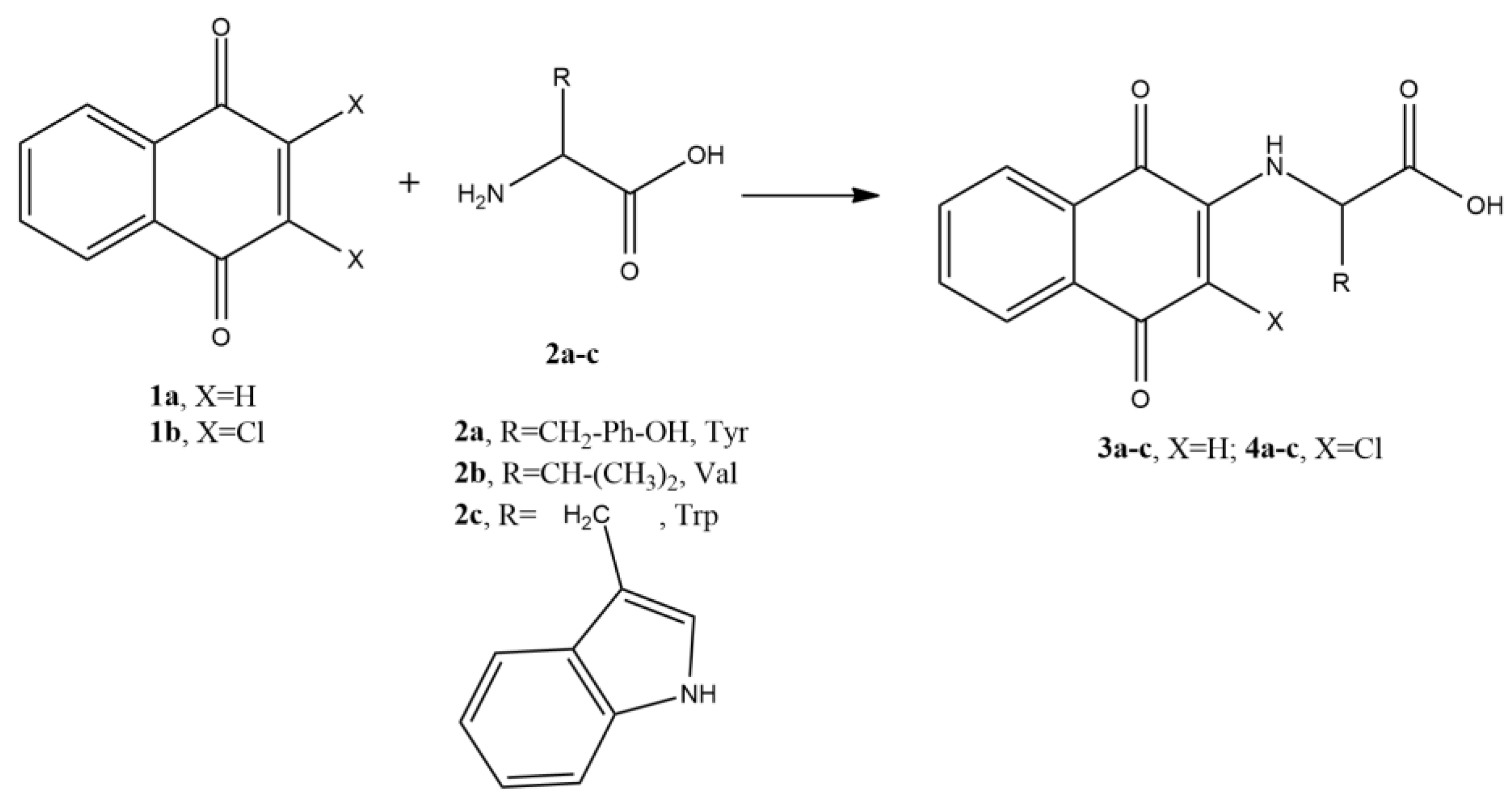
2.1. Chemistry
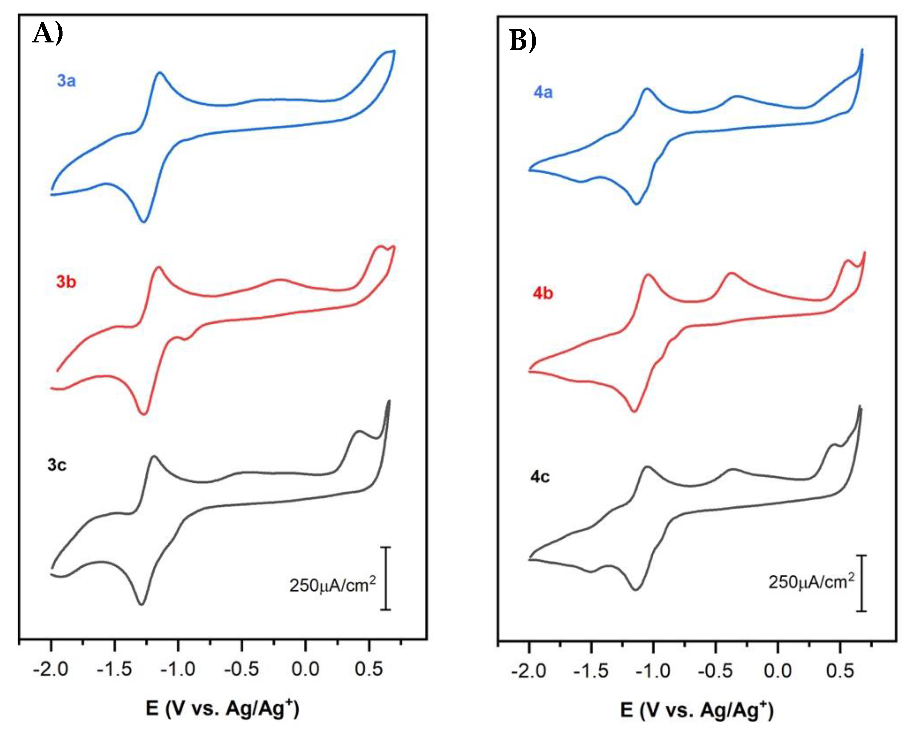
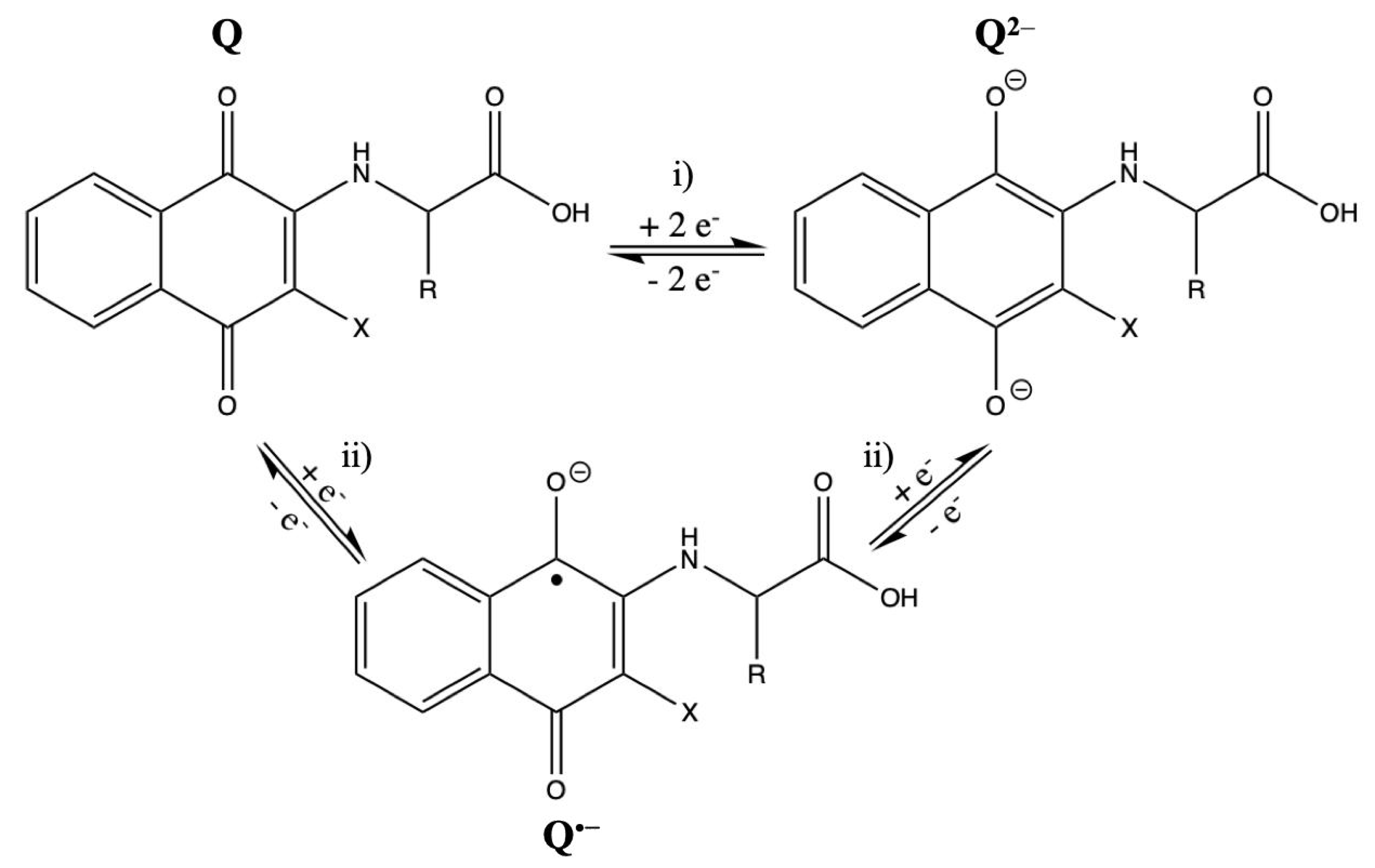
2.2. Electrochemical Studies by Cyclic Voltammetry
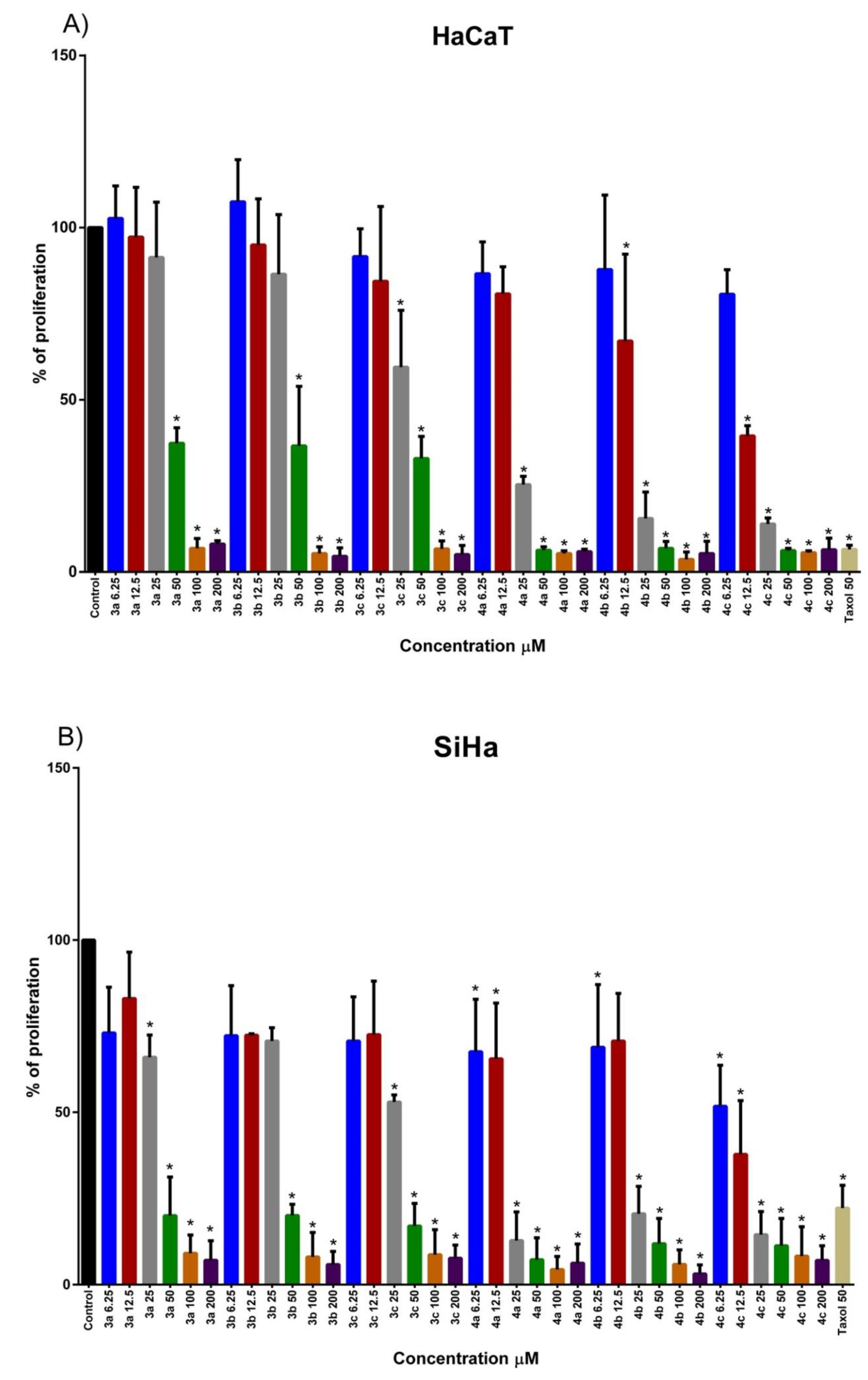
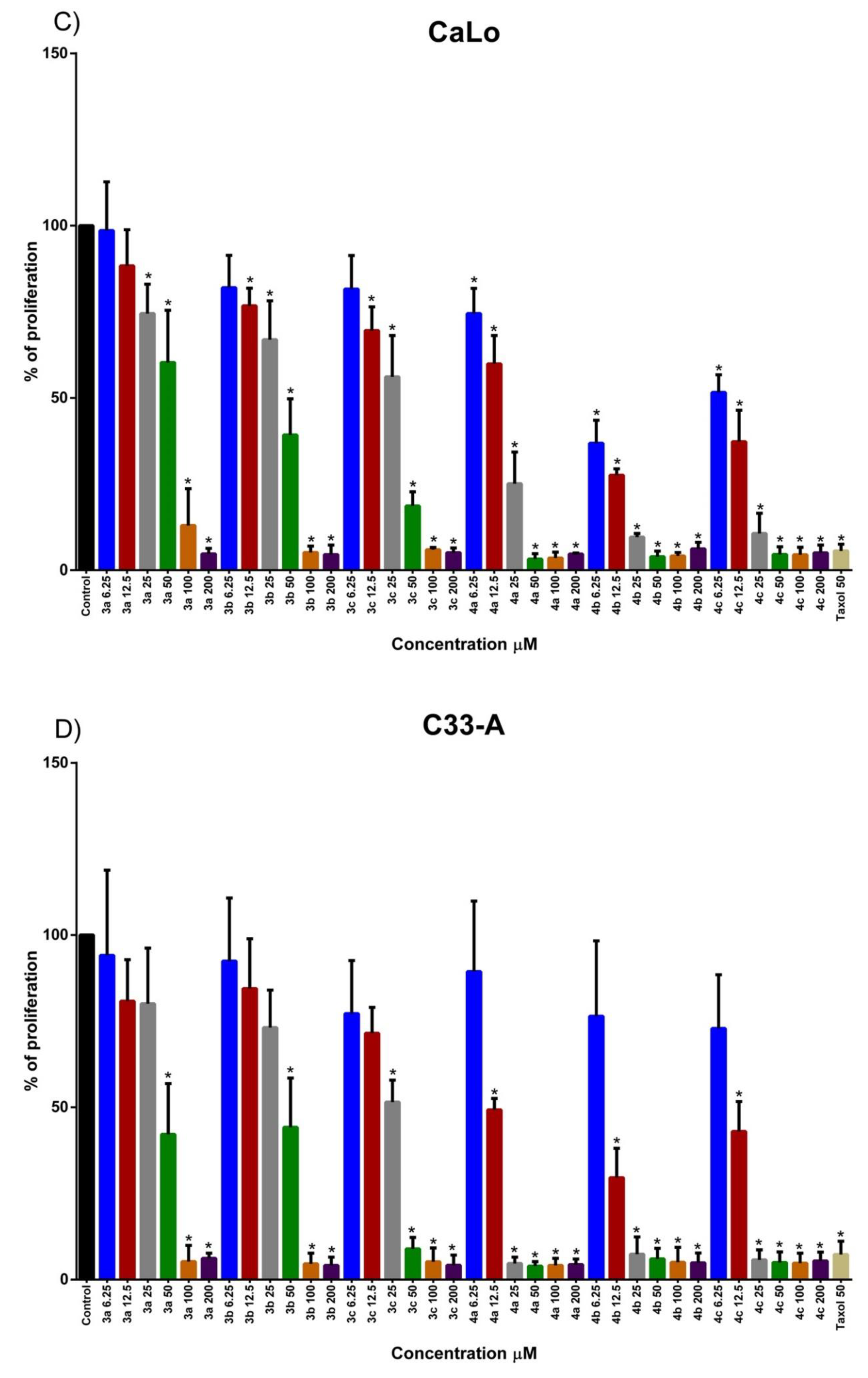
2.3. Effect of Naphthoquinone Amino Acid Derivatives in Cell Proliferation of Tumorigenic Cervical Cancer Cell Lines and Immortal Cell Line
3. Discussion
4. Materials and Methods
4.1. General
4.2. General Procedure for the Synthesis of Compounds 3a–c and 4a–c
4.3. Spectroscopic Characterization of Amino Acid-1,4-Naphthoquinone Derivatives
4.4. Spectroscopic Characterization of Amino Acid-2,3-Dichloronaphthoquinone Derivatives
4.5. Cyclic Voltammetry
4.6. Cell Lines
4.7. Cell Proliferation Analysis
5. Conclusions
Supplementary Materials
Author Contributions
Funding
Acknowledgments
Conflicts of Interest
References
- Sikka, H.C.; Shimabukuro, R.H.; Zweig, G. Studies on Effect of Certain Quinones. Plant Physiol. 1972, 49, 381–384. [Google Scholar] [CrossRef] [PubMed] [Green Version]
- Kosiha, A.; Parthiban, C.; Elango, K.P.; Koshika, A. Synthesis, characterization and DNA binding/cleavage, protein binding and cytotoxicity studies of Co(II), Ni(II), Cu(II) and Zn(II) complexes of aminonaphthoquinone. J. Photochem. Photobiol. B: Boil. 2017, 168, 165–174. [Google Scholar] [CrossRef] [PubMed]
- Chen, J.; Huang, Y.-W.; Liu, G.; Afrasiabi, Z.; Sinn, E.; Padhye, S.; Ma, Y. The cytotoxicity and mechanisms of 1,2-naphthoquinone thiosemicarbazone and its metal derivatives against MCF-7 human breast cancer cells. Toxicol. Appl. Pharmacol. 2004, 197, 40–48. [Google Scholar] [CrossRef]
- Kongkathip, N.; Kongkathip, B.; Siripong, P.; Sangma, C.; Luangkamin, S.; Niyomdecha, M.; Pattanapa, S.; Piyaviriyagul, S.; Kongsaeree, P. Potent antitumor activity of synthetic 1,2-Naphthoquinones and 1,4-Naphthoquinones. Bioorganic Med. Chem. 2003, 11, 3179–3191. [Google Scholar] [CrossRef]
- Li, C.J.; Li, Y.-Z.; Pinto, A.V.; Pardee, A.B. Potent inhibition of tumor survival in vivo by beta -lapachone plus taxol: Combining drugs imposes different artificial checkpoints. Proc. Natl. Acad. Sci. USA 1999, 96, 13369–13374. [Google Scholar] [CrossRef] [Green Version]
- Kongkathip, N.; Luangkamin, S.; Kongkathip, B.; Sangma, C.; Grigg, R.; Kongsaeree, P.; Prabpai, S.; Pradidphol, N.; Piyaviriyagul, S.; Siripong, P. Synthesis of Novel Rhinacanthins and Related Anticancer Naphthoquinone Esters. J. Med. Chem. 2004, 47, 4427–4438. [Google Scholar] [CrossRef] [PubMed]
- A Pedersen, J. On the application of electron paramagnetic resonance in the study of naturally occurring quinones and quinols. Spectrochim. Acta Part A: Mol. Biomol. Spectrosc. 2002, 58, 1257–1270. [Google Scholar] [CrossRef]
- Coates, C.S.; Ziegler, J.; Manz, K.; Good, J.; Kang, B.; Milikisiyants, S.; Chatterjee, R.; Hao, S.; Golbeck, J.H.; Lakshmi, K.V. The Structure and Function of Quinones in Biological Solar Energy Transduction: A Cyclic Voltammetry, EPR, and Hyperfine Sub-Level Correlation (HYSCORE) Spectroscopy Study of Model Naphthoquinones. J. Phys. Chem. B 2013, 117, 7210–7220. [Google Scholar] [CrossRef]
- Smithson, C.S.; Macdonald, D.J.; Letvenuk, T.M.; Carello, C.E.; Jennings, M.; Lough, A.J.; Britten, J.; Decken, A.; Preuss, K.E. A 1,2,3-dithiazolyl-o-naphthoquinone: A neutral radical with isolable cation and anion oxidation states. Dalton Trans. 2016, 45, 9608–9620. [Google Scholar] [CrossRef]
- Tarábek, J.; Wen, J.; Dron, P.I.; Pospíšil, L.; Michl, J. EPR Spectroscopy of Radical Ions of a 2,3-Diamino-1,4-naphthoquinone Derivative. J. Org. Chem. 2018, 83, 5474–5479. [Google Scholar] [CrossRef]
- Wardman, P. Electron transfer and oxidative stress as key factors in the design of drugs selectively active in hypoxia. Curr. Med. Chem. 2001, 8, 739–761. [Google Scholar] [CrossRef]
- Ham, S.W.; Choe, J.-I.; Wang, M.-F.; Peyregne, V.; Carr, B. Fluorinated quinoid inhibitor: Possible ‘pure’ arylator predicted by the simple theoretical calculation. Bioorganic Med. Chem. Lett. 2004, 14, 4103–4105. [Google Scholar] [CrossRef]
- Kar, S.; Wang, M.; Ham, S.W.; Carr, B. Fluorinated Cpd 5, a pure arylating K-vitamin derivative, inhibits human hepatoma cell growth by inhibiting Cdc25 and activating MAPK. Biochem. Pharmacol. 2006, 72, 1217–1227. [Google Scholar] [CrossRef]
- Park, H.; Carr, B.; Li, M.; Ham, S.W. Fluorinated NSC as a Cdc25 inhibitor. Bioorganic Med. Chem. Lett. 2007, 17, 2351–2354. [Google Scholar] [CrossRef] [PubMed]
- Subramaniya, B.R.; Srinivasan, G.; Sadullah, S.S.M.; Davis, N.; Subhadara, L.B.R.; Halagowder, D.; Devaraj, S.N. Apoptosis Inducing Effect of Plumbagin on Colonic Cancer Cells Depends on Expression of COX-2. PLoS ONE 2011. [Google Scholar] [CrossRef] [PubMed] [Green Version]
- Parkin, D.M.; Bray, F.; Ferlay, J.; Pisani, P. Estimating the world cancer burden: Globocan 2000. Int. J. Cancer 2001, 94, 153–156. [Google Scholar] [CrossRef] [PubMed]
- Woodworth, C.D.; Cheng, S.; Simpson, S.; Hamacher, L.; Chow, L.T.; Broker, T.R.; A Dipaolo, J. Recombinant retroviruses encoding human papillomavirus type 18 E6 and E7 genes stimulate proliferation and delay differentiation of human keratinocytes early after infection. Oncogene 1992, 7, 619–626. [Google Scholar]
- Hausen, H.Z. Immortalization of human cells and their malignant conversion by high risk human papillomavirus genotypes. Semin. Cancer Boil. 1999, 9, 405–411. [Google Scholar] [CrossRef] [PubMed]
- Bittner, S.; Gorohovsky, S.; Levi, O.P.-T.; Becker, J.Y. Synthesis, electrochemical and spectral properties of some ω-N-quinonyl amino acids. Amino Acids 2002, 22, 71–93. [Google Scholar] [CrossRef] [PubMed]
- De Moraes, T.A.P.; Filha, M.J.S.; Camara, C.A.; Da Silva, T.M.S.; Soares, B.M.; Bomfim, I.S.; Pessoa, C.; Ximenes, G.C.; Silva, V.; Junior, V.A.S. Synthesis and Cytotoxic Evaluation of a Series of 2-Amino-Naphthoquinones against Human Cancer Cells. Molecules 2014, 19, 13188–13199. [Google Scholar] [CrossRef]
- Janeczko, M.; Demchuk, O.M.; Strzelecka, D.; Kubiński, K.; Maslyk, M. New family of antimicrobial agents derived from 1,4-naphthoquinone. Eur. J. Med. Chem. 2016, 124, 1019–1025. [Google Scholar] [CrossRef] [PubMed]
- Bittner, S.; Gorohovsky, S.; Lozinsky, E.; Shames, A.I. EPR study of anion radicals of various N-quinonyl amino acids. Amino Acids 2000, 19, 439–449. [Google Scholar] [CrossRef] [PubMed]
- Gorohovsky, S.; Bittner, S. Novel N-quinonyl amino acids and their transformation to 3-substituted p-isoxazinones. Amino Acids 2001, 20, 135–144. [Google Scholar] [CrossRef] [PubMed]
- Rivera-Ávalos, E.; De Loera, D.; Araujo-Huitrado, J.G.; Escalante-García, I.L.; Muñoz-Sánchez, M.A.; Hernández, H.; López, J.A.; López, L. Synthesis of Amino Acid-Naphthoquinones and In Vitro Studies on Cervical and Breast Cell Lines. Mol. 2019. [Google Scholar] [CrossRef] [Green Version]
- Roy, S.; Guin, P.S. Solvation of 1-Amino-4-Hydroxy-9,10-Anthraquinone Governs Its Electrochemical Behavior in Non-Aqueous and Aqueous Media: A Cyclic Voltammetry Study. J. Electrochem. Soc. 2014, 162, 124–131. [Google Scholar] [CrossRef]
- Guin, P.S.; Das, S.; Mandal, P.C. Electrochemical Reduction of Quinones in Different Media: A Review. Int. J. Electrochem. 2011, 2011, 1–22. [Google Scholar] [CrossRef] [Green Version]
- Eggins, B.R.; Chambers, J.Q. Proton Effects in the Electrochemistry of the Quinone Hydroquinone System in Aprotic Solvents. J. Electrochem. Soc. 1970. [Google Scholar] [CrossRef]
- Aguilar-Martínez, M.; Cuevas, G.; Jiménez-Estrada, M.; González, I.; Lotina-Hennsen, B.; Macías-Ruvalcaba, N. An Experimental and Theoretical Study of the Substituent Effects on the Redox Properties of 2-[(R-phenyl)amine]-1,4-naphthalenediones in Acetonitrile. J. Org. Chem. 1999, 64, 3684–3694. [Google Scholar] [CrossRef]
- Cauquis, G.; Marbach, G. The Redox Behavior of Biological Quinones and its Relation With the Mitochondrial Respiratory Chain BT–Biological Aspects of Electrochemistry. In Proceedings of the 1st International Symposium, Istituto Superiore di Sanità, Rome, Italy, 31 May–4 June 1971; Birkhäuser Basel: Basel, Switzerland, 1971; pp. 205–214. [Google Scholar] [CrossRef]
- Staley, P.A.; Lopez, E.M.; Clare, L.A.; Smith, D.K. Kinetic Stabilization of Quinone Dianions via Hydrogen Bonding by Water in Aprotic Solvents. J. Phys. Chem. C 2015, 119, 20319–20327. [Google Scholar] [CrossRef]
- Bard, A.J.; Faulkner, L.R. Electrochemical Methods: Fundamentals and Applications, 2nd ed.; John Wiley & Sons: Hoboken, NJ, USA, 2007. [Google Scholar]
- Song, Y.; Buettner, G.R. Thermodynamic and kinetic considerations for the reaction of semiquinone radicals to form superoxide and hydrogen peroxide. Free. Radic. Boil. Med. 2010, 49, 919–962. [Google Scholar] [CrossRef] [Green Version]
- De Abreu, F.C.; Ferraz, P.A.D.L.; Goulart, M.O.F. Some Applications of Electrochemistry in Biomedical Chemistry. Emphasis on the Correlation of Electrochemical and Bioactive Properties. J. Braz. Chem. Soc. 2002, 13, 19–35. [Google Scholar] [CrossRef]
- Hillard, E.A.; De Abreu, F.C.; Ferreira, D.C.M.; Jaouen, G.; Goulart, M.O.F.; Amatore, C. ChemInform Abstract: Electrochemical Parameters and Techniques in Drug Development, with an Emphasis on Quinones and Related Compounds. ChemInform 2008, 39, 2612–2628. [Google Scholar] [CrossRef]
- Pereyra, C.E.; Dantas, R.F.; Ferreira, S.B.; Gomes, L.P.; Silva-Junior, F.P. The diverse mechanisms and anticancer potential of naphthoquinones. Cancer Cell Int. 2019, 19, 207–220. [Google Scholar] [CrossRef] [PubMed] [Green Version]
- Enache, T.A.; Paquim, A.M.C.; Oliveira-Brett, A.M. Amyloid–β peptides time-dependent structural modifications: AFM and voltammetric characterization. Anal. Chim. Acta 2016, 926, 36–47. [Google Scholar] [CrossRef] [PubMed]
- Enache, T.A.; Brett, C. Alzheimer’s disease amyloid beta peptides in vitro electrochemical oxidation. Bioelectrochemistry 2017, 114, 13–23. [Google Scholar] [CrossRef] [PubMed]
- Dourado, A.H.B.; Pastrian, F.; De Torresi, S.I.C. The long and successful journey of electrochemically active amino acids. From fundamental adsorption studies to potential surface engineering tools. Anais da Academia Brasileira de Ciências 2018, 90, 607–630. [Google Scholar] [CrossRef] [Green Version]
- Marastoni, M.; Trapella, C.; Scotti, A.; Fantinati, A.; Ferretti, V.; Marzola, E.; Eleonora, G.; Gavioli, R.; Preti, D. Naphthoquinone amino acid derivatives, synthesis and biological activity as proteasome inhibitors. J. Enzym. Inhib. Med. Chem. 2017, 32, 865–877. [Google Scholar] [CrossRef] [Green Version]
- Wang, J. Combination Treatment of Cervical Cancer Using Folate-Decorated, pH-Sensitive, Carboplatin and Paclitaxel Co-Loaded Lipid-Polymer Hybrid Nanoparticles. Drug Des. Dev. Ther. 2020, 14, 823–832. [Google Scholar] [CrossRef] [Green Version]
- Chekulaeva, M.; Parker, R.; Filipowicz, W. The GW/WG repeats of Drosophila GW182 function as effector motifs for miRNA-mediated repression. Nucleic Acids Res. 2010, 38, 6673–6683. [Google Scholar] [CrossRef] [Green Version]
- Shrestha-Dawadi, P.B.; Bittner, S.; Fridkin, M.; Rahimipour, S. On the Synthesis of Naphthoquinonyl Heterocyclic Amino Acids. Synthesis 1996, 1996, 1468–1472. [Google Scholar] [CrossRef]
Sample Availability: Samples of the compounds 3a–c and 4a–c are available from the authors. |


| Compound | Nq-aa | MAS a (%) | |
|---|---|---|---|
| TEA | KOH | ||
| 3a | 1:1.8 | 85 | 90 |
| 3b | 1:1.5 | 80 | 92 |
| 3c | 1:1.2 | 80 | 88 |
| 4a | 1:1.7 | 95 | 90 |
| 4b | 1:2.5 | 92 | 87 |
| 4c | 1:1.5 | 91 | 89 |
| Compound | Epa | Epc | ΔEp b | E1/2 c | ipa | ipc | |ipa/ipc| |
|---|---|---|---|---|---|---|---|
| (V) | (V) | (V) | (V) | (mA cm−2) | |||
| 3a | −1.15 | −1.27 | 0.13 | −1.21 | 0.28 | −0.30 | 0.95 |
| 3b | −1.16 | −1.27 | 0.12 | −1.21 | 0.28 | −0.30 | 0.95 |
| 3c | −1.19 | −1.30 | 0.11 | −1.24 | 0.26 | −0.27 | 0.95 |
| 4a | −1.04 | −1.15 | 0.11 | −1.10 | 0.22 | −0.24 | 0.92 |
| 4b | −1.05 | −1.19 | 0.14 | −1.12 | 0.21 | −0.25 | 0.83 |
| 4c | −1.05 | −1.17 | 0.12 | −1.11 | 0.23 | −0.27 | 0.85 |
| Cell line | Naphthoquine Amino acid Derivatives | IC50 |
|---|---|---|
| SiIHa | 3a | 40.98 µM |
| 3b | 47.22 µM | |
| 3c | 28.8 µM | |
| 4a | 6.830 µM | |
| 4b | 11.34 µM | |
| 4c | 3.209 µM | |
| CaLo | 3a | 81.08 µM |
| 3b | 56.96 µM | |
| 3c | 25.20 µM | |
| 4a | 7.028 µM | |
| 4b | 2.878 µM | |
| 4c | 2.697 µM | |
| C33-A | 3a | 55.64 µM |
| 3b | 55.56 µM | |
| 3c | 21.36 µM | |
| 4a | ~0.001577 µM | |
| 4b | ~0.0003566 µM | |
| 4c | ~0.003454 µM | |
| HaCaT | 3a | 61.6 µM |
| 3b | 55.29 µM | |
| 3c | 40.82 µM | |
| 4a | 9.882 µM | |
| 4b | 7.970 µM | |
| 4c | ~0.3311 µM |
© 2020 by the authors. Licensee MDPI, Basel, Switzerland. This article is an open access article distributed under the terms and conditions of the Creative Commons Attribution (CC BY) license (http://creativecommons.org/licenses/by/4.0/).
Share and Cite
Córdova-Rivas, S.; Araujo-Huitrado, J.G.; Rivera-Avalos, E.; Escalante-García, I.L.; Durón-Torres, S.M.; López-Hernández, Y.; Hernández-López, H.; López, L.; de Loera, D.; López, J.A. Differential Proliferation Effect of the Newly Synthesized Valine, Tyrosine and Tryptophan–Naphthoquinones in Immortal and Tumorigenic Cervical Cell Lines. Molecules 2020, 25, 2058. https://0-doi-org.brum.beds.ac.uk/10.3390/molecules25092058
Córdova-Rivas S, Araujo-Huitrado JG, Rivera-Avalos E, Escalante-García IL, Durón-Torres SM, López-Hernández Y, Hernández-López H, López L, de Loera D, López JA. Differential Proliferation Effect of the Newly Synthesized Valine, Tyrosine and Tryptophan–Naphthoquinones in Immortal and Tumorigenic Cervical Cell Lines. Molecules. 2020; 25(9):2058. https://0-doi-org.brum.beds.ac.uk/10.3390/molecules25092058
Chicago/Turabian StyleCórdova-Rivas, Sergio, Jorge Gustavo Araujo-Huitrado, Ernesto Rivera-Avalos, Ismailia L. Escalante-García, Sergio M. Durón-Torres, Yamilé López-Hernández, Hiram Hernández-López, Lluvia López, Denisse de Loera, and Jesús Adrián López. 2020. "Differential Proliferation Effect of the Newly Synthesized Valine, Tyrosine and Tryptophan–Naphthoquinones in Immortal and Tumorigenic Cervical Cell Lines" Molecules 25, no. 9: 2058. https://0-doi-org.brum.beds.ac.uk/10.3390/molecules25092058