
Recent Advances in Metal Organic Frameworks Based Surface Enhanced Raman Scattering Substrates: Synthesis and Applications


Abstract
:
1. Introduction
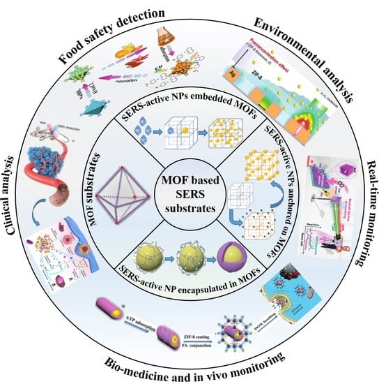
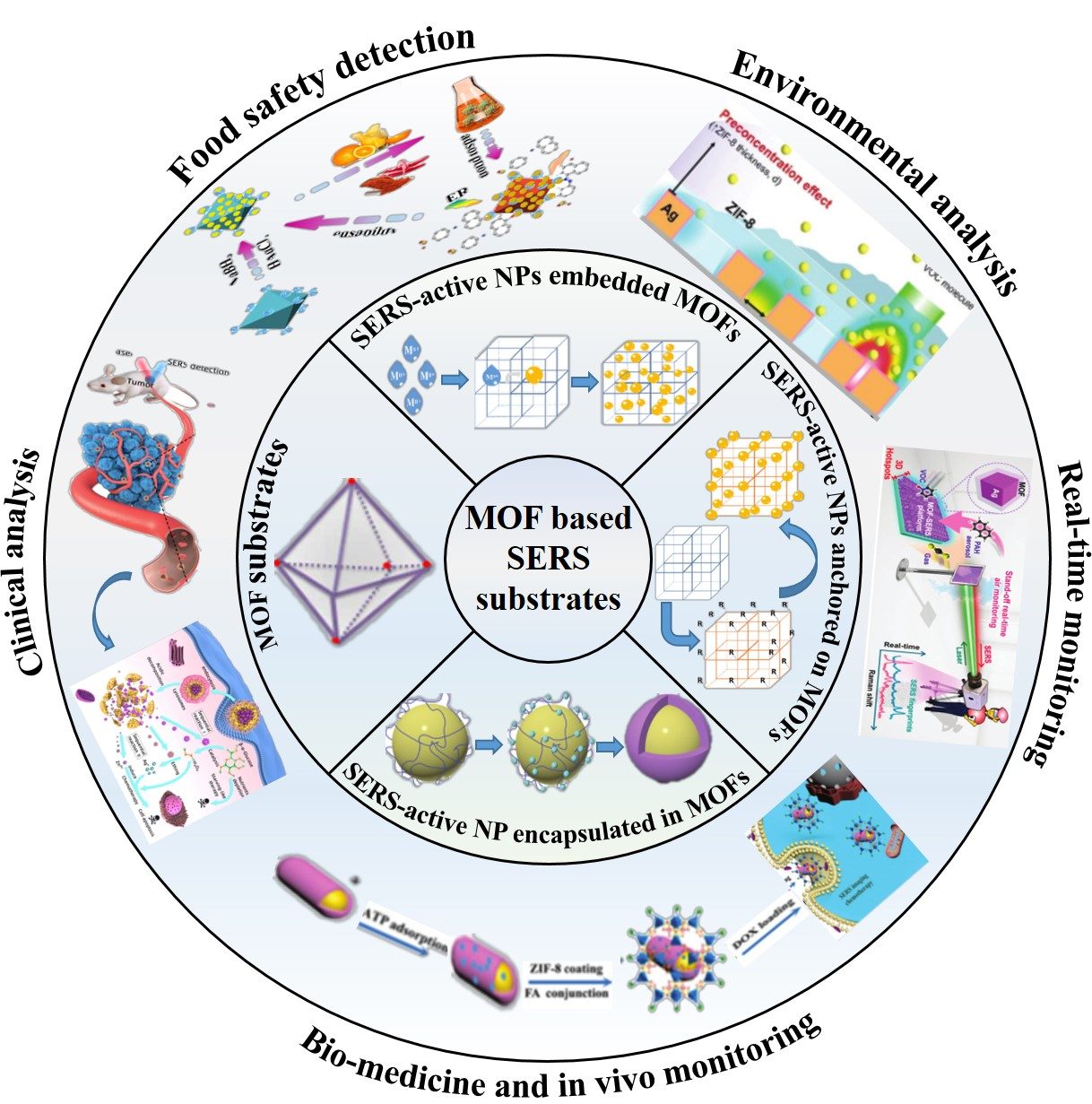
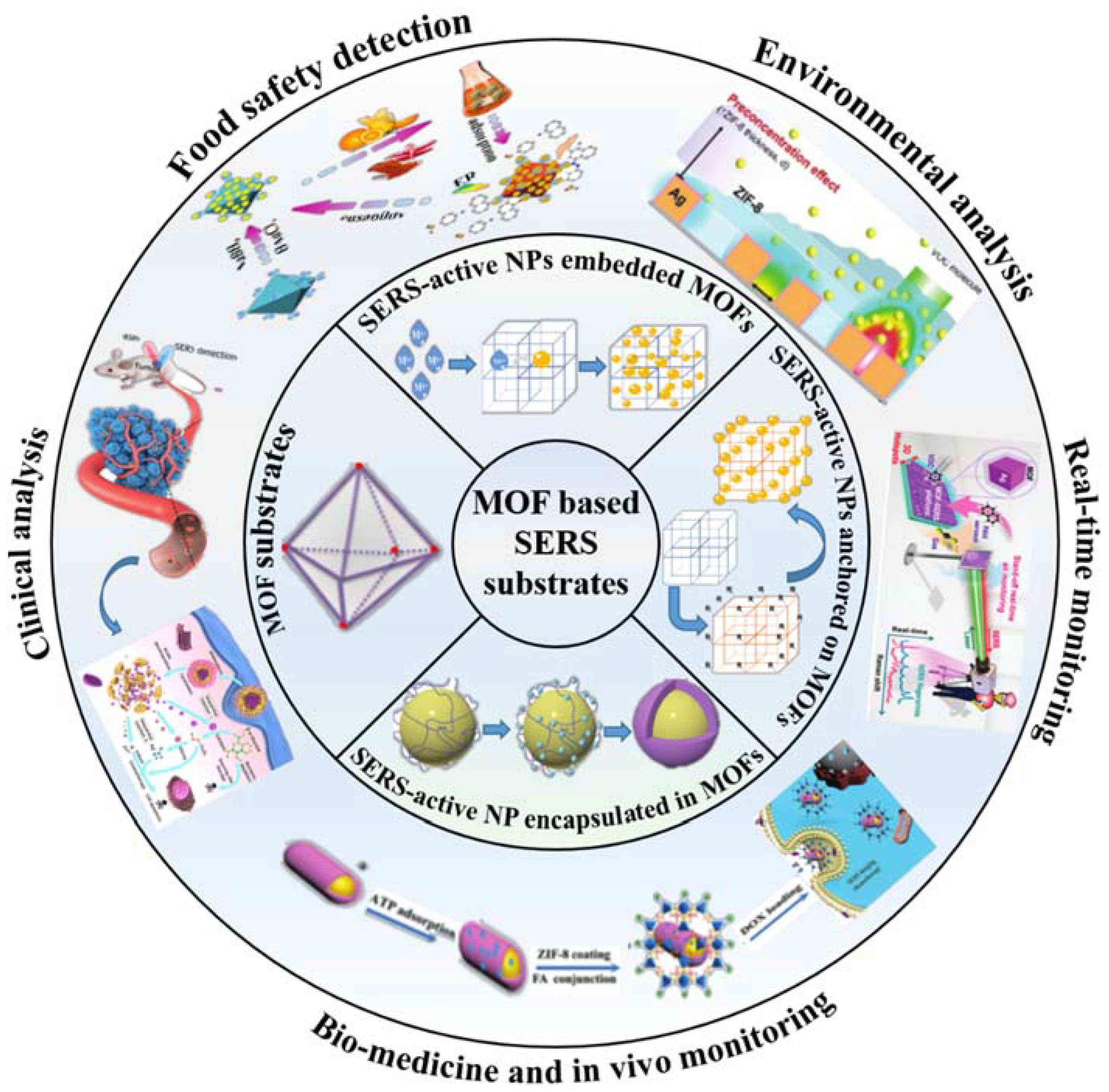
2. MOF Substrates
3. Nano-Composites of MOFs and SERS-Active Metal Substrates
3.1. SERS-Active NPs Embedded MOFs (SERS-Active NPs@ MOFs)
3.1.1. Synthesis
Solution Impregnation Method
Chemical Vapor Deposition (CVD)
Solid Grinding
In Situ Growth of MOFs in the Presence of SERS-Active NPs
3.1.2. Applications
Detection of Chemicals with Low Affinity toward Bare Metal Substrates
Environmental and Clinical Analysis
Catalytic and Biomedical Applications
3.2. SERS-Active NPs Anchored on the Surface of MOFs (SERS-Active NPs/MOFs)
3.2.1. Synthesis
In Situ Synthesis Method
Assembling As-Prepared SERS-Active Metal NPs on the Surface of MOFs
3.2.2. Application
Food Safety Detection
Environmental Analysis
Detection of Biomarkers
Bio-Medicine and In Vivo Monitoring
3.3. SERS-Active NP Encapsulated in MOFs (SERS-Active NP@MOFs)
3.3.1. Synthesis
Seed Growth Method
One-Pot Method
3.3.2. Applications
Food Safety Detection
Detection of Gas and VOCs
Real-Time Monitoring of Catalytic Activity
Drug Carriers
4. Conclusions and Future Perspectives
Author Contributions
Funding
Conflicts of Interest
Abbreviations
| AFP | Alpha-fetoprotein |
| CTAB | Hexadecyl trimethylammonium bromide |
| ELISA | Enzyme linked immunosorbent assay |
| GOx | Glucose oxidase |
| LOD | Limit of detection |
| MBN | 4-mercaptobenzonitrile |
| MG | Malachite green |
| MOF | Metal–organic framework |
| NP | Nanoparticle |
| NR | Nanorod |
| NT-proBNP | N-terminal pro-brain natriuretic peptide |
| PAH | Polycyclic aromatic hydrocarbon |
| PDMS | Polydimethylsiloxane |
| PEG | Poly(ethylene glycol) |
| PVP | Polyvinylpyrrolidone |
| SERS | Surface-enhanced Raman scattering |
| TBZ | Thiabendazole |
| TMB | 3,3′,5,5′-tetramethylbenzidine |
| TP | Tetrapod |
| VOC | Volatile organic compound |
References
- Langer, J.; Dorleta, J.D.A.; Javier, A.; Ramon, A.A.-P.; Baptiste, A.; Jeremy, J.B.; Guillermo, C.B.; Steven, E.J.B.; Anja, B.; Alexandre, G.B.; et al. Present and Future of Surface-Enhanced Raman Scattering. ACS Nano 2020, 14, 28–117. [Google Scholar] [CrossRef] [PubMed] [Green Version]
- Graham, D.; van Duyne, R.; Ren, B. Surface-enhanced Raman scattering. Analyst 2016, 141, 4995. [Google Scholar]
- Fan, M.; Andrade, G.F.S.; Brolo, A.G. A review on recent advances in the applications of surface-enhanced Raman scattering in analytical chemistry. Anal. Chim. Acta 2020, 1097, 1–29. [Google Scholar] [CrossRef] [PubMed]
- Efremov, E.V.; Ariese, F.; Gooijer, C. Achievements in resonance Raman spectroscopy. Review of a technique with a distinct analytical chemistry potential(Review). Anal. Chim. Acta 2008, 606, 119–134. [Google Scholar] [CrossRef]
- Stöckel, S.; Kirchhoff, J.; Neugebauer, U.; Rösch, P.; Popp, J. The application of Raman spectroscopy for the detection and identification of microorganisms(Review). J. Raman Spectrosc. 2016, 47, 89–109. [Google Scholar] [CrossRef]
- Fan, M.; Andrade, G.F.S.; Brolo, A.G. A review on the fabrication of substrates for surface enhanced Raman spectroscopy and their applications in analytical chemistry. Anal. Chim. Acta 2011, 693, 7–25. [Google Scholar] [CrossRef] [PubMed]
- Pilot, R.; Signorini, R.; Durante, C.; Orian, L.; Bhamidipati, M.; Fabris, L. A review on surface-enhanced Raman scattering. Biosensors 2019, 9, 57. [Google Scholar] [CrossRef] [Green Version]
- Cardinal, M.F.; Ende, E.V.; Hackler, R.A.; McAnally, M.O.; Stair, P.C.; Schatz, G.C.; Duyne, R.P. Van Expanding applications of SERS through versatile nanomaterials engineering. Chem. Soc. Rev. 2017, 46, 3886–3903. [Google Scholar] [CrossRef] [Green Version]
- Moneo, A.; Gonzalez-Orive, A.; Bock, S.; Fenero, M.; Herrer, I.L.; Milan, D.C.; Lorenzoni, M.; Nichols, R.J.; Cea, P.; Perez-Murano, F.; et al. Towards molecular electronic devices based on “all-carbon” wires. Nanoscale 2018, 10, 14128–14138. [Google Scholar] [CrossRef] [Green Version]
- Du, S.; Yu, C.; Tang, L.; Lu, L. Applications of SERS in the detection of stress-related substances(Review). Nanomaterials 2018, 8, 757. [Google Scholar] [CrossRef] [Green Version]
- Sharma, B.; Frontiera, R.R.; Henry, A.-I.; Ringe, E.; Duyne, R.P. Van SERS: Materials, applications, and the future. Mater. Today 2012, 15, 16–25. [Google Scholar] [CrossRef]
- Mosier-Boss, P.A. Review of SERS substrates for chemical sensing. Nanomaterials 2017, 7, 142. [Google Scholar] [CrossRef] [PubMed] [Green Version]
- Maier, S.A. Plasmonics: Fundamentals and Applications; Springer: New York, NY, USA, 2007. [Google Scholar]
- Ling, H.K.L.; Lee, Y.H.; Koh, C.S.L.; Phan-Quang, G.C.; Han, X.; Lay, C.L.; Sim, H.Y.F.; Kao, Y.-C.; An, Q.; Yi, X. Designing surface-enhanced Raman scattering (SERS) platforms beyond hotspot engineering: Emerging opportunities in analyte manipulations and hybrid materials. Chem. Soc. Rev. 2019, 48, 731–756. [Google Scholar]
- Wang, X.; Guo, L. SERS Activity of Semiconductors: Crystalline and Amorphous Nanomaterials. Angew. Chemie Int. Ed. 2020, 59, 4231–4239. [Google Scholar] [CrossRef] [PubMed]
- Bernatová, S.; Donato, M.G.; Ježek, J.; Pilát, Z.; Samek, O.; Magazzù, A.; Maragò, O.M.; Zemánek, P.; Gucciardi, P.G. Wavelength-Dependent Optical Force Aggregation of Gold Nanorods for SERS in a Microfluidic Chip. J. Phys. Chem. C 2019, 123, 5608–5615. [Google Scholar] [CrossRef]
- Huang, C.; Li, A.; Chen, X.; Wang, T. Understanding the Role of Metal–Organic Frameworks in Surface-Enhanced Raman Scattering Application. Small 2020, 16, 2004802. [Google Scholar] [CrossRef] [PubMed]
- Lai, H.; Li, G.; Xu, F.; Zhang, Z. Metal-organic frameworks: Opportunities and challenges for surface-enhanced Raman scattering-a review. J. Mater. Chem. C 2020, 8, 2952–2963. [Google Scholar] [CrossRef]
- Zhou, H.-C.J.; Kitagawa, S. Metal–Organic Frameworks (MOFs). Chem. Soc. Rev. 2014, 43, 5415–5418. [Google Scholar] [CrossRef] [Green Version]
- Wang, S.; McGuirk, C.M.; D’Aquino, A.; Mason, J.A.; Mirkin, C.A. Metal-Organic Framework Nanoparticles. Adv. Mater. 2018, 30, e1800202. [Google Scholar] [CrossRef]
- Qiu, S.; Xue, M.; Zhu, G. Metal-organic framework membranes: From synthesis to separation application. Chem. Soc. Rev. 2014, 43, 6116–6140. [Google Scholar] [CrossRef]
- Zhao, X.; Wang, Y.; Li, D.-S.; Bu, X.; Feng, P. Metal-Organic Frameworks for Separation. Adv. Mater. 2018, 30, e1705189. [Google Scholar] [CrossRef] [PubMed]
- He, Y.; Chen, F.; Li, B.; Qian, G.; Zhou, W. Banglin Chen Porous metal-organic frameworks for fuel storage. Coord. Chem. Rev. 2019, 373, 167–198. [Google Scholar] [CrossRef]
- Xu, Y.; Li, Q.; Xue, H.; Pang, H. Metal-organic frameworks for direct electrochemical applications. Coord. Chem. Rev. 2018, 376, 292–318. [Google Scholar] [CrossRef]
- Hu, Y.; Liao, J.; Wang, D.; Li, G. Fabrication of gold nanoparticle-embedded metal-organic framework for highly sensitive surface-enhanced raman scattering detection. Anal. Chem. 2014, 86, 3955–3963. [Google Scholar] [CrossRef] [PubMed]
- Campbell, M.G.; Dinca, M. Metal-Organic Frameworks as Active Materials in Electronic Sensor Devices. Sensors 2017, 17, 1108. [Google Scholar] [CrossRef] [PubMed]
- Horcajada, P.; Gref, R.; Baati, T.; Allan, P.K.; Maurin, G.; Couvreur, P.; Férey, G.; Morris, R.E.; Serre, C. Metal–Organic Frameworks in Biomedicine. Chem. Rev. 2011, 112, 1232–1268. [Google Scholar] [CrossRef] [PubMed]
- Xianlong, Z.; Guoliang, L.; Di, W.; Xiuling, L.; Na, H.; Jian, C.; Guang, C.; Yongning, W. Recent progress in the design fabrication of metal-organic frameworks-based nanozymes and their applications to sensing and cancer therapy. Biosens. Bioelectron. 2019, 137, 178–198. [Google Scholar]
- Chen, S.; Song, Z.; Lyu, J.; Guo, Y.; Lucier, B.E.G.; Luo, W.; Workentin, M.S.; Sun, X.; Huang, Y. Anhydride Post-Synthetic Modification in a Hierarchical Metal-Organic Framework. J. Am. Chem. Soc. 2020. [Google Scholar] [CrossRef]
- Yu, Q.; Li, Z.; Cao, Q.; Qu, S.; Jia, Q. Advances in luminescent metal-organic framework sensors based on post-synthetic modification. Trends Anal. Chem. TRAC 2020, 129. [Google Scholar] [CrossRef]
- Yang, K.; Zong, S.; Zhang, Y.; Qian, Z.; Liu, Y.; Zhu, K.; Li, L.; Li, N.; Wang, Z.; Cui, Y. Array-Assisted SERS Microfluidic Chips for Highly Sensitive and Multiplex Gas Sensing. ACS Appl. Mater. Interfaces 2020, 12, 1395–1403. [Google Scholar] [CrossRef]
- Li, D.; Cao, X.; Zhang, Q.; Ren, X.; Jiang, L.; Li, D.; Deng, W.; Liu, H. Facile: In situ synthesis of core-shell MOF@Ag nanoparticle composites on screen-printed electrodes for ultrasensitive SERS detection of polycyclic aromatic hydrocarbons. J. Mater. Chem. A 2019, 7, 14108–14117. [Google Scholar] [CrossRef]
- Kreno, L.E.; Greeneltch, N.G.; Farha, O.K.; Hupp, J.T.; Van Duyne, R.P. SERS of molecules that do not adsorb on Ag surfaces: A metal-organic framework-based functionalization strategy. Analyst 2014, 139, 4073–4080. [Google Scholar] [CrossRef] [PubMed]
- Ding, Q.; Wang, J.; Chen, X.; Liu, H.; Li, Q.; Wang, Y.; Yang, S. Quantitative and Sensitive SERS Platform with Analyte Enrichment and Filtration Function. Nano Lett. 2020. [Google Scholar] [CrossRef] [PubMed]
- Sim, H.Y.F.; Lee, H.K.; Han, X.; Koh, C.S.L.; Phan-Quang, G.C.; Lay, C.L.; Kao, Y.; Phang, I.Y.; Yeow, E.K.L.; Ling, X.Y. Concentrating Immiscible Molecules at Solid@MOF Interfacial Nanocavities to Drive an Inert Gas–Liquid Reaction at Ambient Conditions. Angew. Chemie Int. Ed. 2018, 57, 17058–17062. [Google Scholar] [CrossRef] [PubMed]
- Guselnikova, O.; Postnikova, P.; Kolska, Z.; Zaruba, K.; Kohoute, M.; Elashnikova, R.; Svorcika, V.; Lyutakova, O. Homochiral metal-organic frameworks functionalized SERS substrate for atto-molar enantio-selective detection. Appl. Mater. Today 2020, 20, 100666. [Google Scholar] [CrossRef]
- Ma, X.; Wen, S.; Xue, X.; Guo, Y.; Jin, J.; Song, W.; Zhao, B. Controllable Synthesis of SERS-Active Magnetic Metal-Organic Framework-Based Nanocatalysts and Their Application in Photoinduced Enhanced Catalytic Oxidation. ACS Appl. Mater. Interfaces 2018, 10, 25726–25736. [Google Scholar] [CrossRef]
- Yu, T.H.; Ho, C.H.; Wu, C.Y.; Chien, C.H.; Lin, C.H.; Lee, S. Metal-organic frameworks: A novel SERS substrate. J. Raman Spectrosc. 2013, 44, 1506–1511. [Google Scholar] [CrossRef]
- Fu, J.H.; Zhong, Z.; Xie, D.; Guo, Y.J.; Kong, D.X.; Zhao, Z.X.; Zhao, Z.X.; Li, M. SERS-Active MIL-100(Fe) Sensory Array for Ultrasensitive and Multiplex Detection of VOCs. Angew. Chemie Int. Ed. 2020. [Google Scholar] [CrossRef]
- Sun, H.; Cong, S.; Zheng, Z.; Wang, Z.; Chen, Z.; Zhao, Z. Metal-Organic Frameworks as Surface Enhanced Raman Scattering Substrates with High Tailorability. J. Am. Chem. Soc. 2019, 141, 870–878. [Google Scholar] [CrossRef]
- Cao, X.; Hong, S.; Jiang, Z.; She, Y.; Wang, S.; Zhang, C.; Li, H.; Jin, F.; Jin, M.; Wang, J. SERS-active metal-organic frameworks with embedded gold nanoparticles. Analyst 2017, 142, 2640–2647. [Google Scholar] [CrossRef]
- Yang, Q.; Xu, Q.; Jiang, H.-L. Metal-organic frameworks meet metal nanoparticles: Synergistic effect for enhanced catalysis. Chem. Soc. Rev. 2017, 46, 4774–4808. [Google Scholar] [CrossRef] [PubMed]
- Hermes, S.; Schröter, M.K.; Schmid, R.; Khodeir, L.; Muhler, M.; Tissler, A.; Fischer, R.W.; Fischer, R.A. Metal@MOF: Loading of highly porous coordination polymers host lattices by metal organic chemical vapor deposition. Angew. Chem. Int. Ed. 2005, 44, 6237–6241. [Google Scholar] [CrossRef] [PubMed]
- Jiang, H.-L.; Liu, B.; Akita, T.; Haruta, M.; Sakurai, H.; Xu, Q. Au@ZIF-8: CO Oxidation over Gold Nanoparticles Deposited to Metal-Organic Framework. J. Am. Chem. Soc. 2009, 131, 11302–11303. [Google Scholar] [CrossRef] [PubMed]
- Ishida, T.; Kawakita, N.; Akita, T.; Haruta, M. One-potN-alkylation of primary amines to secondary amines by gold clusters supported on porous coordination polymers. Gold Bull. 2009, 42, 267–274. [Google Scholar] [CrossRef] [Green Version]
- Ishida, T.; Nagaoka, M.; Akita, T.; Haruta, M. Deposition of Gold Clusters on Porous Coordination Polymers by Solid Grinding and Their Catalytic Activity in Aerobic Oxidation of Alcohols. Chem. A Eur. J. 2008, 14, 8456–8460. [Google Scholar] [CrossRef]
- Sugikawa, K.; Furukawa, Y.; Sada, K. SERS-active metal-organic frameworks embedding gold nanorods. Chem. Mater. 2011, 23, 3132–3134. [Google Scholar] [CrossRef]
- Sugikawa, K.; Nagata, S.; Furukawa, Y.; Kokado, K.; Sada, K. Stable and functional gold nanorod composites with a metal-organic framework crystalline shell. Chem. Mater. 2013, 25, 2565–2570. [Google Scholar] [CrossRef]
- Hu, Y.; Cheng, H.; Zhao, X.; Wu, J.; Muhammad, F.; Lin, S.; He, J.; Zhou, L.; Zhang, C.; Deng, Y.; et al. Surface-Enhanced Raman Scattering Active Gold Nanoparticles with Enzyme-Mimicking Activities for Measuring Glucose and Lactate in Living Tissues. ACS Nano 2017, 11, 5558–5566. [Google Scholar] [CrossRef]
- Jiang, Z.; Gao, P.; Yang, L.; Huang, C.; Li, Y. Facile in Situ Synthesis of Silver Nanoparticles on the Surface of Metal-Organic Framework for Ultrasensitive Surface-Enhanced Raman Scattering Detection of Dopamine. Anal. Chem. 2015, 87, 12177–12182. [Google Scholar] [CrossRef]
- Sun, Z.J.; Jiang, Z.W.; Li, Y.F. Poly(dopamine) assisted in situ fabrication of silver nanoparticles/metal-organic framework hybrids as SERS substrates for folic acid detection. RSC Adv. 2016, 6, 79805–79810. [Google Scholar] [CrossRef]
- Wu, L.; Pu, H.; Huang, L.; Sun, D.W. Plasmonic nanoparticles on metal-organic framework: A versatile SERS platform for adsorptive detection of new coccine and orange II dyes in food. Food Chem. 2020, 328, 127105. [Google Scholar] [CrossRef] [PubMed]
- Wang, Q.; Shi, Z.; Wang, Z.; Zhao, Y.; Li, J.; Hu, H.; Bai, Y.; Xu, Z.; Zhangsun, H.; Wang, L. Rapid simultaneous adsorption and SERS detection of acid orange II using versatile gold nanoparticles decorated NH2-MIL-101(Cr). Anal. Chim. Acta 2020, 1129, 126–135. [Google Scholar] [CrossRef] [PubMed]
- He, Y.; Wang, Y.; Yang, X.; Xie, S.; Yuan, R.; Chai, Y. Metal Organic Frameworks Combining CoFe2O4 Magnetic Nanoparticles as Highly Efficient SERS Sensing Platform for Ultrasensitive Detection of N-Terminal Pro-Brain Natriuretic Peptide. ACS Appl. Mater. Interfaces 2016, 8, 7683–7690. [Google Scholar] [CrossRef] [PubMed]
- Sun, D.; Qi, G.; Ma, K.; Qu, X.; Xu, W.; Xu, S.; Jin, Y. Tumor Microenvironment-Activated Degradable Multifunctional Nanoreactor for Synergistic Cancer Therapy and Glucose SERS Feedback. iScience 2020, 23, 101274. [Google Scholar] [CrossRef] [PubMed]
- Xuan, T.; Gao, Y.; Cai, Y.; Guo, X.; Wen, Y.; Yang, H. Fabrication and characterization of the stable Ag-Au-metal-organic-frameworks: An application for sensitive detection of thiabendazole. Sens. Actuators B Chem. 2019, 293, 289–295. [Google Scholar] [CrossRef]
- Kuang, X.; Ye, S.; Li, X.; Ma, Y.; Zhang, C.; Tang, B. A new type of surface-enhanced Raman scattering sensor for the enantioselective recognition of d/l-cysteine and d/l-asparagine based on a helically arranged Ag NPs@homochiral MOF. Chem. Commun. 2016, 52, 5432–5435. [Google Scholar] [CrossRef] [PubMed]
- Zhang, Y.; Hu, Y.; Li, G.; Zhang, R. A composite prepared from gold nanoparticles and a metal organic framework (type MOF-74) for determination of 4-nitrothiophenol by surface-enhanced Raman spectroscopy. Microchim. Acta 2019, 186. [Google Scholar] [CrossRef]
- Li, Q.; Gong, S.; Zhang, H.; Huang, F.; Zhang, L.; Li, S. Tailored necklace-like Ag@ZIF-8 core/shell heterostructure nanowires for high-performance plasmonic SERS detection. Chem. Eng. J. 2019, 371, 26–33. [Google Scholar] [CrossRef]
- Liao, J.; Wang, D.; Liu, A.; Hu, Y.; Li, G. Controlled stepwise-synthesis of core-shell Au@MIL-100 (Fe) nanoparticles for sensitive surface-enhanced Raman scattering detection. Analyst 2015, 140, 8165–8171. [Google Scholar] [CrossRef]
- Guselnikova, O.; Postnikov, P.; Elashnikov, R.; Miliutina, E.; Svorcik, V.; Lyutakov, O. Metal-organic framework (MOF-5) coated SERS active gold gratings: A platform for the selective detection of organic contaminants in soil. Anal. Chim. Acta 2019, 1068, 70–79. [Google Scholar] [CrossRef]
- Lai, H.; Shang, W.; Yun, Y.; Chen, D.; Wu, L.; Xu, F. Uniform arrangement of gold nanoparticles on magnetic core particles with a metal-organic framework shell as a substrate for sensitive and reproducible SERS based assays: Application to the quantitation of Malachite Green and thiram. Microchim. Acta 2019, 186, 1–9. [Google Scholar] [CrossRef] [PubMed]
- Men, D.; Feng, S.; Liu, G.; Hang, L.; Zhang, T. A Sensitive “Optical Nose” for Detection of Volatile Organic Molecules Based on Au@MOFs Nanoparticle Arrays through Surface-Enhanced Raman Scattering. Part. Part. Syst. Charact. 2020, 37, 1–8. [Google Scholar] [CrossRef]
- Phan-Quang, G.C.; Yang, N.; Lee, H.K.; Sim, H.Y.F.; Koh, C.S.L.; Kao, Y.C.; Wong, Z.C.; Tan, E.K.M.; Miao, Y.E.; Fan, W.; et al. Tracking airborne molecules from afar: Three-dimensional metal-organic framework-surface-enhanced raman scattering platform for stand-off and real-time atmospheric monitoring. ACS Nano 2019, 13, 12090–12099. [Google Scholar] [CrossRef] [PubMed]
- He, J.; Dong, J.; Hu, Y.; Li, G.; Hu, Y. Design of Raman tag-bridged core-shell Au@Cu3(BTC)2 nanoparticles for Raman imaging and synergistic chemo-photothermal therapy. Nanoscale 2019, 11, 6089–6100. [Google Scholar] [CrossRef] [PubMed]
- Osterrieth, J.W.M.; Wright, D.; Noh, H.; Kung, C.W.; Vulpe, D.; Li, A.; Park, J.E.; Van Duyne, R.P.; Moghadam, P.Z.; Baumberg, J.J.; et al. Core-Shell Gold Nanorod@Zirconium-Based Metal-Organic Framework Composites as in Situ Size-Selective Raman Probes. J. Am. Chem. Soc. 2019, 141, 3893–3900. [Google Scholar] [CrossRef] [PubMed]
- Zheng, G.; de Marchi, S.; López-Puente, V.; Sentosun, K.; Polavarapu, L.; Pérez-Juste, I.; Hill, E.H.; Bals, S.; Liz-Marzán, L.M.; Pastoriza-Santos, I.; et al. Encapsulation of Single Plasmonic Nanoparticles within ZIF-8 and SERS Analysis of the MOF Flexibility. Small 2016, 12, 3935–3943. [Google Scholar] [CrossRef] [PubMed]
- Ma, X.; Liu, H.; Wen, S.; Xie, Q.; Li, L.; Wang, X.; Zhao, B.; Song, W. Ultra-sensitive SERS detection, rapid selective adsorption and degradation of cationic dyes on multifunctional magnetic metal-organic framework-based composite. Nanotechnology 2020, 31, 465705. [Google Scholar] [CrossRef] [PubMed]
- Chen, L.; Duan, B.; Luo, Q.; Gu, Z.; Liu, J.; Duan, C. Facet-dependent catalytic activity of ZIF-8 nanocubes and rhombic dodecahedra based on tracing substrate diffusion in pores by SERS: A case study for surface catalysis of MOFs. Catal. Sci. Technol. 2016, 6, 1616–1620. [Google Scholar] [CrossRef]
- Yusran, Y.; Xu, D.; Fang, Q.; Zhang, D.; Qiu, S. MOF-derived Co@N-C nanocatalyst for catalytic reduction of 4-nitrophenol to 4-aminophenol. Microporous Mesoporous Mater. 2017, 241, 346–354. [Google Scholar] [CrossRef]
- Zhang, Z.; Shi, H.; Wu, Q.; Bu, X.; Yang, Y.; Zhang, J.; Huang, Y. MOF-derived CeO2/Au@SiO2 hollow nanotubes and their catalytic activity toward 4-nitrophenol reduction. New J. Chem. 2019, 43, 4581–4589. [Google Scholar] [CrossRef]
- De Marchi, S.; Vázquez-Iglesias, L.; Bodelón, G.; Pérez-Juste, I.; Fernández, L.Á.; Pérez-Juste, J.; Pastoriza-Santos, I. Programmable Modular Assembly of Functional Proteins on Raman-Encoded Zeolitic Imidazolate Framework-8 (ZIF-8) Nanoparticles as SERS Tags. Chem. Mater. 2020, 32, 5739–5749. [Google Scholar] [CrossRef]
- Carrillo-Carrión, C.; Martínez, R.; Navarro Poupard, M.F.; Pelaz, B.; Polo, E.; Arenas-Vivo, A.; Olgiati, A.; Taboada, P.; Soliman, M.G.; Catalán, Ú.; et al. Aqueous Stable Gold Nanostar/ZIF-8 Nanocomposites for Light-Triggered Release of Active Cargo Inside Living Cells. Angew. Chemie Int. Ed. 2019, 58, 7078–7082. [Google Scholar] [CrossRef] [PubMed]


| Type | MOF Based Substrates | Synthesis | Target | LOD (μM) | Matrices | Reference |
|---|---|---|---|---|---|---|
| NPs@MOF | AuNPs/MOF-199, AuNPs/Uio-66, and AuNPs/Uio-67 | Solution impregnation method | Acetamiprid | 2 × 10⁻², 9.0 × 10⁻³, and 2.0 × 10⁻² | - | [41] |
| NPs@MOF | AuNPs/MIL-101 | Solution impregnation method | Benzadine | 9.208 × 10⁻⁴ | Environmental water, serum | [25] |
| NPs@MOF | AuNPs@MIL-101 | Solution impregnation method | Glucose, lactate | 4.2, 5.0 | - | [49] |
| Type | MOF Based Substrates | Synthesis | Target | LOD (μM) | Matrices | Reference |
|---|---|---|---|---|---|---|
| NPs anchored on MOF | Ag NPs/MIL-101 (Fe) | In situ synthesis | Dopamine | 3.2 × 10⁻⁷ | Urine | [50] |
| NPs anchored on MOF | Ag NPs/MIL-101(Cr) | In situ synthesis | Folic acid | 0.3 ± 0.02 | Urine | [51] |
| NPs anchored on MOF | Au NPs/UiO-66(NH2) | In situ synthesis | New coccine, orange II | 0.664, 0.1668 | Soft drink, paprika | [52] |
| NPs anchored on MOF | Au NPs/NH2-MIL-101(Cr) | In situ synthesis | Acid orange II | 0.1527 | Orange juice, Chili powder | [53] |
| NPs anchored on MOF | Au TPs/IRMOF-3 | Covalent bond | NT-proBNP | 0.75 fg/mL | - | [54] |
| NPs anchored on MOF | Ag NPs/HKUST-1(Cu) | Electrodeposition | PAHs | 1.5 × 10⁻⁴ to 2 × 10⁻² | Environmental water | [32] |
| NPs anchored on MOF | Ag NPs/ZIF8 | Electrostatic affinity | Glucose | - | Cell | [55] |
| NPs anchored on MOF | Ag@Au NPs/Mil-101(Fe) | In situ synthesis | Thiabendazole | 0.2484 | Juice | [56] |
| Type | MOF Based Substrates | Synthesis | Target | LOD (μM) | Matrices | Reference |
|---|---|---|---|---|---|---|
| NP@MOF | Au NP@MOF-74 | One-pot method | 4-nitrothiophenol | 6.9 × 10⁻² | - | [58] |
| NP@MOF | Ag NP@ZIF8 | Seed growth method | Crystal violet | 10−2 | ppb level | [59] |
| NP@MOF | Au NP@MIL-100(Fe) | step-by-step method | MG | 8.0 × 10⁻³ | Aquaculture water, fish | [60] |
| NP@MOF | Au NP@MOF-5 | Seed growth method | Organophosphorus pesticides | 10−8 | Soil | [61] |
| NP@MOF | Fe3O4@Au NPs @MIL-100(Fe) | Three-step method | MG, thiram | 4.4 × 10⁻³, 1.5 × 10⁻² | Water | [62] |
| NP@MOF | Ag NP@ZIF8 | Seed growth method | Benzene | 6.917 × 10³ | - | [33] |
| NP@MOF | Au NP@ZIF8 | Seed growth method | Toluene | 1.085 × 10³ | - | [63] |
| NP@MOF | Au@Ag NP@ZIF8 | Seed growth method | Benzaldehyde, 3-ethylbenzaldehyde | 9.423 × 10⁻³ | - | [31] |
| NP@MOF | Ag NP@ZIF8 | Seed growth method | PAHs, CO2 | - | Air/gaseous environment | [64] |
| NP@MOF | Au NP@Cu3(BTC)2 | step-by-step method | Load drug | - | Cell | [65] |
| Type | Advantages | Disadvantages |
|---|---|---|
| MOFs | Diverse structure, molecular selectivity, good for mechanism research | Numbers are limited |
| SERS-active NPs embedded in MOFs | Easy to synthesis | The morphology, quantity, and location of NPs inside MOFs are hard to control, the structure of MOFs might be destroyed |
| SERS-active NPs anchored on MOFs | Good SERS performance, easy to synthesis | Difficult to maintaining the structure of MOFs |
| SERS-active NP encapsulated in MOFs | Good stability, selectivity, and adsorption capacity | Weak SERS enhancement capacity |
Publisher’s Note: MDPI stays neutral with regard to jurisdictional claims in published maps and institutional affiliations. |
© 2021 by the authors. Licensee MDPI, Basel, Switzerland. This article is an open access article distributed under the terms and conditions of the Creative Commons Attribution (CC BY) license (http://creativecommons.org/licenses/by/4.0/).
Share and Cite
Wang, P.; Sun, Y.; Li, X.; Wang, L.; Xu, Y.; Li, G. Recent Advances in Metal Organic Frameworks Based Surface Enhanced Raman Scattering Substrates: Synthesis and Applications. Molecules 2021, 26, 209. https://0-doi-org.brum.beds.ac.uk/10.3390/molecules26010209
Wang P, Sun Y, Li X, Wang L, Xu Y, Li G. Recent Advances in Metal Organic Frameworks Based Surface Enhanced Raman Scattering Substrates: Synthesis and Applications. Molecules. 2021; 26(1):209. https://0-doi-org.brum.beds.ac.uk/10.3390/molecules26010209
Chicago/Turabian StyleWang, Panxue, Yan Sun, Xiang Li, Li Wang, Ying Xu, and Guoliang Li. 2021. "Recent Advances in Metal Organic Frameworks Based Surface Enhanced Raman Scattering Substrates: Synthesis and Applications" Molecules 26, no. 1: 209. https://0-doi-org.brum.beds.ac.uk/10.3390/molecules26010209