
Epilobium angustifolium L. Extracts as Valuable Ingredients in Cosmetic and Dermatological Products


 , , ,
, , ,
Abstract
: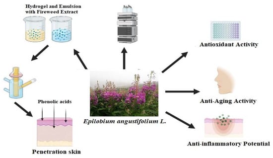
1. Introduction
2. Results
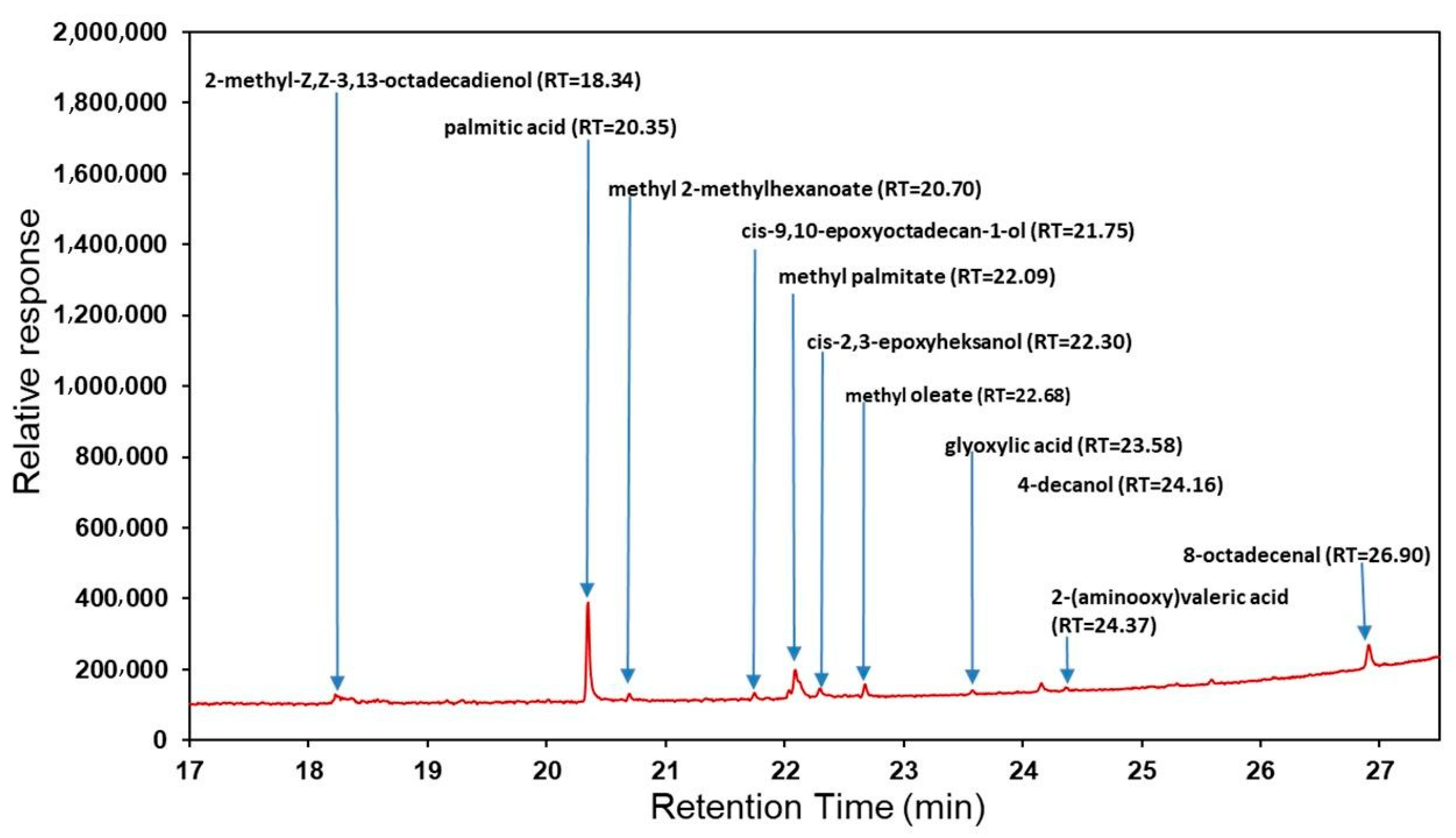
2.1. Determination of Biologically Active Compounds
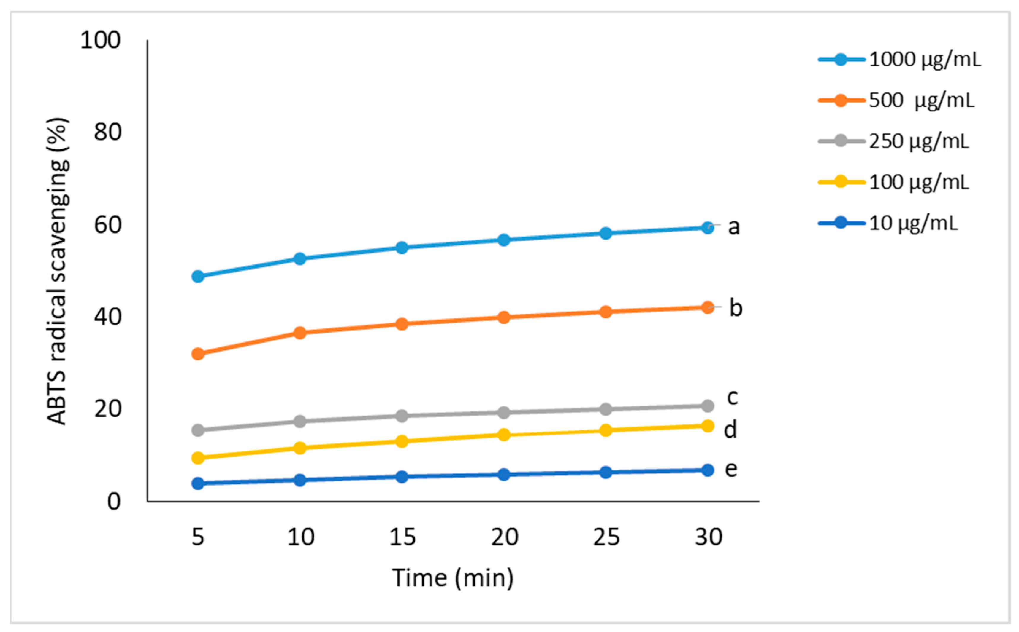
2.2. Assessment of Antioxidant Activity
DPPH Radical Scavenging Assay
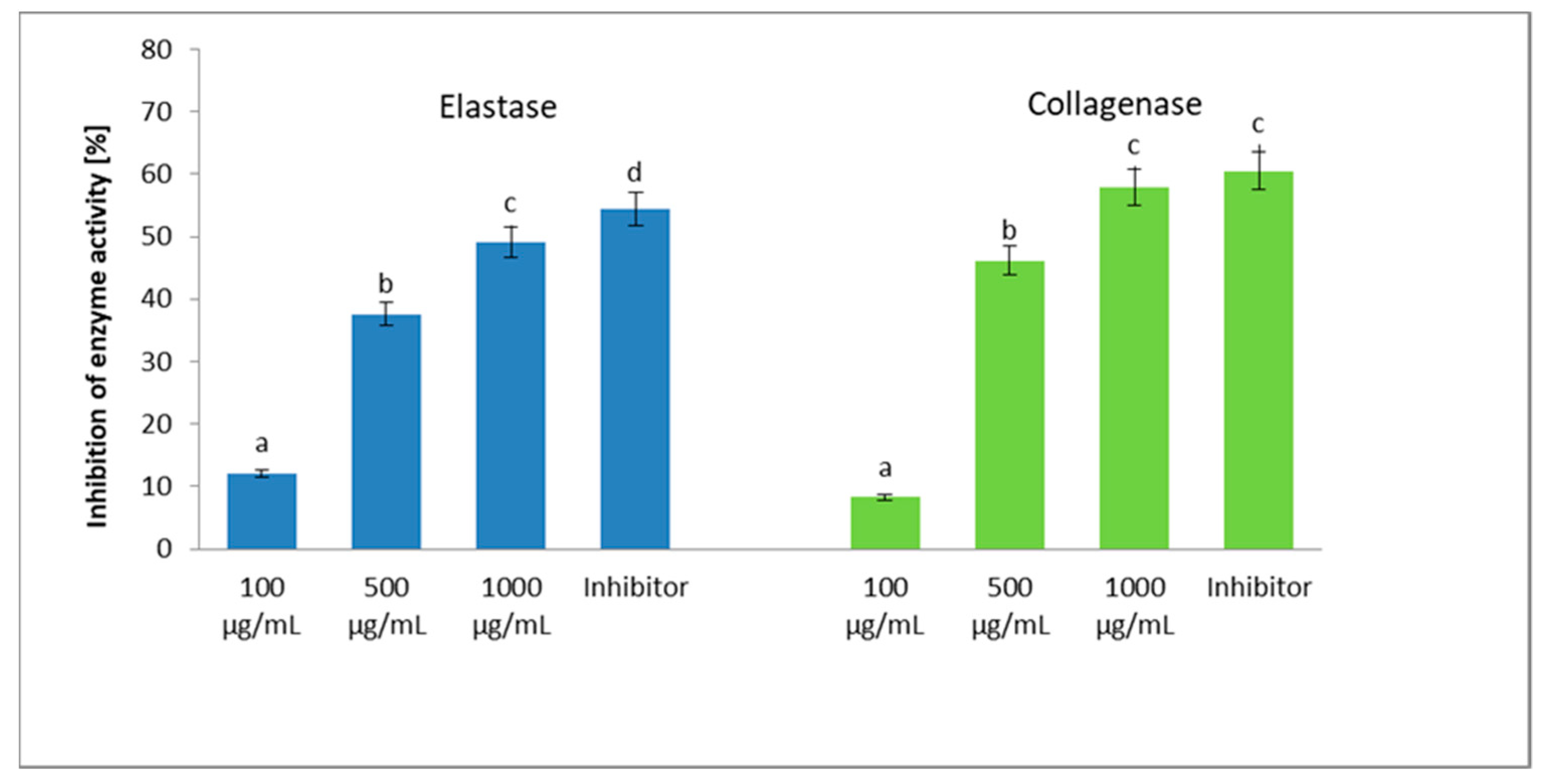
2.3. Assessment of Anti-Collagenase and Anti-Elastase Activity
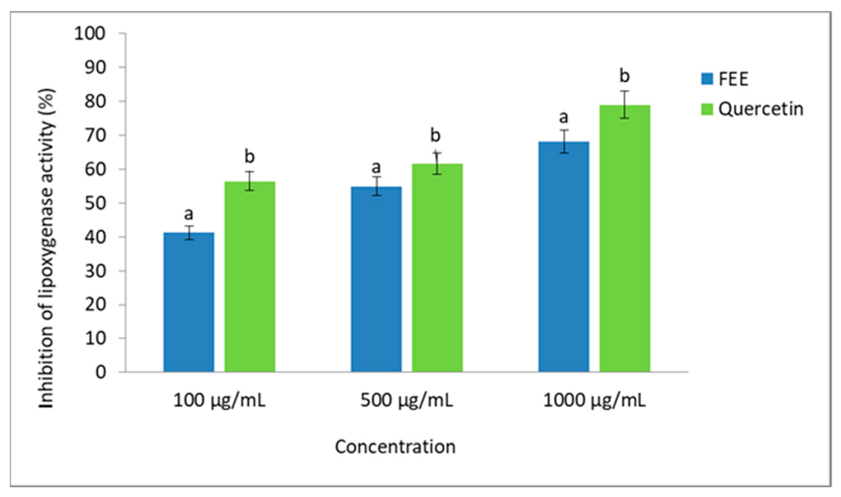
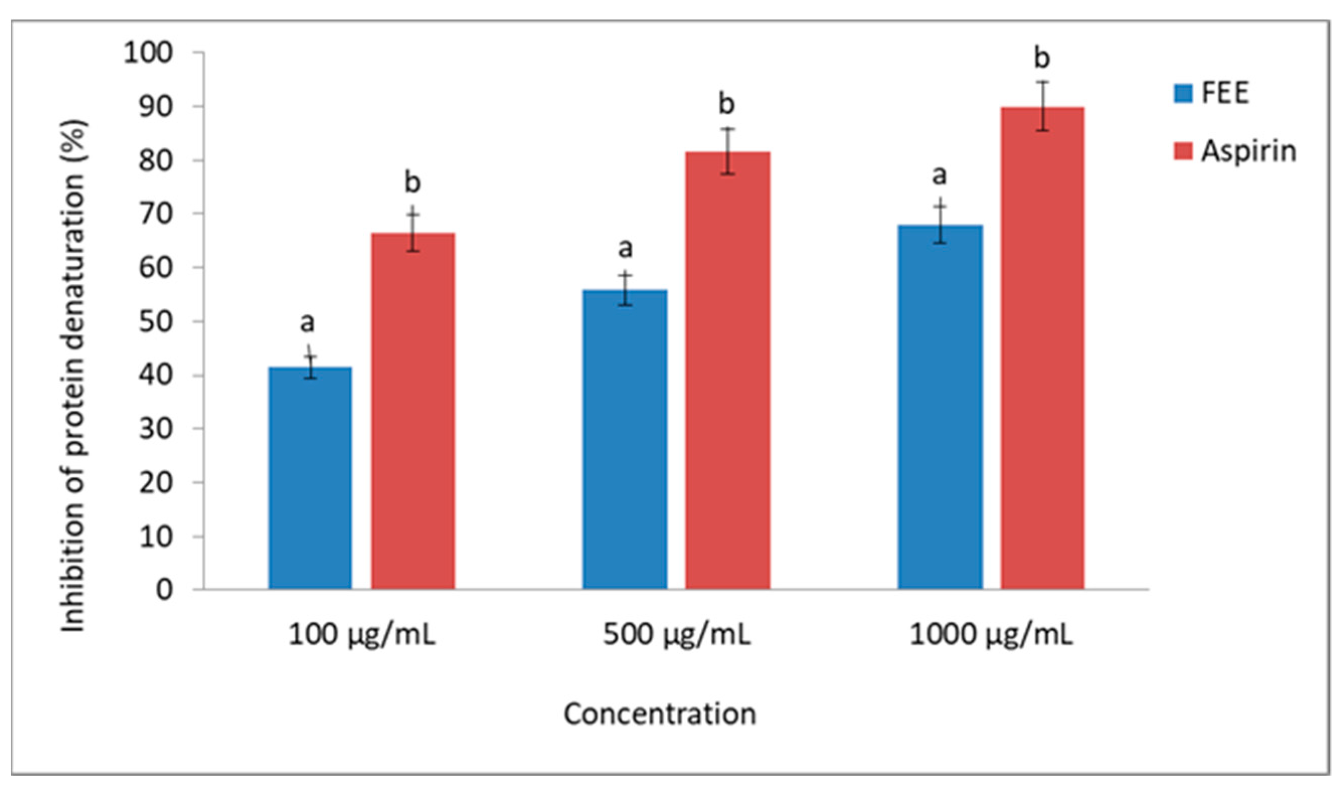
2.4. Assessment of Anti-Inflammatory Potential
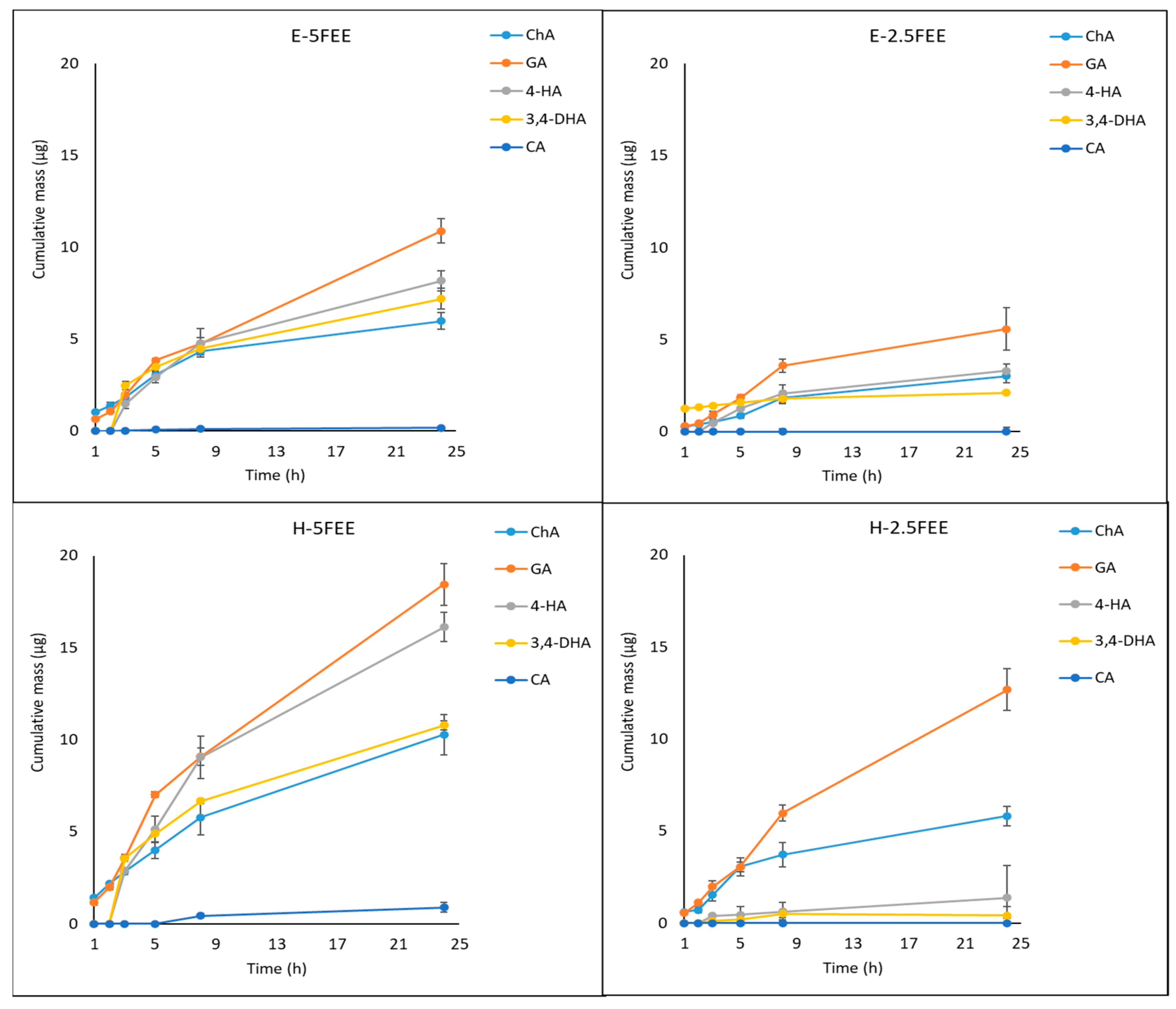
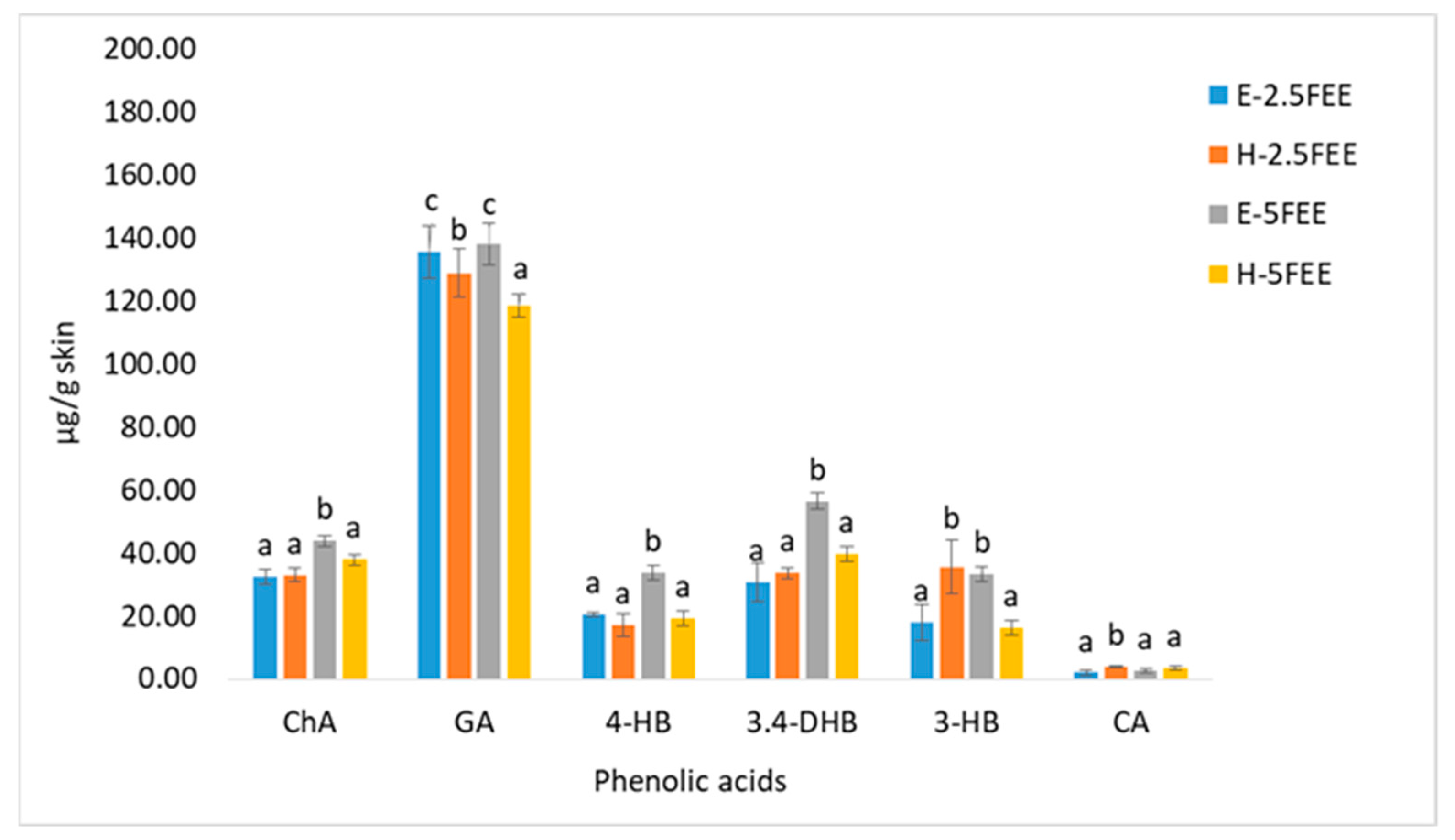
2.5. Skin Penetration
3. Discussion
3.1. Chemical Characterization of the FEE and Its Antioxidant Capacity
3.2. Anti-Collagenase and Anti-Elastase Activity
3.3. Anti-Inflammatory Potential
3.4. Penetration Skin
4. Materials and Methods
4.1. Chemicals
4.2. Plant Materials
4.3. Determination of Biologically Active Compounds
4.3.1. Determination of Bioactive Compounds by GC-MS and HPLC
4.3.2. Total Phenolic Content Determination (TPC)
4.3.3. Total Flavonoids Content Determination (TFC)
4.3.4. Determination of Chlorophyll Content
Chlorophyll b = 22.9 (A645) − 4.68 (A663),
Total chlorophyll (a + b) = 20.2 (A645) + 8.02 (A663),
- A663—absorbance measured at wavelength 663;
- A645—absorbance measured at wavelength 645.
4.4. Evaluation of the Antioxidant Activity
4.4.1. DPPH Radical Scavenging Assay
- As—absorbance of the tested sample;
- Ac—absorbance of the control sample.
4.4.2. ABTS Radical Scavenging Assay
- As—absorbance of the tested sample;
- Ac—absorbance of the control sample.
4.5. Assessment of Antiaging Properties
4.5.1. Determination of Anti-Elastase Activity
4.5.2. Determination of Anti-Collagenase Activity
4.6. Determination of Anti-Inflammatory Properties
4.6.1. Inhibition of Lipoxygenase Activity
- As—absorbance of the tested sample;
- Ac—absorbance of the control sample.
4.6.2. Inhibition of Protein Denaturation
- As—absorbance of the tested sample;
- Ac—absorbance of the negative control.
4.7. Hydrogel End Emulsion Preparation
4.8. In Vitro Skin Permeation Studies
4.9. Statistical Analysis
5. Conclusions
Author Contributions
Funding
Institutional Review Board Statement
Informed Consent Statement
Data Availability Statement
Conflicts of Interest
Sample Availability
References
- Nicolai, M.; Mota, J.; Fernandes, A.S.; Pereira, F.; Pereira, P.; Reis, C.P.; Velasco, M.V.R.; Baby, A.R.; Rosado, C.; Rijo, P. Assessment of the potential skin application of plectranthus ecklonii benth. Pharmaceuticals 2020, 13, 120. [Google Scholar] [CrossRef]
- Ribeiro, A.S.; Estanqueiro, M.; Oliveira, B.M.; Sousa Lobo, J.M. Main benefits and applicability of plant extracts in skin care products. Cosmetics 2015, 2, 48–65. [Google Scholar] [CrossRef] [Green Version]
- Zagórska-Dziok, M.; Ziemlewska, A.; Bujak, T.; Nizioł-Łukaszewska, Z.; Hordyjewicz-Baran, Z. Cosmetic and dermatological properties of selected ayurvedic plant extracts. Molecules 2021, 26, 614. [Google Scholar] [CrossRef] [PubMed]
- Bickers, D.B.; Athar, M. Oxidative stress in the pathogenesis of skin disease. J. Invest. Derm. 2006, 126, 2565–2575. [Google Scholar] [CrossRef] [Green Version]
- Esposito, S.; de Simone, G.; Pan, A.; Brambilla, P.; Gattuso, G.; Mastroianni, C.; Kertusha, B.; Contini, C.; Massoli, L.; Francisci, D. Epidemiology and microbiology of skin and soft tissue infections: Preliminary results of a national registry. J. Chemother. 2018, 31, 9–14. [Google Scholar] [CrossRef] [PubMed]
- Sõukand, R.; Mattalia, G.; Kolosova, V.; Stryamets, N.; Prakofjewa, J.; Belichenko, O.; Kuznetsova, N.; Minuzzi, P.; Keedus, L.; Prūse, B. Inventing a herbal tradition: The complex roots of the current popularity of Epilobium angustifolium in Eastern Europe. J. Ethnopharmacol. 2020, 247, 112254. [Google Scholar] [CrossRef] [PubMed]
- Kalle, R.; Belichenko, O.; Kuznetsova, N.; Kolosova, V.; Prakofjewa, J.; Stryamets, N.; Mattalia, G.; Šarka, P.; Simanova, A.; Prūse, B.; et al. Gaining momentum: Popularization of Epilobium angustifolium as food and recreational tea on the Eastern edge of Europe. Appetite 2020, 150, 104638. [Google Scholar] [CrossRef] [PubMed]
- Battinelli, L.; Tita, B.; Evandri, M.G.; Mazzanti, G. Antimicrobial activity of Epilobium spp. extracts. Il Farm. 2001, 56, 345–348. [Google Scholar] [CrossRef]
- Karakaya, S.; Süntar, I.; Yakinci, O.F.; Sytar, O.; Ceribasi, S.; Dursunoglu, B.; Ozbek, H.; Guvenalp, Z. In vivo bioactivity assessment on Epilobium species: A particular focus on Epilobium angustifolium and its components on enzymes connected with the healing process. J. Ethnopharmacol. 2020, 262, 113207. [Google Scholar] [CrossRef]
- Nowak, A.; Cybulska, K.; Makuch, E.; Kucharski, Ł.; Różewicka-Czabańska, M.; Prowans, P.; Czapla, N.; Bargiel, P.; Petriczko, J.; Klimowicz, A. In vitro human skin penetration, antioxidant and antimicrobial activity of ethanol-water extract of fireweed (Epilobium angustifolium L.). Molecules 2021, 26, 329. [Google Scholar] [CrossRef]
- Ferrante, C.; Chiavaroli, A.; Angelini, P.; Venanzoni, R.; Flores, G.A.; Brunetti, L.; Petrucci, M.; Politi, M.; Menghini, L.; Leone, S. Phenolic content and antimicrobial and antiinflammatory effects of Solidago virga-aurea, Phyllanthus niruri, Epilobium angustifolium, Peumus boldus, and Ononis spinosa extracts. Antibiotics 2020, 9, 783. [Google Scholar] [CrossRef] [PubMed]
- Bazargani, M.M.; Falahati-Anbaran, M.; Rohloff, J. Comparative analyses of phytochemical variation within and between congeneric species of willow herb, Epilobium hirsutum and E. parviflorum: Contribution of environmental factors. Front. Plant. Sci. 2021, 11, 595190. [Google Scholar] [CrossRef] [PubMed]
- Sroka, Z.; Cisowski, W. Hydrogen peroxide scavenging, antioxidant and anti-radical activity of some phenolic acids. Food Chem. Texicol. 2003, 41, 753–758. [Google Scholar] [CrossRef]
- Dacrema, M.; Sommellab, E.; Santarcangeloa, C.; Brunoc, B.; Maranoc, M.G.; Insoliac, V.; Saviano, A.; Campiglia, P.; Stornaiuolo, M.; Daglia, M. Metabolic profiling, in vitro bioaccessibility and in vivo bioavailability of a commercial bioactive Epilobium angustifolium L. extract. Biomed. Pharm. 2020, 131, 110670. [Google Scholar] [CrossRef] [PubMed]
- Zillich, O.V.; Schweiggert-Weisz, U.; Hasenkopf, K.; Eisner, P.; Kerscher, M. Release and in vitro skin permeation of polyphenols from cosmetic emulsions. Int. J. Cosmet. Sci. 2013, 35, 491–501. [Google Scholar] [CrossRef]
- Kaškonienė, V.; Stankevičius, M.; Drevinskas, T.; Akuneca, I.; Kaškonas, P.; Bimbiraitė-Survilienė, K.; Maruška, A.; Ragažinskienė, O.; Kornyšova, O.; Briedis, V. Evaluation of phytochemical composition of fresh and dried raw material of introduced Chamerion angustifolium L. using chromatographic, spectrophotometric and chemometric techniques. Phytochemistry 2015, 115, 184–193. [Google Scholar] [CrossRef] [PubMed]
- Zeng, Q.Y.; Wu, J.; Lin, P. Chemical composition and antimicrobial activity of the essential oil from Epilobium angustifolium. Chem. Nat. Compd. 2016, 52, 1113–1115. [Google Scholar] [CrossRef]
- Bajer, T.; Šilhab, D.; Ventura, K.; Bajerová, P. Composition and antimicrobial activity of the essential oil, distilled aromatic water and herbal infusion from Epilobium parviflorum. Schreb. Ind. Crops Prod. 2017, 100, 95–105. [Google Scholar] [CrossRef]
- Canlı, K.; Yetgin, A.; Akata, I.; Altuner, E.M. Antimicrobial activity and chemical composition screening of Epilobium montanum Root. Indian J. Pharm. Educ. Res. 2017, 51, 239–243. [Google Scholar] [CrossRef]
- Adeyemi, M.A.; Ekunseitan, D.A.; Abiola, S.S.; Dipeolu, M.A.; Egbeyale, L.T.; Sogunle, O.M. Phytochemical analysis and GC-MS determination of Lagenaria breviflora R. fruit. Int. J. Pharmacogn. Phytochem. Res. 2017, 9, 1045–1050. [Google Scholar]
- Aguoru, C.U.; Bashayi, C.G.; Ogbonna, I.O. Phytochemical profile of stem bark extracts of Khaya senegalensis by gas chromatography—Mass spectrometry (GC-MS) analysis. J. Pharmacogn. Phytother. 2017, 9, 35–43. [Google Scholar]
- Jariene, E.; Lasinskas, M.; Danilcenko, H.; Vaitkeviciene, N.; Slepetiene, A.; Najman, K. Polyphenols, antioxidant activity and volatile compounds in fermented leaves of medicinal plant rosebay willowherb (Chamerion angustifolium (L.) Holub). Plants 2020, 9, 1683. [Google Scholar] [CrossRef]
- Lasinskas, M.; Jariene, E.; Vaitkeviciene, N.; Hallmann, E.; Najman, K. Effect of different durations of solid-phase fermentation for fireweed (Chamerion angustifolium (L.) Holub) leaves on the content of polyphenols and antioxidant activity in vitro. Molecules 2020, 25, 1011. [Google Scholar] [CrossRef] [Green Version]
- Ruszová, E.; Cheel, J.; Pávek, S.; Moravcová, M.; Hermannová, M.; Matějková, I.; Spilková, J.; Velebný, V.; Kubala, L. Epilobium angustifolium extract demonstrates multiple effects on dermal fibroblasts in vitro and skin photo-protection in vivo. Gen. Physiol. Biophys. 2013, 32, 347–359. [Google Scholar] [CrossRef] [Green Version]
- Shikov, A.N.; Poltanov, E.A.; Dorman, H.J.D.; Makarov, V.G.; Tikhonov, V.P.; Hiltunen, R. Chemical composition and in vitro antioxidant evaluation of commercial water-soluble willow herb (Epilobium angustifolium L.) extracts. Food J. Agric Chem. 2006, 54, 3617–3624. [Google Scholar] [CrossRef]
- Cando, D.; Morcuende, D.; Utrera, M.; Estévez, M. Phenolic-rich extracts from willowherb (Epilobium hirsutum L.) inhibit lipid oxidation but accelerate protein carbonylation and discoloration of beef patties. Eur. Food Res. Technol. 2014, 238, 741–751. [Google Scholar] [CrossRef]
- Remmel, I.; Vares, L.; Toom, L.; Matto, V.; Raal, A. Phenolic compounds in five Epilobium species collected from Estonia. Nat. Prod. Commun. 2012, 7, 1323–1324. [Google Scholar] [CrossRef] [PubMed] [Green Version]
- Onar, H.C.; Yusufoglu, A.; Turker, G.; Yanardag, R. Elastase, tyrosinase and lipoxygenase inhibition and antioxidant activity of an aqueous extract from Epilobium angustifolium L. leaves. J. Med. Plants Res. 2012, 6, 716–726. [Google Scholar]
- Adamska-Szewczyk, A.; Zgórka, G. Plant polyphenols in cosmetics—A review. Eur. J. Med. Technol. 2019, 3, 1–10. [Google Scholar]
- Liu, J.; Du, C.; Beaman, H.T.; Monroe, M.B.B. Characterization of phenolic acid antimicrobial and antioxidant structure—Property relationships. Pharmaceutics 2020, 12, 419. [Google Scholar] [CrossRef]
- Efenberger-Szmechtyk, M.; Nowak, A.; Czyzowska, A. Plant extracts rich in polyphenols: Antibacterial agents and natural preservatives for meat and meat products. Crit. Rev. Food Sci. Nutr. 2020, 61, 1–30. [Google Scholar] [CrossRef] [PubMed]
- Ikram, M.; Ali, A.; Jan, G.; Jan, F.G.; Romman, M.; Ishaq, M.; Islam, Y.; Khan, N. Antimicrobial and antioxidant activities of methanolic extract and fractions of Epilobium roseum (Schreb.) against Bacterial Strains. American J. Plant Sci. 2021, 12, 275–284. [Google Scholar] [CrossRef]
- Działo, M.; Mierziak, J.; Korzun, U.; Preisner, M.; Szopa, J.; Kulma, A. The potential of plant phenolics in prevention and therapy of skin disorders. Int. J. Mol. Sci. 2016, 17, 160. [Google Scholar] [CrossRef] [PubMed] [Green Version]
- Nowak, A.; Zielonka-Brzezicka, J.; Pechaiko, D.; Tkacz, M.; Klimowicz, A. Ocena właściwości antyoksydacyjnych liści Ginkgo biloba L. po zakończeniu wegetacji (The evaluation of the antioxidant properties of Ginkgo biloba L. leaves after the end of the growing season). Pomeranian J. Life Sci. 2017, 63, 9–15. [Google Scholar] [CrossRef] [Green Version]
- Kalisz, S.; Oszmiański, J.; Kolniak-Ostek, J.; Grobelna, A.; Kieliszek, M.; Cendrowski, A. Effect of a variety of polyphenols compounds and antioxidant properties of rhubarb (Rheum rhabarbarum). LWT 2020, 118, 108775. [Google Scholar] [CrossRef]
- Piluzza, G.; Bullitta, S. Correlations between phenolic content and antioxidant properties in twenty-four plant species of traditional ethnoveterinary use in the Mediterranean area. Pharm. Biol. 2010, 49, 240–247. [Google Scholar] [CrossRef]
- Kadam, P.; Patil, M.; Yadav, K. A review on phytopharmacopial potential of Epilobium angustifolium. Pharmacogn. J. 2018, 10, 1076–1078. [Google Scholar] [CrossRef] [Green Version]
- Nowak, A.; Klimowicz, A.; Duchnik, W.; Kucharski, Ł.; Florkowska, K.; Muzykiewicz, A.; Wira, D.; Zielonka-Brzezicka, J.; Siedłowska, A.; Nadarzewska, K. Application of green-extraction technique to evaluate of antioxidative capacity of wild population of fireweed (Epilobium angustifolium). Herba Pol. 2019, 65, 18–30. [Google Scholar] [CrossRef]
- Maruška, A.; Ragažinskienė, O.; Vyšniauskas, O.; Kaškonienė, V.; Bartkuvienė, V.; Kornysova, O. Flavonoids of willow herb (Chamerion angustifolium (L.) Holub) and their radical scavenging activity during vegetation. Adv. Med. Sci. 2014, 59, 136–141. [Google Scholar] [CrossRef] [PubMed]
- Schepetkin, I.A.; Ramstead, A.G.; Kirpotina, L.N.; Voyich, J.M.; Jutila, M.A.; Quinn, M.T. Therapeutic potential of polyphenols from Epilobium angustifolium (Fireweed). Phytother. Res. 2016, 30, 1287–1297. [Google Scholar] [CrossRef] [Green Version]
- Granica, S.; Piwowarski, J.P.; Czerwińska, M.E.; Kiss, A.K. Phytochemistry, pharmacology and traditional uses of different Epilobium species (Onagraceae): A review. J. Ethnopharmacol. 2014, 156, 316–346. [Google Scholar] [CrossRef]
- Tóth, H.B.; Blazics, B.; Kéry, Á. Polyphenol composition and antioxidant capacity of Epilobium species. J. Pharm. Biomed. Anal. 2009, 49, 26–31. [Google Scholar] [CrossRef]
- Arct, J.; Pytkowska, K. Flavonoids as component of biologically active cosmeceuticals. Clin. Dermatol. 2008, 26, 347–357. [Google Scholar] [CrossRef]
- Zagórska-Dziok, M.; Bujak, T.; Ziemlewska, A.; Nizioł-Łukaszewska, Z. Positive effect of Cannabis sativa L. herb extracts on skin cells and assessment of cannabinoid-based hydrogels properties. Molecules 2021, 26, 802. [Google Scholar] [CrossRef]
- Pittayapruek, P.; Meephansan, J.; Prapapan, O.; Komine, M.; Ohtsuki, M. Role of matrix metalloproteinases in photoaging and photocarcinogenesis. Int. J. Mol. Sci. 2016, 17, 868. [Google Scholar] [CrossRef] [PubMed] [Green Version]
- Gonulalan, E.M.; Nemutlu, E.; Demirezer, L.O. A new perspective on evaluation of medicinal plant biological activities: The correlation between phytomics and matrix metalloproteinases activities of some medicinal plants. Saudi Pharm. J. 2019, 27, 446–452. [Google Scholar] [CrossRef] [PubMed]
- Doungsaard, P.; Chansakaow, S.; Sirithunyalug, J.; Shang-Chian, L.; Wei-Chao, L.; Chia-Hua, L.; Kuan-Ha, L.; Leelapornpisid, P. In vitro biological activities of the anti-aging potential of Dimocarpus longan leaf extracts. Chiang Mai Univ. J. Nat. Sci. 2020, 19, 235–251. [Google Scholar] [CrossRef]
- Thring, T.S.; Hili, P.; Naughton, D.P. Anti-collagenase, anti-elastase and anti-oxidant activities of extracts from 21 plants. BMC Complement. Altern. Med. 2009, 9, 1–27. [Google Scholar] [CrossRef] [Green Version]
- Sin, B.Y.; Kim, H.P. Inhibition of collagenase by naturally-occurring flavonoids. Arch. Pharm. Res. 2005, 28, 1152–1155. [Google Scholar] [CrossRef]
- Geeta, G.; Widodo, W.S.; Widowati, W.; Ginting, C.N.; Lister, I.N.E.; Armansyah, A.; Girsang, E. Comparison of antioxidant andanti-collagenase activity of genistein and epicatechin. Pharm. Sci. Res. 2019, 6, 111–117. [Google Scholar] [CrossRef]
- Lim, H.; Kim, H.P. Inhibition of mammalian collagenase, matrix metalloproteinase-1, by naturally-occurring flavonoids. Planta Med. 2007, 73, 1267–1274. [Google Scholar] [CrossRef]
- Nizioł-Łukaszewska, Z.; Zagórska-Dziok, M.; Ziemlewska, A.; Bujak, T. Comparison of the antiaging and protective properties of plants from the Apiaceae family. Oxid. Med. Cell. Longev. 2020, 2020, 5307614. [Google Scholar]
- Gronski, T.J.; Martin, R.L.; Kobayashi, D.K.; Walsh, B.C.; Holman, M.C.; Huber, M.; Van Wart, H.E.; Shapiro, S.D. Hydrolysis of a broad spectrum of extracellular matrix proteins by human macrophage elastase. J. Biol. Chem. 1997, 272, 12189–12194. [Google Scholar] [CrossRef] [PubMed] [Green Version]
- Pientaweeratch, S.; Panapisal, V.; Tansirikongkol, A. Antioxidant, anti-collagenase and antielastase activities of Phyllanthusemblica, Manilkarazapota and silymarin: An in vitro comparative study for anti-aging applications. Pharm. Biol. 2016, 54, 1865–1872. [Google Scholar] [CrossRef] [Green Version]
- German-Baez, L.J.; Valdez-Flores, M.; Figueroa-Perez, M.G.; Garduno-Felix, K.G.; Valdez-Ortiz, R.; Meza-Ayala, K.A.; Valdez-Ortiz, A. Anti-aging and nutraceutical characterization of plant infusions used in traditional medicine. Pak. J. Nutr. 2017, 16, 285–292. [Google Scholar] [CrossRef]
- Oguntibeju, O.O. Medicinal plants with anti-inflammatory activities from selected countries and regions of Africa. J. Inflamm. Res. 2018, 11, 307–317. [Google Scholar] [CrossRef] [PubMed] [Green Version]
- Azab, A.; Nassar, A.; Azab, A.N. Anti-inflammatory activity of natural products. Molecules 2016, 21, 1321. [Google Scholar] [CrossRef]
- Wisastra, R.; Dekker, F.J. Inflammation, cancer and oxidative lipoxygenase activity are intimately linked. Cancers 2014, 6, 1500–1521. [Google Scholar] [CrossRef] [Green Version]
- Mashima, R.; Okuyama, T. The role of lipoxygenases in pathophysiology; new insights and future perspectives. Redox Biol. 2015, 6, 297–310. [Google Scholar] [CrossRef] [Green Version]
- Sangeetha, G.; Vidhya, R. In vitro anti-inflammatory activity of different parts of Pedalium murex (L.). Int. J. Herb. Med. 2016, 4, 31–36. [Google Scholar]
- Kwok, C.S.; Loke, Y.K. Critical overview on the benefits and harms of aspirin. Pharmaceuticals 2010, 3, 1491–1506. [Google Scholar] [CrossRef] [Green Version]
- Deharo, E.; Ginsburg, H. Analysis of additivity and synergism in the anti-plasmodial effect of purifiedcompounds from plant extracts. Malar. J. 2011, 10, 5. [Google Scholar] [CrossRef] [PubMed] [Green Version]
- Umar, M.I.; Asmawi, M.Z.; Sadikun, A.; Abdul Majid, A.M.S.; Atangwho, I.J.; Ahamed, M.B.K.; Altaf, R.; Ahmad, A. Multi-constituent synergism is responsible for anti-inflammatory effect of Azadirachta indica leaf extract. Pharm. Biol. 2014, 52, 1411–1422. [Google Scholar] [CrossRef] [Green Version]
- Kroes, B.H.; van den Berg, A.J.; Quarles van Ufford, H.C.; van Dijk, H.; Labadie, R.P. Anti-inflammatory activity of gallic acid. Planta. Med. 1992, 58, 499–504. [Google Scholar] [CrossRef]
- Miguel, M.G. Antioxidant and anti-inflammatory activities of essential oils: A short review. Molecules 2010, 15, 9252–9287. [Google Scholar] [CrossRef] [Green Version]
- Alonso, C.; Rubio, L.; Touriño, S.; Martí, M.; Barba, C.; Fernández-Campos, F.; Coderch, L.; Parra, J.L. Antioxidative effects and percutaneous absorption of five polyphenols. Free. Radic. Biol. Med. 2014, 75, 149–155. [Google Scholar] [CrossRef] [PubMed]
- Bertges, F.S.; Amaral, M.; Pereira, M.; Fonseca, M.J.V.; Sousa, O.V.; Vilela, F.M.P.; Alves, M. Assessment of chemical changes and skin penetration of green Arabica coffee beans biotransformed by Aspergillus oryzae. Biocatal. Agric. Biotechnol. 2020, 23, 101512. [Google Scholar] [CrossRef]
- Nowak, A.; Church, M.K.; Duchnik, W.; Różewicka-Czabańska, M.; Bielecka-Grzela, S.; Prowans, P.; Petriczko, J.; Czapla, N.; Bargiel, P.; Klimowicz, A. Comparison of artificial hydrophilic and lipophilic membranes and human skin to evaluate niacinamide penetration in vitro. Acta Pol. Pharm. 2020, 77, 271–279. [Google Scholar] [CrossRef]
- Dal Belo, S.E.; Gaspar, L.R.; Campos, M.P.; Marty, J.P. Skin penetration of epigallocatechin-3-gallate and quercetin from green tea and ginkgo biloba extracts vehiculated in cosmetic formulations. Skin Pharmacol. Physiol. 2009, 22, 299–304. [Google Scholar] [CrossRef]
- Jankowski, A.; Dyja, R.; Sarecka-Hujar, B. Dermal and transdermal delivery of active substances from semisolid bases. Indian J. Pharm. Sci. 2017, 79, 488–500. [Google Scholar] [CrossRef]
- Žilius, M.; Ramanauskienė, K.; Briedis, V. Release of propolis phenolic acids from semisolid formulations and their penetration into the human skin in vitro. Evid. Based Complement. Alternat. Med. 2013, 2013, 958717. [Google Scholar] [CrossRef] [PubMed] [Green Version]
- Marti-Maestres, G.; Mestres, J.P.; Bres, J.; Martin, S.; Ramos, J.V. The “in vitro” percutaneous penetration of three antioxidant compounds. Int. J. Pharm. 2007, 331, 139–144. [Google Scholar] [CrossRef]
- Tuntiyasawasdikul, S.; Limpongsa, E.; Jaipakdee, N.; Sripanidkulchai, B.O. Effects of Vehicles and enhancers on the skin permeation of phytoestrogenic diarylheptanoids from Curcuma comosa. AAPS Pharm. Sci. Tech. 2016, 18, 895–903. [Google Scholar] [CrossRef] [PubMed]
- Jaworska, M.; Sikora, E.; Ogonowski, J. Factors influencing the percutaneous penetration of active ingredients (Czynniki wpływające na penetrację składników aktywnych przez skórę). Wiad. Chem. 2011, 65, 321–344. [Google Scholar]
- Liang, Y.; Urano, D.; Liao, K.-L.; Hedrick, T.L.; Gao, Y.; Jones, A.M. A nondestructive method to estimate the chlorophyll content of Arabidopsis seedlings. Plant Methods 2017, 13, 26. [Google Scholar] [CrossRef]
- Janus, E.; Ossowicz, P.; Klebeko, J.; Nowak, A.; Duchnik, W.; Kucharski, Ł.; Klimowicz, A. Enhancement of ibuprofen solubility and skin permeation by conjugation with l-valine alkyl esters. RSC Adv. 2020, 10, 7570–7584. [Google Scholar] [CrossRef] [Green Version]
- Monschein, M.; Jaindl, K.; Buzimkić, S.; Bucar, F. Content of phenolic compounds in wild populations of Epilobium angustifolium growing at different altitudes. Pharm. Biol. 2015, 53, 1–7. [Google Scholar] [CrossRef] [PubMed] [Green Version]
- Tomczyk, M.; Sosnowska, K.; Pleszczyńska, M.; Strawa, J.; Wiater, A.; Grochowski, D.M.; Tomczykowa, M.; Winnicka, K. Hydrogel containing an extract of tormentillae rhizoma for the treatment of biofilm-related oral diseases. Nat. Prod. Comm. 2017, 12, 417–421. [Google Scholar] [CrossRef] [Green Version]
- Sarveswaran, R.; Jayasuriya, W.J.A.B.; Suresh, T.S. In vitro assays to investigate the anti-inflammatory activity of herbal extracts: A Review. World J. Pharm. Res. 2017, 6, 131–141. [Google Scholar]
- Suñer-Carbó, J.; Calpena-Campmany, A.; Halbaut-Bellowa, L.; Clares-Naveros, B.; Rodriguez-Lagunas, M.J.; Barbolini, E.; Zamarbide-Losada, J.; Boix- Montañés, A. Biopharmaceutical development of a bifonazole multiple emulsion for enhanced epidermal delivery. Pharmaceutics 2019, 11, 66. [Google Scholar] [CrossRef] [Green Version]
- Badran, M.; Kuntsche, J.; Fahr, A. Skin penetration enhancement by a microneedle device (Dermaroller®) in vitro: Dependency on needle size and applied formulation. Eur. J. Pharm. Sci. 2009, 36, 511–523. [Google Scholar] [CrossRef] [PubMed]
- Haq, A.; Michniak-Kohn, B. Effects of solvents and penetration enhancers on transdermal delivery of thymoquinone: Permeability and skin deposition study. Drug. Deliv. 2018, 25, 1943–1949. [Google Scholar] [CrossRef] [PubMed] [Green Version]
- Kuntsche, J.; Bunjes, H.; Fahr, A.; Pappinen, S.; Rönkkö, S.; Suhonen, M.; Urtii, A. Interaction of lipid nanoparticles with human epidermis and an organotypic cell culture model. Int. J. Pharm. 2008, 354, 180–195. [Google Scholar] [CrossRef] [PubMed]
- Simon, A.; Amaro, M.I.; Healy, A.M.; Cabral, L.M.; De Sousa, V.P. Comparative evaluation of rivastigmine permeation from a transdermal system in the franz cell using synthetic membranes and pig ear skin with in vivo-in vitro correlation. Int. J. Pharm. 2016, 512, 234–241. [Google Scholar] [CrossRef] [PubMed]
- Makuch, E.; Nowak, A.; Günther, A.; Pełech, R.; Kucharski, Ł.; Duchnik, W.; Klimowicz, A. Enhancement of the antioxidant and skin permeation properties of eugenol by the esterification of eugenol to new derivatives. AMB Express 2020, 10, 1–15. [Google Scholar] [CrossRef]
- Kopečná, M.; Macháček, M.; Prchalová, E.; Štěpánek, P.; Drašar, P.; Kotora, M.; Vávrová, K. Galactosyl pentadecene reversibly enhances transdermal and topical drug delivery. Pharm. Res. 2017, 34, 2097–2108. [Google Scholar] [CrossRef]
- Davies, D.; Ward, R.; Heylings, J. Multi-species assessment of electrical resistance as a skin integrity marker for in vitro percutaneous absorption studies. Toxicol. In Vitro 2004, 18, 351–358. [Google Scholar] [CrossRef]


| Chemical Compounds | (mg/100 mL) |
|---|---|
| chlorogenic acid (ChA) | 22.03 ± 0.19 |
| gallic acid (GA) | 113.07 ± 5.03 |
| 4-hydroxybenzoic acid (4-HA) | 29.72 ± 0.25 |
| 3-hydroxybenzoic acid (3-HB) | 1.66 ± 0.50 |
| 3,4-dihydroxybenzoic acid (3,4-DHB) | 5.91 ± 0.36 |
| caffeic acid (CA) | 0.92 ± 0.08 |
| TPC (mg GA/g DWE) | TFC (mg RR/g DWE) | Assimilation Pigments (mg/g DWE) | ||
|---|---|---|---|---|
| Chlorophyll a | Chlorophyll b | Total Chlorophyll | ||
| 22.15 ± 0.13 | 3.37 ± 0.01 | 0.58 ± 0.02 | 0.15 ± 0.00 | 0.74 ± 0.02 |
| Phenolic Acid | Cumulated Mass (µg) | |||
|---|---|---|---|---|
| Emulsion | Hydrogel | |||
| E-2.5FEE | E-5FEE | H-2.5FEE | H-5FEE | |
| ChA | 3.03 ± 0.35 c | 5.98 ± 0.9 b | 5.82 ± 0.53 b | 10.27 ± 1.08 a |
| GA | 5.59 ± 0.44 c | 10.88 ± 0.65 b | 12.69 ± 1.13 b | 18.44 ± 1.13 a |
| 3-HB | nd | nd | nd | nd |
| 4-HB | 3.31 ± 0.36 c | 8.17 ± 0.54 b | 6.21 ± 1.38 c | 16.12 ± 0.80 a |
| 3,4-DHB | 2.13 ± 0.03 d | 7.19 ± 0.57 b | 5.51 ± 0.42 c | 10.78 ± 0.93 a |
| CA | nd | nd | nd | 0.89 ± 0.05 |
| Sum of phenolic acids | 14.06 | 32.24 | 30.23 | 56.52 |
| Emulsion | ||||||
| Phenolic Acid | JSS (μg·cm−2·h−1) | Lag Time (h) | KP·10−8 (cm·h−1) | |||
| E-2.5FEE | E-5FEE | E-2.5FEE | E-5FEE | E-2.5FEE | E-5FEE | |
| ChA | 0.22 | 0.48 | 0.178 | <0.001 | 4.00 | 4.45 |
| GA | 0.48 | 0.93 | 0.844 | 0.870 | 0.55 | 5.32 |
| 3-HB | nd | nd | nd | nd | nd | nd |
| 4-HB | 0.42 | 0.94 | 1.941 | 1.767 | 9.65 | 10.82 |
| 3,4-DHB | 0.08 | 0.40 | <0.001 | <0.001 | 1.51 | 7.95 |
| CA | nd | nd | nd | nd | nd | nd |
| Hydrogel | ||||||
| Phenolic Acid | JSS (μg·cm−2·h−1) | Lag Time (h) | KP·10−8 (cm·h−1) | |||
| H-2.5FEE | H-5FEE | H-2.5FEE | H-5FEE | H-2.5FEE | H-5FEE | |
| ChA | 0.78 | 1.09 | 1.049 | <0.001 | 14.07 | 9.86 |
| GA | 0.77 | 1.19 | 0.500 | <0.001 | 0.88 | 0.68 |
| 3-HB | nd | nd | nd | nd | nd | nd |
| 4-HB | 0.56 | 1.63 | 1.524 | 1.689 | 12.88 | 18.80 |
| 3,4-DHB | 0.34 | 0.65 | 2.000 | <0.001 | 6.64 | 12.75 |
| CA | nd | 0.29 | nd | 5.001 | nd | 5.64 |
| Ingredient | Emulsion | Hydrogel | ||
|---|---|---|---|---|
| E-2.5FEE | E-5FEE | H-2.5FEE | H-5FEE | |
| FEE * | 2.5 | 5.0 | 2.5 | 5.0 |
| HEC * | - | - | 2 | 2 |
| Glycol propanol * | 20 | 20 | 20 | 20 |
| Biobase * | 6 | 6 | - | - |
| Grape seed oil | 20 | 20 | - | - |
| Bee wax * | 7 | 7 | - | - |
| Water up to * | 100 | 100 | 100 | 100 |
Publisher’s Note: MDPI stays neutral with regard to jurisdictional claims in published maps and institutional affiliations. |
© 2021 by the authors. Licensee MDPI, Basel, Switzerland. This article is an open access article distributed under the terms and conditions of the Creative Commons Attribution (CC BY) license (https://creativecommons.org/licenses/by/4.0/).
Share and Cite
Nowak, A.; Zagórska-Dziok, M.; Ossowicz-Rupniewska, P.; Makuch, E.; Duchnik, W.; Kucharski, Ł.; Adamiak-Giera, U.; Prowans, P.; Czapla, N.; Bargiel, P.; et al. Epilobium angustifolium L. Extracts as Valuable Ingredients in Cosmetic and Dermatological Products. Molecules 2021, 26, 3456. https://0-doi-org.brum.beds.ac.uk/10.3390/molecules26113456
Nowak A, Zagórska-Dziok M, Ossowicz-Rupniewska P, Makuch E, Duchnik W, Kucharski Ł, Adamiak-Giera U, Prowans P, Czapla N, Bargiel P, et al. Epilobium angustifolium L. Extracts as Valuable Ingredients in Cosmetic and Dermatological Products. Molecules. 2021; 26(11):3456. https://0-doi-org.brum.beds.ac.uk/10.3390/molecules26113456
Chicago/Turabian StyleNowak, Anna, Martyna Zagórska-Dziok, Paula Ossowicz-Rupniewska, Edyta Makuch, Wiktoria Duchnik, Łukasz Kucharski, Urszula Adamiak-Giera, Piotr Prowans, Norbert Czapla, Piotr Bargiel, and et al. 2021. "Epilobium angustifolium L. Extracts as Valuable Ingredients in Cosmetic and Dermatological Products" Molecules 26, no. 11: 3456. https://0-doi-org.brum.beds.ac.uk/10.3390/molecules26113456